Research Article
Topography and Anatomical Variations of the Axillary Artery
Kiwook Yang ,
1Hyunsu Lee ,
1In-Jang Choi ,
1Woonhyeok Jeong ,
2Hong-Tae Kim ,
3Qun Wei ,
4and Jae-Ho Lee
11
Department of Anatomy, School of Medicine, Keimyung University, Daegu, Republic of Korea
2
Department of Plastic and Reconstructive Surgery, School of Medicine, Keimyung University, Daegu, Republic of Korea
3
Department of Anatomy, School of Medicine, Catholic University of Daegu, Daegu, Republic of Korea
4
Department of Biomedical Engineering, School of Medicine, Keimyung University, Daegu, Republic of Korea Correspondence should be addressed to Qun Wei; [email protected] and Jae-Ho Lee; [email protected] Received 19 August 2020; Revised 7 January 2021; Accepted 19 May 2021; Published 25 May 2021
Academic Editor: Claudia Loardi
Copyright © 2021 Kiwook Yang et al. This is an open access article distributed under the Creative Commons Attribution License, which permits unrestricted use, distribution, and reproduction in any medium, provided the original work is properly cited.
Knowledge of anatomical variations of the limb ’s main arteries is significant for the clinicians. Thus, this study is aimed at examining the branching pattern and anatomical variations of the axillary artery. We conducted research on 59 upper limbs of adult human donated cadavers. All axillary artery branches’ origins were assessed, and the correlations between points of origins and variations of speci fic branches were evaluated. The average length of the axillary artery was found to be 11.22 cm, and this length was de fined as reference line. Based on this reference line, the first, second, and third parts were 37.56%, 39%, and 30.05%, respectively. The STA was originated from 25.11%. The TAA and LTA were 42.67% and 54.82%, respectively. The SSA, ACHA, and PCHA were 64.72%, 83.89%, and 84.53%, respectively. The origin of LTA was correlated with that of SSA (
R = 0:473, P < 0:05) and AHCA (R = 0:307, P < 0:05), respectively. And there was a positive correlation between AHCA andPHCA (
R = 0:705, P < 0:05). The number of branches ranged from 3~6, and 9 types were shown. The most frequent branchingpattern was common origin of the LTA and SSA (22/59). And AHCA and PHCA were originated together in 19 cases, and both patterns were combined in 12 cases. TTA and LTA branched together in 9 cases, and common trunk for the SSA, PHCA, and AHCA was found in 2 cases. According to this pattern, the origin of LTA and PCHA was signi ficantly different. This information is particularly useful for surgeons and clinicians.
1. Introduction
In humans, the axillary artery is the direct continuation of the subclavian artery. It provides the blood supply to the lateral wall of the chest, axilla, and upper extremity. It originates from the side edge of the first rib before it is called the subcla- vian artery [1]. The axillary artery often consists of three parts depending on the artery location relative to the pector- alis minor muscle, which is superficial to the artery. The first part is proximal, the second part is posterior, and the third part is distal to this muscle. In normal anatomy, six branches arise from the axillary artery. The first branch, the superior thoracic artery (STA), originates from the first part. The sec- ond and third branches, the thoracoacromial artery (TAA) and lateral thoracic artery (LTA), originate from the second part. The fourth, fifth, and sixth branches, the subscapular
artery (SSA), anterior circumflex humeral artery (ACHA), and posterior circum flex humeral artery (PCHA), respec- tively, originate from the third part [2]. The origin and mor- phology of the axillary artery branches are diverse, and numerous studies report various variations. In most of these studies, the axillary artery branches were often found to be branched together [3–6].
Details regarding deviation from the normal arterial pat- tern and variations of the axillary artery are crucial for anat- omists, plastic and orthopedic surgeons, vascular radiologists, and interventional cardiologists [7–11]. In addi- tion, injuries of the brachial plexus are quite common and require exploration and repair. The axillary artery variation is a concern during these procedures [12 –14]. For better clin- ical procedures, detailed knowledge of anatomical relation- ships of the axillary artery and distribution of its branches
Volume 2021, Article ID 6393780, 8 pages https://doi.org/10.1155/2021/6393780
axillary artery and its correlation were analyzed in 59 upper limbs of Korean cadavers. This information has practical implications and can be helpful for accurate diagnostic inter- pretation. This study is aimed at determining the branching patterns and variations of the axillary artery.
2. Materials and Methods
2.1. Prevalence and Length of the Axillary Artery. In this study, 59 upper limbs (from 30 donated cadavers, 30 right and 29 left) were dissected. Each cadaver was placed in a supine position with arms abducted and palms facing up.
The skin, super ficial fascia, and adipose tissue were removed to expose the axillary artery. The pectoralis major and pector- alis minor muscles were dissected. After the brachial plexus, axillary vein, teres major muscle, pectoralis major muscle, and pectoralis minor muscle were dissected from each mus- cle and fascia, the axillary artery was identified. After identi- fication, the length between the lateral border of the first rib and the inferior border of the teres major muscle was mea- sured by digital calipers (NA500-300S, Blue Bird, Korea) and de fined as the reference line [1, 2]. The branching pat- terns and variations of the axillary artery were analyzed.
The length from the lateral border of the first rib to the branching points of the axillary artery was compared with the length of the reference line (in percentile).
2.2. Topography of the Axillary Artery. The branching points of the axillary artery were analyzed and divided into three parts. Each branching point was de fined as the length from the lateral border of the first rib to the branching point. All branching points were calculated with respect to the refer- ence line in percentile. Their branching patterns were classi- fied according to the branching combination. The difference in the branching point of each artery was compared accord- ing to the branching pattern, sex, and left and right upper limbs. And the correlation between the branching points and three parts of the axillary artery was analyzed to sex and left and right upper limbs.
2.3. Statistical Analysis. All statistical analyses were con- ducted using SPSS (version 20.0, IBM SPSS®; Chicago, IL).
The Pearson correlation test and Kruskal-Wallis test were used to analyze the relationship between the variations of the axillary artery. P values < 0.05 were considered to indicate statistical signi ficance.
3. Results
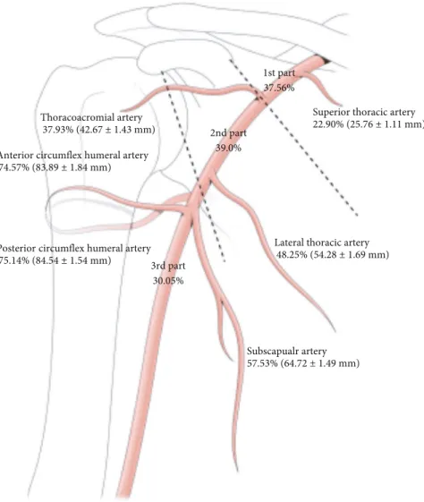
3.1. The Origin and Length of the Axillary Artery. The average length of the axillary artery was found to be 112.50 mm (ref- erence line; median: 110.57 mm; range: 80 –150 mm). With respect to reference line, the length of the starting point of the second part of the axillary artery was 33.39% (median:
36.55%; mean length: 37:56 ± 1:28 mm). The length of the starting point of the third part of the axillary artery was
respectively. SSA, ACHA, and PCHA originated from 57.53% (median: 63.96%; 64:72 ± 1:49 mm), 74.57%
(median: 86.14%; 83:89 ± 1:84 mm), and 75.14% (median:
86.52%; 84:54 ± 1:54 mm), respectively (Figure 1).
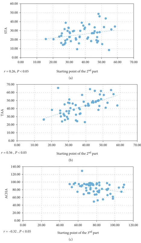
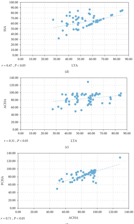
3.2. Correlation among Branches of the Axillary Artery. The correlation between origin points of branches and parts was analyzed. The length of the starting point of the second part of the axillary artery positively correlated with the origin of STA (r = 0:26, P < 0:05, Figure 2(a)) and TAA (r = 0:56, P < 0:05, Figure 2(b)). The length of the starting point of the third part of the axillary artery negatively correlated with ACHA ( r = −0:32, P < 0:05, Figure 2(c)). LTA distri- bution positively correlated with SSA ( r = 0:47, P < 0:05, Figure 2(d)) and AHCA ( r = 0:31, P < 0:05, Figure 2(e)).
There was a positive correlation between AHCA and PHCA (r = 0:71, P < 0:05, Figure 2(f)). Other branches did not have any correlation among themselves.
3.3. Branching Pattern of the Axillary Artery. We examined the branching patterns of the axillary artery to examine the relationship between the origin of the branching point and the distribution pattern. Considering every branch given o ff directly by the axillary artery, the branches were 3 –6 in num- ber. Typical variant of the axillary artery was found in 15 out of 59 cases (25.4%). The most frequent variant of branching pattern of the axillary artery involved 22 cases (37.3%) in which LTA and SSA branched together (Figure 3(a)), followed by a common branch for AHCA and PHCA in 19 cases (Figure 3(b)). One trunk for LTA and SSA and one trunk for ACHA and PCHA were found together in 12 cases. TAA and LTA were branched together in 9 cases (Figure 3(c)).
SSA, PHCA, and AHCA originated from a common trunk in 2 cases. TAA, LTA, and PHCA were branched together in one case. LTA, SSA, and PHCA were branched together in one case. TAA, LTA, and SSA were branched together in one case. SSA and PHCA were branched together in one case.
There was no statistically significant difference with respect to sex and left and right upper limbs.
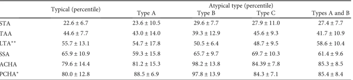
3.4. Topographical Changes in Branches according to the Branching Pattern of the Axillary Artery. Excluding rare pat- terns, the branching pattern was classi fied into 5 types: typi- cal, common branch of LTA and SSA (type A), common branch of ACHA and PCHA (type B), common branch of TAA and LTA (type C), and two common trunks for LTA/SSA and ACHA/PCHA (combined types A and B).
The origin of 6 branches was analyzed according to these
branching patterns (Table 1). Compared to typical pattern,
STA originated distally in atypical types; however, the differ-
ence was not significant (P = 0:330). LTA was originated
more proximally compared to other types ( P = 0:004). And
the origin of PCHA was more distal in atypical pattern, espe-
cially type B ( P = 0:021). Other variable did not have any
correlation.
4. Discussion
In this study, the length of the axillary artery as reference line was found to be 80–90 mm in 4, 90–100 mm in 15, 100–
110 mm in 9, 110–120 mm in 15, and >120 mm in 16 cases.
With respect to this reference line, the axillary artery was divided into three parts. The first, second, and third parts were 33.39%, 36.12%, and 30.49%, respectively. The average length of the origin of the six branches was also investigated.
Interestingly, SSA originated from 57.53%, and it included in the second part of the axillary artery. Most textbooks describe that SSA originates from the third part; however, we showed different results.
The anatomical variation of the axillary artery is clinically important because various blood vessels and nerves pass through it [15–17]. In particular, branches of the axillary artery are quite diverse from the bases, with two or three arteries branching together. These branches correlate with each other based on their origin. As expected, there was a positive correlation between AHCA and PHCA. The origin of LTA was positively correlated with that of SSA. Interest- ingly, the origin of PCHA was negatively associated with the length of the third part. The origin of the branches of
the axillary artery varied according to the branching patterns.
These data indicated hypothesis that these arteries develop simultaneously, and their branching patterns may not be random.
Nine types of branching patterns of the axillary artery were found. This result was comparable with those of previ- ous studies [5, 19, 20]. The patterns and origins of the axillary artery branches have been investigated, but studies about the correlation among these patterns and origins were lacking in other studies [3, 5, 6, 20 –22]. Remarkably, the origins of LTA and PCHA were signi ficantly different according to their branching patterns, suggesting that the morphology of the axillary artery during development in fluences the branching point. When LTA originated with TAA, its origin was more proximal, and when PCHA originated with ACHA, its origin was more distal. Until now, the arterial division was known as random. However, our results proposed hypothesis that the branching pattern and branching point are strongly asso- ciated and have unidentified regulation that one artery can correlate the position of another artery.
Unusual variants of the axillary artery branching or for- mation may also occasionally exist. For instance, the axillary artery may give rise to the radial artery of high origin (known
Superior thoracic artery 22.90% (25.76 ± 1.11 mm) Thoracoacromial artery
37.93% (42.67 ± 1.43 mm)
Lateral thoracic artery 48.25% (54.28 ± 1.69 mm)
Subscapualr artery 57.53% (64.72 ± 1.49 mm) Anterior circumflex humeral artery
74.57% (83.89 ± 1.84 mm)
Posterior circumflex humeral artery 75.14% (84.54 ± 1.54 mm)
1st part 37.56%
2nd part 39.0%
3rd part 30.05%
Figure 1: Topography of the axillary artery.
r = 0.26, P < 0.05 0.00 10.00 20.00 30.00
0.00 10.00 20.00 30.00 40.00 50.00 60.00 70.00
STA
Starting point of the 2nd part (a)
0.00 10.00 20.00 30.00 40.00 50.00 60.00 70.00
0.00 10.00 20.00 30.00 40.00 50.00 60.00 70.00
TAA
Starting point of the 2nd part r = 0.56 , P < 0.05
(b)
r = –0.32 , P < 0.05 0.00 20.00 40.00 60.00 80.00 100.00 120.00 140.00
0.00 20.00 40.00 60.00 80.00 100.00 120.00
ACHA
Starting point of the 3nd part (c)
Figure 2: Continued.
as brachioradial artery) or to the superficial ulnar artery [23–
25]. In our study, we did not find those rare variants. How- ever, knowledge of all unexpected deviations from the typical axillary artery branching pattern is fundamental from the clinical perspective. All variations have an embryological
background since upper limb vasculature develops from the primitive capillary plexus, which gives the potential to vari- ous blood flow pathways formation [20, 25].
Studies about the patterns and origins of the axillary artery branches and their correlation are important for
0.00 10.00 20.00 30.00 40.00 50.00 60.00 70.00 80.00 90.00 100.00
0.00 10.00 20.00 30.00 40.00 50.00 60.00 70.00 80.00 90.00
SSA
r = 0.47 , P < 0.05 LTA
(d)
0.00 20.00 40.00 60.00 80.00 100.00 120.00 140.00
0.00 10.00 20.00 30.00 40.00 50.00 60.00 70.00 80.00 90.00
ACHA
r = 0.31 , P < 0.05 LTA
(e)
0.00 20.00 40.00 60.00 80.00 100.00 120.00 140.00
0.00 20.00 40.00 60.00 80.00 100.00 120.00 140.00
PCHA
r = 0.71 , P < 0.05 ACHA
(f)
Figure 2: Correlation result. (a) Starting point of the 2
ndpart and the origin of superior thoracic artery (STA). (b) Starting point of the
2
ndpart and the origin of thoracoacromial artery (TTA). (c) Starting point of the 3
rdpart and the origin of anterior circum flex humeral
artery (ACHA). (d) The origin of lateral thoracic artery (LTA) and the origin of subscapular artery (SSA). (e) The origin of lateral
thoracic artery (LTA) and the origin of ACHA. (f) The origin of ACHA and the origin of posterior circum flex humeral artery (PCHA).
clinicians [4 –6]. Patients with severe trauma to the shoulder and upper chest should be clinically assessed for vascular damage and musculoskeletal evaluation [12 –17]. In addition, it will be helpful to recognize and understand such a varia- tion, even in radiographic procedures, to increase the accu- racy of the technique and to reduce unnecessary complications [26, 27]. Based on an accurate understanding of the axillary artery branching, as in this study, understand- ing the branching patterns and local anatomical variations
may be helpful in the accurate assessment and proper man- agement of the injured area [28].
This study investigated the patterns and variations of the axillary artery to provide useful information to clinicians especially dealing with the axillary region in the case of reconstructive surgery. Our research involved the axillary artery exclusively. We did not perform a whole-body study of anatomical variations. Kahn et al. [29] suggest that a whole-body study of arterial variants in a single anatomical
TAA
LTA
SSA ACHA
PCHA
(a)
TAA LTA SSA
ACHA PCHA
(b)
STA TAA LTA
SSA
ACHA
PCHA Type C: TAA+LTA
(c)
Figure 3: Frequent branching pattern of the axillary artery. (a) Type A: common origin for lateral thoracic artery (LTA) and subscapular artery (SSA). (b) Type B: common origin for anterior circum flex humeral artery (ACHA) and posterior circumflex humeral artery (PCHA). (c) Type C: common origin for thoracoacomial artery (TAA) and LTA.
Table 1: The origin of the branches of the axillary artery according to the branching pattern.
Typical (percentile) Atypical type (percentile)
Type A Type B Type C Types A and B
STA 22:6 ± 6:7 23:6 ± 10:5 29:6 ± 7:7 27:9 ± 11:0 27:4 ± 7:7
TAA 44:6 ± 7:7 43:0 ± 14:0 39:3 ± 12:9 45:6 ± 9:3 41:7 ± 10:9
LTA
∗∗55:7 ± 13:1 54:7 ± 17:8 50:5 ± 6:4 48:7 ± 9:5 58:6 ± 10:4
SSA 65:9 ± 10:9 59:3 ± 15:8 65:7 ± 9:7 69:7 ± 10:3 61:4 ± 9:6
ACHA 79:6 ± 14:4 81:2 ± 15:3 98:2 ± 13:8 84:39 ± 7:8 85:3 ± 8:5
PCHA
∗80:0 ± 12:8 88:5 ± 6:9 97:8 ± 13:9 84:3 ± 7:1 85:4 ± 8:4
∗P < 0:05,∗∗P < 0:01.