91 https://e-kcj.org
A 66-year-old man who had a history of hypertension and diabetes mellitus presented with severe chest pain and shortness of breath during exercise for 2 weeks. His electrocardiogram showed inverted T waves on the precordial leads, V1–V5, and his cardiac troponin I level was elevated. Coronary angiography revealed a significant stenosis at the ostium of the left main coronary artery (LMCA) (Figure 1A). Intravascular ultrasound (IVUS) showed severe fusiform narrowing of the LMCA lumen without significant atherosclerotic plaque (Figure 1B). Chest computed tomographic angiogram (CTA) showed a 10×11 cm huge aortic arch aneurysm with severe compression of the right main pulmonary artery (PA), left main PA, left main-stem bronchus, as well as the LMCA (Figure 1C and D).
The patient underwent an ascending and aortic arch graft interposition with a 28 mm 4 branch hemashield during heart lung bypass. The cardiac surgeons visually confirmed that the left main artery had no internal stenosis after excision of the huge aortic aneurysm (Figure 2). One-month follow-up angiogram, IVUS, and chest CTA showed a normal-looking LMCA (Figure 3A and B) and focal aortic dilatation at juxta distal to the graft interposition site and focal stenosis in the proximal left PA without evidence of endoleak (Figure 3C-E).
Angiographic assessment of a LMCA stenosis is often difficult and unreliable because of their unique anatomical structures and variable stenosis etiologies.1) Both IVUS and 3-dimensional computed tomography can play an important role in achieving an accurate and rapid diagnosis. Several cases of LMCA compression associated with aneurysm of the sinus Valsalva or ascending aorta have been reported previously,2-4) but aortic arch aneurysm is extremely rare. We describe an uncommon case that presented with an acute coronary syndrome caused by extrinsic compression of the LMCA by a huge aortic arch aneurysm.
Proper surgical correction of the LMCA compression restored the LMCA configuration which was documented by IVUS.
Korean Circ J. 2018 Jan;48(1):91-93 https://doi.org/10.4070/kcj.2017.0313 pISSN 1738-5520·eISSN 1738-5555
Images in
Cardiovascular Medicine
Jeoung-Sook Shin, MD1, So-Yeon Choi , MD, PhD1, Sang Hyun Lim, MD, PhD2, Jun Ho Jung, MD, PhD2, Doo-Kyoung Kang, MD, PhD3,
and Seung-Jea Tahk , MD, PhD1
1Department of Cardiology, Ajou University School of Medicine, Suwon, Korea
2Department of Cardiac Surgery, Ajou University School of Medicine, Suwon, Korea
3Department of Radiology, Ajou University School of Medicine, Suwon, Korea
Extrinsic Compression of the Left Main Coronary Artery by a Huge Aortic Arch Aneurysm Mimicking Acute Coronary Syndrome
Received: Oct 11, 2017 Accepted: Nov 06, 2017 Correspondence to So-Yeon Choi, MD, PhD
Department of Cardiology, Ajou University School of Medicine, 164, World cup-ro, Yeongtong-gu, Suwon 16499, Korea.
E-mail: [email protected] Copyright © 2018. The Korean Society of Cardiology
This is an Open Access article distributed under the terms of the Creative Commons Attribution Non-Commercial License (https://
creativecommons.org/licenses/by-nc/4.0) which permits unrestricted noncommercial use, distribution, and reproduction in any medium, provided the original work is properly cited.
ORCID iDs So-Yeon Choi
https://orcid.org/0000-0001-8299-7892 Seung-Jea Tahk
https://orcid.org/0000-0001-6521-178X Conflict of Interest
The authors have no financial conflicts of interest.
Author Contributions
Conceptualization: Choi SY; Data curation:
Shin JS, Choi SY, Lim SH, Jung JH; Formal analysis: Choi SY, Kang DK; Methodology:
Choi SY, Lim SH; Project administration: Choi SY; Resources: Tahk SJ; Supervision: Tahk SJ; Visualization: Choi SY, Jung JH, Kang DK; Writing - original draft: Shin JS, Choi SY;
Writing - review & editing: Choi SY.
92 https://e-kcj.org https://doi.org/10.4070/kcj.2017.0313
Compressed Left Main due to Aortic Aneurysm
A B
* *
Aorta LMCA Left PA Right PA
Bronchus
C D
Figure 1. Findings of coronary angiography, IVUS and chest CTA. (A) Coronary angiography showing a bird beak-like significant narrowing of the ostium of the LMCA (yellow bold arrow). (B) IVUS demonstrated that the lumen of the LMCA ostium was deformed as a fusiform narrowing with mild atherosclerotic plaque surrounded by decreased echo-signals behind the vessel suspected of extrinsic compression (red arrows). (C, D) Immediate chest CTA revealed a huge 10×11 cm sized aortic arch aneurysm containing a mural thrombus (red asterisk) severely compressed right PA, left PA, left main-stem bronchus, and LMCA.
CTA = computed tomographic angiogram; IVUS = intravascular ultrasound; LMCA = left main coronary artery; PA = pulmonary artery.
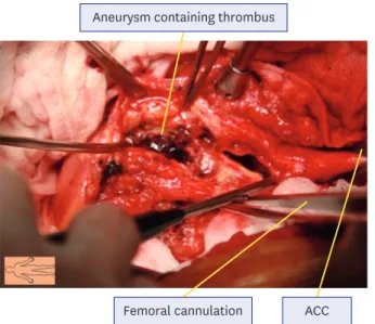
Aneurysm containing thrombus
ACC Femoral cannulation
Figure 2. Surgical finding. Image taken during surgery revealing the giant aortic aneurysm containing a red thrombus.
ACC = aortic cross clamp.
REFERENCES
1. Mintz GS, Painter JA, Pichard AD, et al. Atherosclerosis in angiographically “normal” coronary artery reference segments: an intravascular ultrasound study with clinical correlations. J Am Coll Cardiol 1995;25:1479-85.
PUBMED | CROSSREF
2. Borrello B, Nicolini F, Beghi C, Gherli T. Saccular ascending aorta aneurysm: report of an unusual presentation. Interact Cardiovasc Thorac Surg 2008;7:508-9.
PUBMED | CROSSREF
3. Goto K, Takebayashi H, Mukai S, et al. Acute myocardial infarction caused by left main coronary artery compression as a result of a mycotic aneurysm of the sinus of Valsalva. JACC Cardiovasc Interv 2015;8:e87-9.
PUBMED | CROSSREF
4. Dahya VJ, Chalasani P. Sinus of Valsalva aneurysm causing extrinsic compression of the left main coronary artery. JACC Cardiovasc Interv 2015;8:e99-100.
PUBMED | CROSSREF
93 https://e-kcj.org https://doi.org/10.4070/kcj.2017.0313
Compressed Left Main due to Aortic Aneurysm
Aorta LMCA Left PA Right PA
Bronchus
A B C
D E
Figure 3. Findings of coronary angiography, IVUS and chest CTA after 4-vessel graft interposition. (A) After ascending aortic arch excision and graft interposition, coronary angiogram shows normal-looking appearance of the left coronary artery ostium. (B) IVUS shows the LMCA lumen was patent with minimal
atherosclerotic plaque. (C-E) CTA shows significantly decreased size of aorta with focal dilatation, juxta distal to graft interposition site (4.6 cm) and patent lumens of both left and right PA with focal stenosis in proximal left PA. No definitive evidence of endoleak is shown in the graft interposition site.
CTA = computed tomographic angiogram; IVUS = intravascular ultrasound; LMCA = left main coronary artery; PA = pulmonary artery.