379 https://e-kcj.org
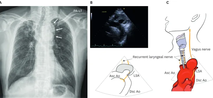
A 73-year-old man presented with new-onset hoarseness for 15 days. His chest X-ray showed an abnormally round shadow above the aortic knob (Figure 1A, arrows) and rightward deviation of the trachea. He underwent a routine echocardiographic examination. The echocardiographic examination showed normal left ventricular systolic function without valvular abnormalities. There was no valvular abnormality. During echocardiographic examinations, about a 55×42 mm sized oval-shaped mass lesion was noted on the aortic arch filled with an echogenic lesion (arrow heads, Figure 1B; Supplementary Video 1). To evaluate the mass lesion, the patient underwent contrast-enhanced computed tomography (CECT).
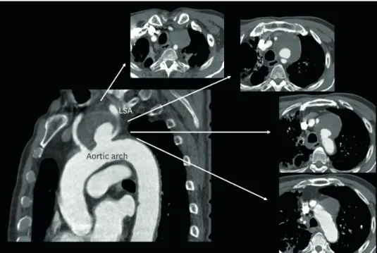
The CECT demonstrated about 65×60 mm sized large saccular aneurysm on the aortic arch filled with mural thrombus near the origin of left subclavian artery (Figure 2). The attending Korean Circ J. 2021 Apr;51(4):379-381
https://doi.org/10.4070/kcj.2020.0529 pISSN 1738-5520·eISSN 1738-5555
Images in
Cardiovascular Medicine
Received: Dec 14, 2020 Revised: Dec 31, 2020 Accepted: Jan 20, 2021 Correspondence to Jae-Hyeong Park, MD, PhD
Division of Cardiology, Department of Internal Medicine, Chungnam National University Hospital, School of Medicine, Chungnam National University, 282 Munhwa-ro, Jung-gu, Daejeon 35015, Korea.
E-mail: [email protected]
Jin-Ok Jeong , MD, PhD, Yun-Seon Park , RDCS, and Jae-Hyeong Park , MD, PhD
Division of Cardiology, Department of Internal Medicine, Chungnam National University Hospital, Chungnam National University College of Medicine, Daejeon, Korea
Ortner's Syndrome Discovered by a
Routine Echocardiographic Examination:
a Huge Aneurysmal Dilatation of the Aortic Arch as a Cause of Hoarseness
B C
A
PA-LT
LSA LSA
Asc Ao Asc Ao
Vagus nerve
Dsc Ao
Dsc Ao Recurrent laryngeal nerve
Figure 1. Chest X-ray shows an abnormally round shadow above the aortic knob (A, arrows) and rightward deviation of the trachea. Echocardiographic examination reveals about 55×42 mm sized oval-shaped mass lesion was noted on the aortic arch filled with an echogenic lesion suggesting thrombus (B, arrow heads). Illustration shows aneurysmal dilatation on the aortic arch with compression of the left recurrent laryngeal nerve (C).
Asc Ao = ascending aorta; LSA = left subclavian artery; Dsc Ao = descending aorta.
Copyright © 2021. The Korean Society of Cardiology
This is an Open Access article distributed under the terms of the Creative Commons Attribution Non-Commercial License (https://
creativecommons.org/licenses/by-nc/4.0) which permits unrestricted noncommercial use, distribution, and reproduction in any medium, provided the original work is properly cited.
ORCID iDs Jin-Ok Jeong
https://orcid.org/0000-0003-0763-4754 Yun-Seon Park
https://orcid.org/0000-0003-0055-695X Jae-Hyeong Park
https://orcid.org/0000-0001-7035-286X Funding
The authors received no financial support for the research, authorship, and/or publication of this article.
Conflict of Interest
The authors have no financial conflicts of interest.
Author Contributions
Data curation: Jeong JO, Park YS, Park JH;
Writing - original draft: Park JH; Writing - review & editing: Park JH.
physician treated the patient with a hybrid repair including the transfer of the branch arteries and thoracic endovascular aortic repair.
Ortner's syndrome is a rare cause of hoarseness due to palsy of the recurrent laryngeal nerve.
1)It is originally described by Ortner, and he described a case with left recurrent laryngeal nerve palsy caused by left atrial dilatation caused by mitral stenosis. This term is now used to describe recurrent laryngeal nerve palsy from cardiovascular causes. In our case, hoarseness came from the palsy of the left recurrent laryngeal nerve by the saccular aneurysm (Figure 1C, illustration). The aortic arch aneurysm can be treated by total arch replacement surgically or hybrid repair including debranching operation with a thoracic endovascular stent graft.
2)3)This patient underwent a hybrid repair successfully without any complication.
ACKNOWLEDGEMENTS
We would like to give special thanks to Sung-Won Park for her drawing a wonderful illustration.
SUPPLEMENTARY MATERIAL
Supplementary Video 1
Echocardiographic examination reveals about 55 × 42 mm sized oval-shaped mass lesion was noted on the aortic arch filled with an echogenic lesion suggesting thrombus.
Click here to view
380 https://e-kcj.org https://doi.org/10.4070/kcj.2020.0529
Aortic Arch Aneurysm Causing Hoarseness
LSA
Aortic arch
Figure 2. Contrast-enahced computerized tomography demonstrates about 65×60 mm sized large saccular aneurysm on the aortic arch filled with mural thrombus near the origin of left subclavian artery.
LSA = left subclavian artery.
REFERENCES
1. Kheok SW, Salkade PR, Bangaragiri A, Koh NS, Chen RC. Cardiovascular hoarseness (Ortner's syndrome): a pictorial review. Curr Probl Diagn Radiol 2020:S0363-0188(20)30190-0.
PUBMED | CROSSREF
2. Rommens KL, Estrera AL. Contemporary management of aortic arch aneurysm. Semin Thorac Cardiovasc Surg 2019;31:697-702.
PUBMED | CROSSREF
3. Choi BK, Lee HC, Lee HW, et al. Successful treatment of a ruptured aortic arch aneurysm using a hybrid procedure. Korean Circ J 2011;41:469-73.
PUBMED | CROSSREF