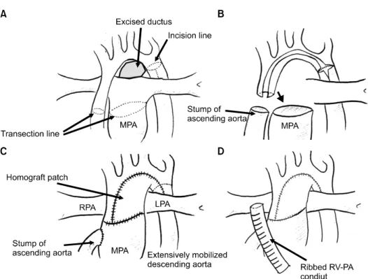
• 검색 결과가 없습니다.

Translocation of the Aortic Arch with Norwood Procedure for Hypoplastic Left Heart Syndrome Variant with Circumflex Retroesophageal Aortic Arch

N/A
N/A
Protected

Academic year: 2021

Share "Translocation of the Aortic Arch with Norwood Procedure for Hypoplastic Left Heart Syndrome Variant with Circumflex Retroesophageal Aortic Arch"

Copied!
5
0
0

로드 중.... (전체 텍스트 보기)

전체 글

(1)

Departments of

1

Thoracic and Cardiovascular Surgery and

2

Radiology, Asan Medical Center, University of Ulsan College of Medicine Received: October 24, 2013, Revised: December 6, 2013, Accepted: December 13, 2013, Published online: August 5, 2014

Corresponding author: Jeong-Jun Park, Department of Thoracic and Cardiovascular Surgery, Asan Medical Center, University of Ulsan College of Medicine, 88 Olympic-ro 43-gil, Songpa-gu, Seoul 138-736, Korea

(Tel) 82-2-3010-3587 (Fax) 82-2-3010-6811 (E-mail) [email protected]

C

The Korean Society for Thoracic and Cardiovascular Surgery. 2014. All right reserved.

CC

This is an open access article distributed under the terms of the Creative Commons Attribution Non-Commercial License (http://creative- commons.org/licenses/by-nc/3.0) which permits unrestricted non-commercial use, distribution, and reproduction in any medium, provided the original work is properly cited.

Retroesophageal aortic arch, in which the aortic arch crosses the midline behind the esophagus to the contralateral side, is a rare form of vascular anomaly. The complete form may cause symptoms by compressing the esophagus or the trachea and need a surgical intervention. We report a rare case of a hypoplastic left heart syndrome var- iant with the left retroesophageal circumflex aortic arch in which the left aortic arch, retroesophageal circumflex aor- ta, and the right descending aorta with the aberrant right subclavian artery encircle the esophagus completely, thus causing central bronchial compression. Bilateral pulmonary artery banding and subsequent modified Norwood proce- dure with extensive mobilization and creation of the neo-aorta were performed. As a result of the successful trans- location of the aorta, the airway compression was relieved. The patient underwent the second-stage operation and is doing well currently.

Key words: 1. Airway

2. Congenital heart disease 3. Hypoplastic left heart syndrome 4. Norwood procedures

5. Retroesophageal circumflex aortic arch

CASE REPORT

A 3.03-kg female neonate prenatally diagnosed with a hy- poplastic left heart syndrome (HLHS) variant, which includes mitral atresia, hypoplastic aortic arch, and muscular ven- tricular septal defect without left ventricular forward flow, was delivered at 39 weeks of gestational age. Initially, a prostaglandin E-1 (PGE-1) infusion was begun immediately, and controlled ventilation was performed to prevent excessive pulmonary blood flow while waiting for an operation. The two-dimensional echocardiography revealed situs solitus, levo-

cardia, and left aortic arch, which mainly originated from the

left ventricle (LV) without a left ventricular outflow tract ob-

struction despite unrestrictive combined perimembranous and

apical muscular ventricular septal defect (VSD) with aortic

overriding. Mitral atresia with LV hypoplasia and a relatively

normal-sized (6.04 mm, z-score: -0.8) ascending aorta with

normal coronary arteries were also detected by echocardio-

graphy. Cardiac computed tomography (CT) revealed tubular

hypoplasia of the aortic arch with a circumflex retro-

esophageal right descending aorta and an aberrant right sub-

clavian artery, which encircled the central airway completely

(2)

Fig. 1. Preoperative chest computed tomography findings. (A) Configura- tion of the aortic arch; (a) aberrant right subclavian artery, (b) left sub- clavian artery, (c) left common car- otid artery, (d) right common carotid artery, and (e) hypoplastic arch. (B) Retroesophageal circumflex aortic arch (red dotted line) crosses the midline behind the trachea, causing airway compression. (C) Airway com- pression by the retroesophageal cir- cumflex arch (red circle); the de- scending aorta crosses the midline behind the left main bronchus. PDA, patent ductus arteriosus.

and thereby led to a marked narrowing of the left main bron- chus by mechanical compression (Fig. 1). Against these le- sions, temporary bilateral pulmonary artery banding (PAB) was performed at 16 days of life. During bilateral PAB, frag- ments of a 3.5-mm Gore-tex graft were secured around the bilateral branch pulmonary arteries. The increase in the sys- temic blood pressure and the peripheral oxygen saturation un- der room air ventilation after PAB were 20 mmHg and 75%

to 80%, respectively. On echocardiography, which was per- formed on postoperative day 2, the internal diameters of the right pulmonary artery and the left pulmonary artery were 2.5 mm and 2.2 mm, respectively. PGE-1 infusion was main- tained until the Norwood procedure was performed. At 30 days of life, a subsequent modified Norwood operation was performed. The operation involved arch repair under selective cerebral perfusion, a 5-mm external ringed conduit from the right ventricle (RV) to the right pulmonary artery (PA) with the ‘Dunk technique’ [1], and atrial septectomy. Before arch reconstruction, the ascending aorta, aortic arch, aortic arch

vessels, and descending aorta were extensively mobilized,

particularly around the descending aorta. A single arterial

cannula was inserted into the right common carotid artery via

the 3.5-mm Gore-tex vascular graft. In the venous drainage,

double venous cannulation via right atrial appendage and in-

ferior vena cava was used. Considering the abnormality of

the arch vessels (aberrant right subclavian artery), the arterial

blood pressure during the selective cerebral perfusion was

monitored via a right temporal artery catheterization. The

schema of the operative procedures is depicted in Fig. 2. The

cardiopulmonary bypass time, aorta clamping time, selective

cerebral perfusion time, and the lowest rectal temperature

were 237 minutes, 89 minutes, 37 minutes, and 24.4

o

C,

respectively. On the intraoperative bronchoscopy, the left

main bronchial stenosis was relieved. Delayed sternal closure

was performed on postoperative day 2. The patient was ex-

tubated on postoperative day 5 and was discharged on post-

operative day 16. A postoperative CT, which was carried out

on postoperative day 8, showed the leftward translocation of

(3)

Fig. 2. Schema of the operation. (A) The left lateral side of the aortic arch is incised beginning at the divided aortic isthmus and extend- ing proximally down to the transected ascending aorta. Further, the MPA was transected. (B) To obtain more space for the airway and the pulmonary vasculature, the repaired arch and the neo-aorta had to translocate leftward as much as possible. Therefore, (C) the as- cending aorta was transected, and the proximal stump of the ascending aorta was anastomosed to the right lateral wall of the MPA. A pulmonary homograft patch was used for the completion of the arch reconstruction and the creation of the neo-aorta. (D) Then, a 5-mm external ringed conduit from the right ventricle (RV) to the right pulmonary artery (PA) with the “Dunk technique”. MPA, main pulmonary artery; RPA, right pulmonary artery; LPA, left pulmonary artery.

the descending aorta that relieved the airway compression (Fig. 3). The patient underwent successful bidirectional cavo-pulmonary shunt at 6 months of age and is now waiting for the Fontan operation.

DISCUSSION

The vascular ring designates the abnormalities caused by a developmental failure of the paired aortic arches during em- bryonic life; these can present with the compression of the adjacent structures, including the central airway, pulmonary arteries, and the esophagus [2,3]. According to the vascular structures of the vascular ring, they have been classified into the following five distinct types: 1) double aortic arch, 2) right aortic arch with left ligamentum, 3) left aortic arch with aberrant right subclavian artery, 4) pulmonary artery sling, and 5) innominate artery compression [2,3]. Unlike these

classifications, the retroesophageal circumflex aortic arch, in which the aortic arch crosses the midline behind the esoph- agus to the contralateral side, is a rare form. Furthermore, the condition of an HLHS variant combined with a vascular ring is extremely rare.

The retroesophageal circumflex aortic arch can cause sev- eral symptoms by compressing the esophagus and the central airway. Even if a concomitant intracardiac anomaly does not present, the treatment of the vascular anomaly can be chal- lenging because excessive tension on the aorta caused by the surgical treatment can constrict the adjacent structures.

Therefore, a compromise of the central airway is a major concern of the surgical treatment of the vascular anomaly.

There is a report on the retroesophageal circumflex aortic

arch, which was combined with the coarctation of aorta

(CoA) and VSD [4]. The authors performed extensive dis-

section around the aorta, including the arch vessels for the re-

(4)

Fig. 3. Postoperative chest compu- ted tomography findings and changes in the location of the descending aorta. (A) Transfer of the native aor- ta to the neo-aorta (red circle);

ring-enforced right ventricle to the pulmonary artery conduit (black ar- row). (B) Relief of airway compre- ssion. (C) Retroesophageal circumflex aortic arch crosses the midline, preoperatively. The white dotted line depicts the virtual line along the trachea. (D) After the operation, the descending aorta was located on the left side of the virtual line along the trachea.

pair of CoA and arch reconstruction by using the trans- location of the descending aorta. In the presented case, how- ever, the HLHS variant was combined with the retro- esophageal circumflex aortic arch, and thus, we had to per- form the Norwood procedure concomitantly. Therefore, we performed the modified Norwood procedure, including the translocation of the aorta combined with the full mobilization of the ascending aorta, aortic arch, arch vessels, and the de- scending aorta to relieve the airway compression.

To date, there have been three options for the Norwood procedure. The first option is the Norwood procedure with a Blalock-Taussig shunt or the RV to PA conduit. The second option is the ‘rapid two-stage Norwood procedure,’ which consists of temporary bilateral PAB and the maintenance of the ductus arteriosus by a ductal stent or PGE-1 infusion with a subsequent Norwood procedure. Temporary bilateral PAB has been performed for high-risk patients as an initial pallia- tion of HLHS until the second-stage operation is undertaken [5,6]. The third option is a ‘hybrid Norwood procedure’ of

bilateral PAB and ductal stenting. In the presented case, to minimize the perioperative risks of a Norwood operation by the stabilization of the pulmonary vasculature, we initially performed bilateral PAB with the maintenance of the PGE-1 infusion. With respect to the type of systemic-to-pulmonary shunt, as numerous previous studies have been reported, we selected the RV to right PA conduit, which was implanted with the ‘Dunk technique’ as the source of the pulmonary blood flow [1,5,7].

Because of the combined HLHS, the main presentations of

our patient were congestive heart failure and excessive pul-

monary blood flow rather than the typical symptoms of the

vascular ring, which involve respiratory symptoms and

dysphagia. Although the patient did not present airway symp-

toms, a markedly narrowed left main bronchus can cause sig-

nificant airway problems, postoperatively. Therefore, efforts

for the achievement of hemodynamic stability as well as the

prevention of postoperative airway compression should be

made during the operation. In this regard, to obtain adequate

(5)

and perioperative CT scans are essential to evaluate the rela- tionship of the airway and the surrounding vascular structures [8].

CONFLICT OF INTEREST

No potential conflict of interest relevant to this article was reported.

REFERENCES

1. Tweddell JS, Mitchell ME, Woods RK, Spray TL, Quinte- ssenza JA. Construction of the right ventricle-to-pulmonary artery conduit in the Norwood: the “Dunk” technique. Oper

4. Song J, Kim WH, Kim H, et al. Repair of coarctation in right circumflex retroesophageal arch. Ann Thorac Surg 2009;87:307-9.

5. Barron DJ. The Norwood procedure: in favor of the RV-PA conduit. Semin Thorac Cardiovasc Surg Pediatr Card Surg Annu 2013;16:52-8.

6. Guleserian KJ, Barker GM, Sharma MS, et al. Bilateral pul- monary artery banding for resuscitation in high-risk, sin- gle-ventricle neonates and infants: a single-center experi- ence. J Thorac Cardiovasc Surg 2013;145:206-13.

7. Ohye RG, Sleeper LA, Mahony L, et al. Comparison of shunt types in the Norwood procedure for single-ventricle lesions. N Engl J Med 2010;362:1980-92.

8. Jhang WK, Park JJ, Seo DM, Goo HW, Gwak M. Periope-

rative evaluation of airways in patients with arch obstruction

and intracardiac defects. Ann Thorac Surg 2008;85:1753-8.

수치

Fig. 1. Preoperative chest computed  tomography findings. (A)  Configura-tion of the aortic arch; (a) aberrant  right subclavian artery, (b) left  sub-clavian artery, (c) left common  car-otid artery, (d) right common carcar-otid  artery, and (e) hypoplast
Fig. 2. Schema of the operation. (A) The left lateral side of the aortic arch is incised beginning at the divided aortic isthmus and extend- extend-ing proximally down to the transected ascendextend-ing aorta
Fig. 3. Postoperative chest compu- compu-ted tomography findings and changes  in the location of the descending  aorta

참조

관련 문서

We report a rare case of hemoptysis from a fistula between an aortic arch aneurysm and the left main bronchus in a patient who had undergone an

Chest radiographic features of the DAA patients include a deviation or compression of the trachea, or identification of a right aortic arch contour. 15) We could see not only

DA: Descending Aorta, L: Left, VSD: Ventricular Septal Defect, (-): Not obtained, TA: Transverse Arch, located between left common carotid and subclavian arteries, *: Aortic

Chest computed tomographic angiogram (CTA) showed a 10×11 cm huge aortic arch aneurysm with severe compression of the right main pulmonary artery (PA), left main PA, left main-stem

tal aortic arch. Intentional coverage of the left subclavian artery carries a low risk according to clinical experiences, if stenoses and abnormalities of the supra-aortic

In the surgery of distal aortic arch and proximal descending aortic lesions through the left thoracotomy incision, right atrium-retrograde cerebral perfusion

Distal type of aortopulmonary septal defect with aortic origin of right pulmonary artery and interrupted aortic arch A case of successful surgical

This case is reported in order to inform the readers of a rare case where floating thrombi in the aortic arch of patients with embolization were successfully