© 2011 The Korean Academy of Medical Sciences.
This is an Open Access article distributed under the terms of the Creative Commons Attribution Non-Commercial License (http://creativecommons.org/licenses/by-nc/3.0) which permits unrestricted non-commercial use, distribution, and reproduction in any medium, provided the original work is properly cited.
pISSN 1011-8934 eISSN 1598-6357
Unusual Cause of Acute Right Ventricular Dysfunction: Rapid Progression of Superior Vena Cava Aneurysm Complicated by Thrombosis and Pulmonary Thromboembolism
Aneurysms of the major thoracic veins are rare. They are usually asymptomatic and thus treated conservatively. We report an extremely rare case of rapidly progressing superior vena cava (SVC) aneurysm complicated by thrombosis and acute pulmonary
thromboembolism (PTE) with right ventricular dysfunction. Thrombolytic therapy for hemodynamically significant acute PTE was harmful to the patient in the present case, because it induced further thrombosis and mobilization of the thrombi within the aneurysm, subsequently causing de novo PTE. Surgical aneurysmectomy combined with pulmonary artery embolectomy would be a treatment of choice in patients with SVC aneurysm complicated by acute PTE.
Key Words: Aneurysm; Vena Cava, Superior; Pulmonary Embolism Sang Gi Oh1, Kye Hun Kim2,
Hyun Ju Seon3, Hyun Ju Yoon2, Youngkeun Ahn2, Myung Ho Jeong2, Jeong Gwan Cho2, Jong Chun Park2, and Jung Chaee Kang2
Departments of 1Cardiothoracic Surgery,
2Cardiovascular Medicine, and 3Radiology, Chonnam National University Hospital, Gwangju, Korea Received: 23 July 2010
Accepted: 9 February 2011 Address for Correspondence:
Kye Hun Kim, MD
Director of Echocardiography Laboratory, The Heart Center of Chonnam National University Hospital, 671 Jebong-ro, Dong-gu, Gwangju 501-757, Korea
Tel: +82.62-220-6978, Fax: +82.62-227-4760 E-mail: [email protected]
DOI: 10.3346/jkms.2011.26.5.690 • J Korean Med Sci 2011; 26: 690-693
CASE REPORT
Cardiovascular Disorders
INTRODUCTION
Aneurysms of the major thoracic veins are rare. Because they are usually asymptomatic, they are diagnosed incidentally dur- ing the evaluation of mediastinal widening on chest roentgen- ography and treated conservatively (1-3). Development of com- plications such as rupture, venous obstruction, and thrombosis has been described and may require surgery (4-6). We report a 55-yr old male patient with superior vena cava (SVC) aneurysm which showed rapid progression and complicated by thrombo- sis and hemodynamically significant acute pulmonary throm- boembolism (PTE) with review of the literature.
CASE DESCRIPTION
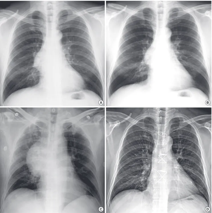
A 55-yr-old male presented with acute chest pain, dyspnea, and hypotension on December 8, 2009. The patient has been followed without any medications for incidentally detected asymptom- atic SVC aneurysm for last 2 yr (Fig. 1A), and the size of the SVC aneurysm did not change (Fig. 1B). Chest radiography at admis- sion showed marked widening of the right middle mediastinum due to abnormal soft-tissue density suggesting the rapid expan- sion of previously noted SVC aneurysm (Fig. 1C). Echocardiog- raphy revealed marked enlargement and hypokinesia of the right ventricle (RV) with moderate pulmonary hypertension suggest-
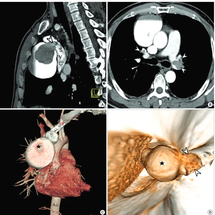
ing acute PE. Chest computed tomographic angiography (CTA) showed about 6.9 × 6.1 × 9.9 cm sized lobulated giant saccular aneurysm originating from mainly SVC and partially involving left brachiocephalic vein with internal thrombi (Fig. 2A). Con- siderable amounts of thromboembolic filling defects in left pul- monary artery, both left and right lobar, segmental, and subseg- mental arteries were also noted (Fig. 2B). Three-dimensional reconstruction with endovascular views showed the giant sac- cular SVC aneurysm with 4.1 × 3.7 cm sized internal thrombi, and the thrombi protruded into the SVC through the narrow an- eurysmal neck (Fig. 2C, D). Venous CTA of the lower extremities and abdominal CTA showed no evidence of deep vein throm- bosis or other venous aneurysms. To exclude the possible hy- percoagulable status as a cause of pulmonary thromboembo- lism, the laboratory tests for hypercoagulability was done and revealed no abnormalities. Surgical exclusion of SVC aneurysm with pulmonary embolectomy was planned, but the patient and family refused surgery. Therefore, thrombolytic therapy using recombinant tissue plasminogen activator with the dose of 200 mg was administered intravenously for 2 hr for hemodynami- cally significant PTE, and the RV function and pulmonary artery pressure was normalized on FU echocardiography. However, the patient complained recurrent chest pain and dyspnea on the next day. FU CTA revealed more decreased amounts of thrombi in the left pulmonary artery and its branches, but the amounts
Oh SG, et al. • Venous Aneurysm and Pulmonary Thromboembolism
http://jkms.org 691
DOI: 10.3346/jkms.2011.26.5.690
of thrombi in the SVC aneurysm, SVC, right pulmonary artery and its branches were increased. Mobilization of thrombi from the SVC aneurysm due to thrombolytic therapy was the possi- ble cause of recurrent PTE, and it was expected that further em- bolization of thrombi would be fatal to the patient. Therefore, surgical treatment was strongly recommended. Resection of SVC aneurysm and pulmonary artery embolectomy were performed successfully. At surgery, femoral arterial and venous cannula- tion was done to prepare for accidental rupture of aneurysm during sternotomy. After dissection of the aneurysm, the other
venous cannulation at the innominate vein and isolation of the proximal and distal portion of the aneurysm was done. Under cardiopulmonary bypass, aneurysmal wall was opened longi- tudinally after snaring of proximal and distal portion of the an- eurysm and innominate vein. Aenurysm which contained fresh thrombi was thin-walled and had relatively normal venous en- dothelium grossly (Fig. 3A), and the histopathologic examina- tion showed compatible findings of true aneurysm (Fig. 3B).
Thin-walled aneurysmal wall was completely resected and sub- sequent reconstruction of the superior vena cava was performed
A B
C D
Fig. 1. Serial radiographic changes of the superior vena cava (SVC) aneurysm. Chest radiography at 2 yr ago (A) and 1 month (B) ago revealed the SVC aneurysm without any changes in size. The SVC aneurysm was markedly increased in size on chest radiography at admission (C) and disappeared after surgery (D).
Oh SG, et al. • Venous Aneurysm and Pulmonary Thromboembolism
692 http://jkms.org DOI: 10.3346/jkms.2011.26.5.690
A B
C D
Fig. 2. Multiplanar reformat images (A, B) and three-dimensional volume rendered images (C, D) of chest computed tomographic angiography demonstrated the superior vena cava (SVC) aneurysm with internal thrombi (★) protruding into the SVC through the narrow neck (arrow head in A and D) and pulmonary arterial embolism (arrow heads in B).
with glutaraldehyde fixed autologuous pericardium that was already harvested. Outer surface of the pericardium was but- tressed with Dacron to prevent potential formation of pseudoa- neurysm. A transverse arteriotomy was made in the pulmonary trunk to the left main pulmonary artery and another incision was made in the right main pulmonary artery, and the fresh and organized thrombi were gently extracted. Following the success- ful surgical management, the mediastinal widening on chest ra- diography was normalized (Fig. 1D) and the patient was recov- ered without any postoperative events. The patient was discharg-
ed and treated by anticoagulation with warfarin for 6 months to achieve complete resolution of the possible remained pulmo- nary arterial thrombi.
DISCUSSION
Aneurysms of the major thoracic veins are rare and usually as- ymptomatic, even though several complications including an- eurysmal rupture, thrombosis, or venous obstructions have been reported (4-6). However, the development of acute PTE associ-
Oh SG, et al. • Venous Aneurysm and Pulmonary Thromboembolism
http://jkms.org 693
DOI: 10.3346/jkms.2011.26.5.690
A B
Fig. 3. A giant saccular aneurysm with internal thrombi formation arising from the superior vena cava was shown on surgical fields (A). On histopathologic examination, the aneurysmal wall contained all three layers of vascular wall, indicating true aneurysm (B).
ated with thrombosed venous aneurysms, like the present case, is extremely rare. There had been only 2 cases of acute PTE caused by thrombosis of the major intrathoracic venous aneurysms in the literature (7, 8).
The present case gives several important messages in the treat- ment of asymptomatic or complicated intrathoracic venous an- eurysms. First, although the SVC aneurysm did not produce any kind of symptoms or size change for 2 yr, the present case showed sudden rapid growing of the aneurysms with internal thrombus formation. Therefore, the benefit of surgical therapy to prevent the developments of fatal complications should be considered in saccular SVC aneurysm at the time of diagnosis, even in as- ymptomatic cases. In case of fusiform SVC aneurysm, conser- vative management is recommended in the general consensus.
Second, long-term anticoagulation with warfarinization might be considered to prevent aneurysmal thrombosis and subse- quent PTE, especially in a fusiform aneurysm. In case of saccu- lar aneurysm, however, rupture of the aneurysm is the major complication in addition to thromboembolism. In case of rup- ture during anticoagulation, it can be a disaster. Therefore, the benefit of anticoagulation should be counter-balanced by the risk of rupture. Third, surgical aneurysmectomy and pulmonary artery embolectomy would be a treatment of choice in patients with SVC aneurysm complicated by thrombosis and massive PE as discussed in the previous reports (4-8).
In conclusion, we report a very rare case of SVC aneurysm which showed rapid expansion and complicated by thrombo- sis. The acute RV dysfunction may be caused by massive PTE from the thrombosed SVC aneurysm and improved by surgical aneurysmectomy and embolectomy.
REFERENCES
1. Schatz IJ, Fine G. Venous aneurysms. N Engl J Med 1962; 266: 1310-2.
2. Rappaport DC, Ros PR, Moser RP Jr. Idiopathic dilatation of the thorac- ic venous system. Can Assoc Radiol J 1992; 43: 385-7.
3. Koga S, Ikeda S, Sanuki Y, Ninomiya A, Izumikawa T, Miyahara Y, Kohno S. A case of asymptomatic fusiform aneurysm of the superior vena cava detected by magnetic resonance imaging. Int J Cardiol 2006; 113: e39-41.
4. Taira A, Akita H. Ruptured venous aneurysm of the persistent left superi- or vena cava. Angiology 1981; 32: 656-9.
5. Pasic M, Schöpke W, Vogt P, von Segesser L, Schneider J, Turina M. An- eurysm of the superior mediastinal veins. J Vasc Surg 1995; 21: 505-9.
6. Gomez MA, Delhommais A, Presicci PF, Besson M, Roger R, Alison D.
Partial thrombosis of an idiopathic azygos vein aneurysm. Br J Radiol 2004; 77: 342-3.
7. Ream CR, Giardina A. Congenital superior vena cava aneurysm with com- plications caused by infectious mononucleosis. Chest 1972; 62: 755-7.
8. Nakamura Y, Nakano K, Nakatani H, Fukuda T, Honda K, Homma N.
Surgical exclusion of a thrombosed azygos vein aneurysm causing pul- monary embolism. J Thorac Cardiovasc Surg 2007; 133: 834-5.