pISSN: 0378-6471 eISSN: 2092-9374
http://dx.doi.org/10.3341/jkos.2012.53.12.1864
= 증례보고 =
산소로 유도된 망막증 모델에서 트리암시놀론이 망막 혈관 관련 인자에 미치는 영향
유웅선1⋅서성욱1,2⋅박종문1,2⋅유지명1,2⋅정인영1,2 경상대학교 의학전문대학원 안과학교실1, 경상대학교 건강과학연구원2
목적: 트리암시놀론 아세토나이드(TA)의 Oxygen-induced retinopathy (OIR) 쥐 모델에서 혈관 관련 인자 및 혈관 누출에 대한 효과를 알아보고자 한다.
대상과 방법: Sprague-Dawley계 흰쥐를 생후 2일부터 14일까지 고농도 산소에 노출한 후 생후 15일부터 30일까지 정상산소에 노출시 켜 대조군과 비교하였다. TA (4 mg/kg) 2 μl를 생후 16일에 유리체내로 주입하였다. 망막 혈관 누출 현상은 Evans blue method를 이용하였고 RT-PCR을 이용하여 혈관관련인자들의 mRNA 변화를 보았다.
결과: OIR 모델 흰쥐는 상대적 저산소로 인한 혈관누출이 나타났으며 TA을 주입하였을 때 혈관 투과성이 대조군에 비해 현저히 감소하 였다. OIR 모델에서 HIF-1α, VEGF, SDF-1, ICAM-1의 mRNA 증가를 보였으며 TA 처리시 감소하였다. 또한 Occludin mRNA는 감소를 보였으며 TA 처리시 증가하였다.
결론: OIR 모델에서 TA처리시 망막혈관누출의 감소를 보이며 이는 TA가 혈관누출억제에 작용하는 것으로 생각한다. 또한 허혈망막증 에 증가하며 트리암시놀론 처리시 감소되는 것으로 이미 보고된 HIF-1α 및 VEGF 뿐만 아니라, SDF-1, ICAM-1, Occludin과 같은 망막혈관 관련 인자에도 트리암시놀론이 작용하여 망막혈관파괴에 방지 효과가 있을 것으로 생각하며 추후 각 인자들의 단백질 변화 를 통한 확인이 필요할 것이다.
<대한안과학회지 2012;53(12):1864-1869>
■ 접 수 일: 2010년 5월 10일 ■ 심사통과일: 2012년 7월 9일
■ 게재허가일: 2012년 10월 19일
■ 책 임 저 자: 정 인 영
경상남도 진주시 강남로 79 경상대학교병원 안과
Tel: 055-750-8170, Fax: 055-753-4158 E-mail: [email protected]
미숙아 망막증은 미숙아에서 발생하는 주된 안과 질환으 로 특히 1,000 g 이하의 출생 체중이거나 29주 이내의 미 숙아에서 잘 발생하며1주변부의 망막은 불완전한 혈액 공 급 및 신생혈관의 생성으로 상대적으로 저산소 상태가 된 다.2 미숙아 망막증에서 비정상적인 혈관형성은 유리체 출 혈이나 견인성 망막박리와 같은 위험한 결과를 초래할 수 있다. 망막 신생혈관 생성은 당뇨망막병증,3 나이관련황반 변성4등과 같은 여러 심각한 안과 질환에서 주된 기전으로 알려졌다. 눈에서 신생혈관 형성은 주로 혈관의 많은 누출 과 연관되고 이는 망막 형태의 손상을 일으킨다.
혈관내피세포형성인자(VEGF)가 증식성 당뇨병성 망막 병증에서 중요한 혈관생성 인자라는 사실은 잘 알려졌다.5-7 또한 HIF-1α (Hypoxia-inducible factor-1α)가 저산소 환경에서 VEGF의 생성을 증가시켜 망막혈관의 구조를 변화 시킨다고 알려졌으며,8-11 최근에는 SDF-1 (Stromal de-
rived factor-1),12,13 occludin,14,15 ICAM-1이나 E-se- lectin16-23의 혈관에 대한 영향에 대해서도 많은 연구가 이 루어지고 있다. 저자들은 선행연구에서 미숙아 망막증 모델 을 이용하여 트리암시놀론이 VEGF와 허혈반응 proin- flammation 유전자들의 억제에 작용함을 발표하였다.24
트리암시놀론 아세토나이드(Triamcinolone acetonide, TA)는 포도막염이나 망막혈관 질환에 따른 망막부종, 증식 당뇨망막병증, 나이관련황반변성에서 맥락막신생혈관을 장 기적으로 억제하는 효과적이고 안전한 스테로이드 제재이 다.25-30
저자들은 본 연구에서 TA의 oxygen-induced retinop- athy (OIR) 쥐 모델에서 혈관 누출 관련된 인자들 및 혈관 누출에 미치는 영향에 대해 알아보고자 한다.
대상과 방법
1. 실험동물
실험용 쥐(Samtako Co Ltd, Korea)는 임신한 암컷 Sprague-Dawley를 사용하였다. 모든 쥐는 각각 사육하였 으며, 실험실 내 불빛은 12시간 간격으로 등화되도록 하였
다. 모든 실험과정은 안과학 및 시기능 연구에서의 동물 사 용에 대한 ARVO (Association for Research in Vision and Ophthalmology) 규정을 준수하였다.
2. Oxygen-induced retinopathy (OIR) 모델 생성 및 트리암시놀론(TA)의 주입
새로 출생한 쥐는 이미 보고된 방법대로24 출생 후 어미 쥐로부터 격리하여 출생 후 2일(P2)부터 14일(P14)까지 고 농도의 산소(80 ±1.5%)에 22시간, 실내공기(21 ±1.3%
산소)에 2시간 노출시켰으며, 15일(P15)부터 30일(P30) 까지 실내공기에 노출시켰다. 대조군 쥐는 출생 후(P0)부 터 30일(P30)까지 실내공기에 두었다.
대조군 및 OIR 쥐 모두 마취 시킨 후 TA (Sigma, 4 mg/kg)를 현미경하에서 30게이지 주사를 이용하여 생후 16일(P16)에 유리체강 내 주입(2 μl/eye)하였다. 좌안은 대조군에서와 같이 같은 양의 평형염액을 주입하였다.
쥐를 생후 14일(P14), 16일(P16), 18일(P18), 22일 (P22), 26일(P26), 30일(P30) 군으로 나누어 희생시켰으 며 체중을 측정하였다. 각 군마다 7마리에서 10마리의 쥐 를 사용하였다.
3. Evans blue를 이용한 혈관 투과성의 측정
각 시점(P14, P16, P18, P20, P22, P26, P30)에서 망막 혈관 누출에 대한 트리암시놀론의 효과는 Evans-blue- albumin 누출 방법을 이용하여 정량 하였으며 이는 이미 발 표된 프로토콜을 수정 없이 따랐다.31 측두 동맥혈을 채취 하여 원심분리(20분 동안 4℃, 10,000 ×g) 후 포름아마이 이드 형태의 Evans blue의 표준곡선을 통해서 상층액의 염 색약의 농도를 계산하였다. 망막에서 추출된 염색약의 농도 는 포름아마이드 형태의 Evans blue의 표준 곡선에서 계산 한 후 혈장 내의 Evans blue의 농도를 건조된 망막무게 및 시간 평균의 농도를 이용하여 전체 단백질 mg당 발견된 Evans blue μg으로 표현하였다.
4. RNA 추출 및 RT-PCR (Reverse transcriptase polymerase chain reaction)
생후 16일된 control 및 생후 16일, 18일, 20일된 OIR 쥐 에서 망막RNA 추출 및 RT-PCR을 이미 보고된 방법대로 시행하였다.32HIF-1α, VEGF, SDF-1, occludin, ICAM-1 cDNA와 대조군으로 사용된 β-actin cDNA는 역전사효소 (reverse transcriptase, MMLV, 200U, Promega, Madison,
WI, USA)와 합성하였고, 각각의 시발체(primer)로 이용하 였다(HIF-1α : 5’-GGACAAGTCACCACAGGA-3’, 5’- GGAGTTTCAGAGGCAGGT-3’, VEGF : 5’-CACTGGAC CCTGGCTTT-3’, 5’-GCTGGCTTTGGTGAGGT-3’, SD F-1 : 5’-GAGAGCCACATCGCCAGAG-3’, 5’-TTTCG GGTCAATGCACACTTG-3’, occludin : 5′-CAGCCCAG TTCCTCCAGTAG-3′, 5′-GCTCATCCAGCTCTCTGTC C-3’, ICAM-1 : 5'-CAACTGGAAGCTGTTTGAGCTG -3', 5'-GTGTTCACAGTCTTGCTCCAT-3', β-actin : 5’-GGTGAAGATGACCCGACAGT-3’, 5’-GGTGAAGA TGACCCGACAGT-3’). 열순환기에서 94℃에서 2분, 60℃
에서 1분, 72℃에서 2분간 배양하였다. β-actin cDNA을 대조군으로 사용하였으며 증폭된 cDNA는 1.5% agarose 젤에 녹여 전기영동하였다.
5. 통계분석
밀도분석은 SigmaGel 1.0 (Jandel Scientific, Germany) 과SigmaPlot 4.0 (SPSS Inc., USA)을 이용하였으며 통계 분석은 Mann-Whitney U-test (SPSS Inc., USA)를 이용 하였으며 p<0.05를 통계학적 의미를 가지는 것으로 보았다.
결 과
1. OIR 쥐의 망막에서 혈관 투과성의 변화
대조군에서는 정상산소 분압 하에서(P14-P30) 망막의 혈관 투과성이 낮게 나타났으나 변화를 보이지 않았다 (p>0.1, n=7). 이는 정상 혈액-망막 장벽을 반영하는 것 으로 보인다. 그러나 OIR 쥐에서는 고농도의 산소분압 노 출 후(P2-P14) 정상 분압으로 돌아온 지 2일부터(P16) 망막의 혈관 투과성이 3.2배 증가하였다(p<0.05, n=7).
혈관 투과성은 P18에서 최고점을 보였으며(8.7배, p<
0.001, n=10), P18부터 P22까지 감소하는 양상(7.6배, p<0.05, n=7)을 보였으며 P30에서 가장 낮은 수치를 보였 으나 여전히 대조군에 비해 높은 수치(2.52배, p<0.05, n=7)를 보였다(Fig. 1).
2. OIR 쥐의 혈관 투과성에 대한 TA의 영향
P16에 TA을 유리체강 내 주입한 후 P14, P16, P18, P20, P22, P26, P30에서 혈관 투과성을 측정하였다. TA는 주입 후 2일 후(P18)에 가장 강력한 영향을 보였으며 1.8 배의 혈관 투과성 감소를 관찰할 수 있었다(p<0.001,
Figure 1. Time course of vascular permeability changes in the retina of OIR rats. Vascular permeability in the retina was meas- ured at P14, P16, P18, P20, P22, P26 and P30. The Evans blue in the tissues was normalized by total protein concentration and expressed as μg of dye per mg of protein in the tissue. Values are mean ± SD (n = 7 to 10). (*p < 0.05: compared with OIR and control group) (OIR: Oxygen-induced retinopathy).
Figure 2. Time-dependent effects of TA on vascular hyper- permeability in the retinas of OIR rats. At P16 the right eye re- ceived an intravitreal injection of TA (4 mg per eye) and the left eye received saline. Vascular permeability was measured 2 and 0, 2, 4, 6, 10 and 14 days before and after the injection.
The Evans blue in the tissues was normalized by total protein concentration and expressed as μg of dye per mg of protein in the tissue. Values are mean ± SD (n = 7 to 10). (*p < 0.05:
compared with TA- and saline-treated group) (TA: tri- amcinolone acetonide).
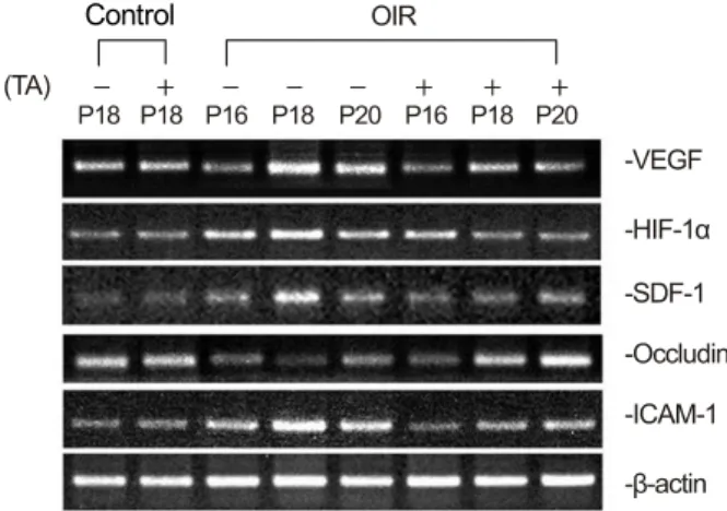
Figure 3. Expression of mRNAs analyzed by RT-PCR in the retina of both control and OIR rats and changes after TA treatment. This image is representative from six rats, showing PCR-amplified products of VEGF, HIF-1α, SDF-1, Occludin and ICAM-1, and β-actin as a loading control (TA: tri- amcinolone acetonide, OIR: oxygen-induced retinopathy).
n=10). TA의 혈관 누출에 대한 보호적인 효과는 주입 후 6일간 지속되었으며 주입 후 14일(P30)에 대조군과 유사 한 수준으로 돌아왔다(p>0.1, n=7)(Fig. 2).
3. OIR 쥐의 망막에서 TA 주입 후 혈관형성 관련 유 전자의 발현
RT-PCR 결과에서 HIF-1α, VEGF, SDF-1, ICAM-1
각각의 mRNA들이 대조군에 비해 OIR 쥐에서 P16부터 증 가하여 P18에서 가장 많이 발현되었다. 하지만 occludin은 감소하는 소견을 보였다. TA의 효과 판정에서 TA 주입 후 HIF-1α, VEGF, SDF-1, ICAM-1 각각의 mRNA발현이 OIR 쥐의 망막에서 TA를 주입하지 않은 쥐에 비하여 감소 되었으며 occludin은 증가하는 소견을 보였다(Fig. 3).
고 찰
OIR 모델은 미숙아 망막증에서 혈관-망막 장벽 파괴나 혈관 누출을 연구하는 모델로 주로 사용된다.33-36초기 합 병증으로 신생혈관의 혈성이나 황반부종이 생기며 이는 심 각한 시력 상실로도 이어질 수 있다. 최근 anti-VEGF를 이 용한 치료가 시도되고 있으나 아직까지 병리적 상태의 만 족할만한 회복을 유도하는 치료는 없는 상태이다.37-41
TA는 효과적인 혈관생성 억제제로 당뇨망막병증이나 연 령관련황반변성에서 신생혈관 형성의 억제 및 황반부종의 치료로 인간과 동물에서 사용되었다.42-46저자들은 이미 임 상에서 널리 사용되고 있는 트리암시놀론을 이용하여 허혈 성 망막증에서 혈관인자로서는 대표적인 VEGF와 여러 proinflammatory regulatory systems관련 유전자들의 변 화에 직접적인 억제작용이 있음을 확인하였고24 혈관병리 와 관련한 연구의 필요성을 가지게 되었다. 본 연구에서 저 자들은 OIR 쥐에서 TA가 망막의 혈관 관련 인자 및 혈관 누출에 보호적인 효과가 있는지 알아보고자 하였다.
TA의 유리체강 내 주입은 시간이 흐름에 따라 혈관 누출 을 효과적으로 방지하였으며 신생혈관 생성을 억제하였다.
Control
가장 흥미로운 점은 주입 후 6일까지 효과가 지속되었다는 점이다. 이전까지 보고된 연구에서는 endostatin이나 an- giostatin 등 강력한 혈관생성 억제 인자의 주입 후 약 2일 간만 혈관 과누출에 억제 효과를 강하게 보였다고 보고되
었다.47,48따라서 이번 연구 결과로 OIR 쥐의 망막 혈관 누
출 억제에 TA 유리체강 내 주입술이 효과적인 치료임을 알 수 있다.
이 연구에서 OIR을 형성하기 위해 P2부터 P14까지 고농 도 산소 분압(80%)에 노출시켰으며 P15부터 P30까지 정 상 산소 분압(실내공기)에 노출시켰다. 이 시기에 HIF-1α 나 VEGF의 발현이 증가됨을 통해 상대적 저산소 상태임을 알 수 있다. 저산소 상태에서 HIF-1α는 VEGF의 발현을 증가시키고 이는 직접적으로 망막 신생혈관 형성을 자극하 며 이들이 안내의 증식성 혈관 질환에서 망막 신생혈관 생 성 작용을 한다는 것은 널리 연구되었다.8-11이 연구에서도 역시 OIR 모델에서 HIF-1α과 VEGF가 상대적인 저산소 분압 상태에서 발현이 증가됨을 보여주었으며 특히 혈관 누출 및 혈관내피세포의 증식이 증가 되는 곳에 많이 발현 되었다. TA를 주입한 경우 이러한 HIF-1α나 VEGF의 변 화를 유발하였으며 이 결과는 상대적 저산소 농도에서 OIR 의 HIF-1α나 VEGF를 통한 신생혈관 형성에 TA가 보호적 인 역할을 알 수 있었다.
SDF-1은 저산소 상태의 간이나 심장에서 증가하는 인 자로 알려져 왔으며 CXCR4 리간드를 통해 발현하는 것으 로 알려졌으며 신생혈관형성에 작용하는 것으로 보고되고 있다.12Lima e Silva et al13은 당뇨망막증과 맥락막신생혈 관에서 SDF-1과 CXCR4가 발현이 증가됨을 보고하였고 이는 VEGF의 발현 증가를 일으킨다고 보고하였다. 본 연 구에서도 OIR 모델의 저산소상태에서 SDF-1이 증가됨을 알 수 있었다. 게다가 TA을 유리체강에 주입한 모델에서는 SDF-1이 감소하였다.
VEGF는 혈관내피세포의 ICAM-1과 E-selectin의 발현 을 증가시킴으로써 염증세포의 이동을 촉진시키고 나아가 신생혈관생성을 조장한다.16-22Tatar et al23은 맥락막신생 혈관에서 TA의 주입이 신생혈관 형성 억제나 염증반응을 떨어뜨리고 이는 ICAM-1의 발현 억제가 개입이 됨을 보 고하였다. 따라서 최근에 나이관련황반변성의 신생혈관 치 료에 ICAM-1과 대식세포를 대상으로 작용하는 것이 연구 되고 있다. 저자들의 연구에서 OIR 모델에서도 저산소상태 에서 ICAM-1의 발현이 증가됨을 알 수 있었으며, TA를 유리체강에 주입 시 발현 감소가 나타났다.
본 연구에서 OIR 모델에서 TA를 주입한 후 SDF-1과 ICAM-1의 감소는 이전에 VEGF와 HIF-1α의 발현 감소 에 더불어 신생혈관생성 억제에 관여하는 것으로 생각이
되며 이는 나아가 치료에도 TA가 효과가 있음을 알 수 있 었다.
Occludin은 혈관내피세포의 치밀이음부(tight junction) 에서 중요한 역할을 하는 인자로 VEGF에 의한 혈관 과누 출 시 감소되는 것으로 알려져 왔다.14McAllister et al15은 분지정맥폐쇄 환자에서 occludin 감소가 혈관 과누출에 작 용한다고 하였으며 유리체강내 TA 주입 시 VEGF의 발현 감소로 occludin의 감소를 막아준다고 보고하였다.
저자들은 OIR 모델에서 역시 저산소상태에서 occludin이 감소됨을 알 수 있었으며 TA 주입 시 occludin 감소가 방지 됨을 확인하였다.
지금까지 TA의 망막혈관에 대한 정확한 기전과 부작용 이 많은 해결점으로 남아 있다. 그러나 앞의 여러 연구에서 TA는 당뇨병성망막증과 연령관련황반변성, 망막혈관폐쇄 에 신생혈관형성을 억제함이 밝혀졌으며 시력의 호전에도 효과가 있음이 보고되었다. 이번 연구에서 저자들은 OIR 모델에서 TA가 망막혈관 관련 인자에 도움이 됨을 알 수 있었다.
결론적으로 본 연구는 TA가 이전에 보고된 HIF-1α 및 VEGF 뿐만 아니라, SDF-1, ICAM-1, occludin에 작용하 여 저산소증에서 망막혈관파괴의 방지 효과가 있음을 알 수 있었다. 나아가 TA가 사람의 미숙아망막증에서 상대적 저농도 산소로 인한 혈관변화의 치료에도 기여할 수 있을 것이라 생각하며 여러 인자들의 단백질의 변화에 관한 연 구가 추가적으로 필요할 것이다.
참고문헌
1) Hebbandi SB, Bowen JR, Hipwell GC, et al. Ocular sequelae in ex- tremely premature infants at 5 years of age. J Paediatr Child Health 1997;33:339-42.
2) Moore A. Retinopathy of prematurity. Boston: Blackwell Scientific, 1990;365-75.
3) Noma H, Funatsu H, Yamashita H, et al. Regulation of angio- genesis in diabetic retinopathy: possible balance between vascular endothelial growth factor and endostatin. Arch Ophthalmol 2002;120:1075-80.
4) Raisler BJ, Berns KI, Grant MB, et al. Adeno-associated virus type-2 expression of pigmented epithelium-derived factor or Kringles 1-3 of angiostatin reduce retinal neovascularization. Proc Natl Acad Sci U S A 2002;99:8909-14.
5) Adamis AP, Miller JW, Bernal MT, et al. Increased vascular endo- thelial growth factor levels in the vitreous of eyes with pro- liferative diabetic retinopathy. Am J Ophthalmol 1994;118:445- 50.
6) Aiello LP, Avery RL, Arrigg PG, et al. Vascular endothelial growth factor in ocular fluid of patients with diabetic retinopathy and other retinal disorders. N Engl J Med 1994;331:1480-7.
7) Malecaze F, Clamens S, Simorre-Pinatel V, et al. Detection of vas-
cular endothelial growth factor messenger RNA and vascular en- dothelial growth factor-like activity in proliferative diabetic retinopathy. Arch Ophthalmol 1994;112:1476-82.
8) Brafman A, Mett I, Shafir M, et al. Inhibition of oxygen-induced retinopathy in RTP801-deficient mice. Invest Ophthalmol Vis Sci 2004;45:3796-805.
9) Treins C, Giorgetti-Peraldi S, Murdaca J, Van Obberghen E.
Regulation of vascular endothelial growth factor expression by ad- vanced glycation end products. J Biol Chem 2001;276:43836-41.
10) Poulaki V, Qin W, Joussen AM, et al. Acute intensive insulin ther- apy exacerbates diabetic blood-retinal barrier breakdown via hy- poxia-inducible factor-1alpha and VEGF. J Clin Invest 2002;
109:805-15.
11) Grimm C, Wenzel A, Groszer M, et al. HIF-1-induced eryth- ropoietin in the hypoxic retina protects against light-induced reti- nal degeneration. Nat Med 2002;8:718-24.
12) Grunewald M, Avraham I, Dor Y, et al. VEGF-induced adult neo- vascularization: recruitment, retention, and role of accessory cells.
Cell 2006;124:175-89.
13) Lima e Silva R, Shen J, Hackett SF, et al. The SDF-1/CXCR4 li- gand/receptor pair is an important contributor to several types of ocular neovascularization. FASEB J 2007;21:3219-30.
14) Antonetti DA, Barber AJ, Khin S, et al. Vascular permeability in experimental diabetes is associated with reduced endothelial oc- cludin content: vascular endothelial growth factor decreases occlu- din in retinal endothelial cells. Penn State Retina Research Group.
Diabetes 1998;47:1953-9.
15) McAllister IL, Vijayasekaran S, Chen SD, Yu DY. Effect of tri- amcinolone acetonide on vascular endothelial growth factor and occludin levels in branch retinal vein occlusion. Am J Ophthalmol 2009;147:838-46, 846. e1-2.
16) Kim I, Moon SO, Kim SH, et al. Vascular endothelial growth factor expression of intercellular adhesion molecule 1 (ICAM-1), vas- cular cell adhesion molecule 1 (VCAM-1), and E-selectin through nuclear factor-kappa B activation in endothelial cells. J Biol Chem 2001;276:7614-20.
17) Joussen AM, Poulaki V, Qin W, et al. Retinal vascular endothelial growth factor induces intercellular adhesion molecule-1 and endo- thelial nitric oxide synthase expression and initiates early diabetic retinal leukocyte adhesion in vivo. Am J Pathol 2002;160:501-9.
18) Clauss M, Gerlach M, Gerlach H, et al. Vascular permeability fac- tor: a tumor-derived polypeptide that induces endothelial cell and monocyte procoagulant activity, and promotes monocyte migration. J Exp Med 1990;172:1535-45.
19) Barleon B, Sozzani S, Zhou D, et al. Migration of human mono- cytes in response to vascular endothelial growth factor (VEGF) is mediated via the VEGF receptor flt-1. Blood 1996;87:3336-43.
20) Sunderkötter C, Steinbrink K, Goebeler M, et al. Macrophages and angiogenesis. J Leukoc Biol 1994;55:410-22.
21) Ferrara N. Vascular endothelial growth factor: basic science and clinical progress. Endocr Rev 2004;25:581-611.
22) Sakurai E, Taguchi H, Anand A, et al. Targeted disruption of the CD18 or ICAM-1 gene inhibits choroidal neovascularization.
Invest Ophthalmol Vis Sci 2003;44:2743-9.
23) Tatar O, Adam A, Shinoda K, et al. Early effects of intravitreal tri- amcinolone acetonide on inflammation and proliferation in human choroidal neovascularization. Arch Ophthalmol 2009;127:275-81.
24) Kim YH, Chung IY, Choi MY, et al. Triamcinolone suppresses reti-
nal vascular pathology via a potent interruption of proin- flammatory signal-regulated activation of VEGF during a relative hypoxia. Neurobiol Dis 2007;26:569-76.
25) Antcliff RJ, Spalton DJ, Stanford MR, et al. Intravitreal tri- amcinolone for uveitic cystoid macular edema: an optical coher- ence tomography study. Ophthalmology 2001;108:765-72.
26) Young S, Larkin G, Branley M, Lightman S. Safety and efficacy of intravitreal triamcinolone for cystoid macular oedema in uveitis.
Clin Experiment Ophthalmol 2001;29:2-6.
27) Jonas JB, Söfker A. Intraocular injection of crystalline cortisone as adjunctive treatment of diabetic macular edema. Am J Ophthalmol 2001;132:425-7.
28) Jonas JB, Hayler JK, Panda-Jonas S. Intravitreal injection of crys- talline cortisone as adjunctive treatment of proliferative vitreoretinopathy. Br J Ophthalmol 2000;84:1064-7.
29) Danis RP, Ciulla TA, Pratt LM, Anliker W. Intravitreal tri- amcinolone acetonide in exudative age-related macular degeneration.
Retina 2000;20:244-50.
30) Challa JK, Gillies MC, Penfold PL, et al. Exudative macular de- generation and intravitreal triamcinolone: 18 month follow up.
Aust N Z J Ophthalmol 1998;26:277-81.
31) Xu Q, Qaum T, Adamis AP. Sensitive blood–retinal barrier break- down quantitation using Evans blue. Invest. Ophthalmol Vis Sci 2001;42:789-94.
32) Kim YS, Kim YH, Cheon EW, et al. Retinal expression of clusterin in the streptozotocin-induced diabetic rat. Brain Res 2003;976:
53-9.
33) Shih SC, Ju M, Liu N, Smith LE. Selective stimulation of VEGFR-1 prevents oxygen-induced retinal vascular degeneration in retinopathy of prematurity. J Clin Invest 2003;112:50-7.
34) Leske DA, Wu J, Fautsch MP, et al. The role of VEGF and IGF-1 in a hypercarbic oxygen-induced retinopathy rat model of ROP. Mol Vis 2004;10:43-50.
35) Zhang SX, Ma JX, Sima J, et al. Genetic difference in suscepti- bility to the blood-retina barrier breakdown in diabetes and oxy- gen-induced retinopathy. Am J Pathol 2005;166:313-21.
36) Gao G, Shao C, Zhang SX, et al. Kallikrein-binding protein in- hibits retinal neovascularization and decreases vascular leakage.
Diabetologia 2003;46:689-98.
37) Ozaki H, Hayashi H, Vinores SA, et al. Intravitreal sustained re- lease of VEGF causes retinal neovascularization in rabbits and breakdown of the blood-retinal barrier in rabbits and primates. Exp Eye Res 1997;64:505-17.
38) Saishin Y, Saishin Y, Takahashi K, et al. VEGF-TRAP(R1R2) sup- presses choroidal neovascularization and VEGF-induced break- down of the blood-retinal barrier. J Cell Physiol 2003;195:241-8.
39) Nambu H, Nambu R, Oshima Y, et al. Angiopoietin 1 inhibits ocu- lar neovascularization and breakdown of the blood-retinal barrier.
Gene Ther 2004;11:865-73.
40) Houle JM, Strong HA. Duration of skin photosensitivity and in- cidence of photosensitivity reactions after administration of verteporfin. Retina 2002;22:691-7.
41) Brooks HL Jr, Caballero S Jr, Newell CK, et al. Vitreous levels of vascular endothelial growth factor and stromal-derived factor 1 in patients with diabetic retinopathy and cystoid macular edema be- fore and after intraocular injection of triamcinolone. Arch Ophthalmol 2004;122:1801-7.
42) Danis RP, Bingaman DP, Yang Y, Ladd B. Inhibition of preretinal
=ABSTRACT=
Effect of Triamcinolone on Retinal Vessel-Related Factors in Oxygen-Induced Retinopathy Rats
Woong Sun Yoo, MD1, Seong Wook Seo, MD, PhD1,2, Jong Moon Park, MD, PhD1,2, Ji Myong Yoo, MD, PhD1,2, In Young Chung, MD, PhD1,2
Department of Ophthalmology, Gyeongsang National University School of Medicine1, Jinju, Korea Gyeongsang Institute of Health Science, Gyeongsang National University2, Jinju, Korea
Purpose: The present study investigated the effects of triamcinolone acetonide (TA) on retinal vessel-related factors and retinal vascular leakage in the retina of oxygen-induced retinopathy (OIR) rats.
Methods: Sprague-Dawley rats used for OIR were exposed to hyperoxia from postnatal day 2 to day 14, then returned to normoxia from day 15 to day 30 and compared with control rats. On postnatal day 16, 2 μl of TA (4 mg/kg) was injected into the vitreous of the right eye in control and OIR rats. The Evans blue method was used for evaluating vascular leakage and RT-PCR was performed for confirmation of mRNA expression.
Results: In OIR rats exposed to hyperoxia, retinal vascular permeability increased when returned to normoxia. After intra- vitreal injection of TA, vascular permeability was decreased in OIR rats, but no changes were found in control rats. In OIR rats, mRNAs of HIF-1α, VEGF, SDF-1 and ICAM-1 were more expressed and down-regulated after intravitreal injection of TA. Occludin mRNA level was decreased in the hypoxic condition and up-regulated after TA treatment.
Conclusions: The present study suggests the ability of TA to inhibit retinal vascular leakage in OIR rats and a possibility that TA controls expression of the HIF-1, VEGF, SDF-1, ICAM-1 and Occludin, which may protect retinal vascular destruc- tion from hypoxic conditions; further studies are necessary to confirm changes in protein levels.
J Korean Ophthalmol Soc 2012;53(12):1864-1869
Key Words: Hypoxia inducible factor, Oxygen-induced retinopathy (OIR), Triamcinolone acetonide, Vascular endothelial growth factor
Address reprint requests to In Young Chung, MD, PhD
Department of Ophthalmology, Gyeongsang National University Hospital
#79 Gangnam-ro, Jinju 660-702, Korea
Tel: 82-55-750-8170, Fax: 82-55-753-4158, E-mail: [email protected] and optic nerve head neovascularization in pigs by intravitreal tri- amcinolone acetonide. Ophthalmology 1996;103:2099-104.
43) Jonas JB, Kreissig I, Kamppeter B, Degenring RF. [Intravitreal tri- amcinolone acetonide for the treatment of intraocular edematous and neovascular diseases]. Ophthalmologe 2004;101:113-20.
44) Lai DR, Chen HR, Lin LM, et al. Clinical evaluation of different treatment methods for oral submucous fibrosis. A 10-year experi- ence with 150 cases. J Oral Pathol Med 1995;24:402-6.
45) Matsuda S, Gomi F, Oshima Y, et al. Vascular endothelial growth factor reduced and connective tissue growth factor induced by tri- amcinolone in ARPE19 cells under oxidative stress. Invest
Ophthalmol Vis Sci 2005;46:1062-8.
46) Lipworth BJ. Systemic adverse effects of inhaled corticosteroid therapy: A systematic review and meta-analysis. Arch Intern Med 1999;159:941-55.
47) Takahashi K, Saishin Y, Saishin Y, et al. Intraocular expression of endostatin reduces VEGF-induced retinal vascular permeability, neovascularization, and retinal detachment. FASEB J 2003;17:
896-8.
48) Sima J, Zhang SX, Shao C, et al. The effect of angiostatin on vas- cular leakage and VEGF expression in rat retina. FEBS Lett 2004;564:19-23.