DOI : 10.3341/jkos.2009.50.11.1645
= 증례보고 = 접수번호 : 50-11-03-25
건강 검진을 위해 내원한 50세 이상 한국 성인에서 유리체망막 질환의 유병률
염동주1⋅오현섭1⋅유형곤2⋅송수정1
성균관대학교 의과대학 강북삼성병원 안과학교실1, 서울대학교 의과대학 안과학교실2
목적: 건강 검진을 위해 내원한 50세 이상의 한국 성인에서 유리체망막 질환의 유병률을 알아보고자 하였다.
대상과 방법: 2006년 1월부터 2006년 12월까지 강북삼성병원 종합검진센터에서 건강 검진을 받은 50세 이상의 성인 11,180명의 안저 사진을 대상으로 하였다. 유리체망막 질환의 유병률을 2005년 주민등록인구를 기준으로 직접표준화법으로 연령 및 성별을 보정하여 산출하였다.
결과: 유리체망막 질환의 연령 성별 보정 후 유병률은 9.9%였다. 유리체망막 질환의 유병률은 유의하게 나이가 많을수록 증가하였으나 (p=0.000), 성별에 따른 차이는 없었다(p=0.553). 가장 흔한 유리체망막 질환은 나이관련황반변성으로 연령 성별 보정 후 유병률은 5.2%
였다. 다음으로 흔한 질환은 망막앞막, 망막정맥폐쇄, 당뇨망막병증 순이었으며, 각각의 유병률은 1.5%, 1.1%, 0.9%였다.
결론: 유리체망막 질환의 유병률은 9.9%로 유리체망막 질환은 한국에서 주요 안 질환 중 하나로 생각된다. 한국의 고령사회 진입 속도와 당뇨병 유병률의 증가 속도를 생각할 때 유리체망막 질환의 역학과 예방에 대한 연구가 중요할 것으로 생각된다.
<대한안과학회지 2009:50(11):1645-1651>
■ 접 수 일: 2009년 3월 27일 ■ 심사통과일: 2009년 8월 4일
■ 책 임 저 자: 송 수 정
서울시 종로구 평동 108 성균관대학교 강북삼성병원 안과 Tel: 02-2001-2250, Fax: 02-2001-2262 E‐mail: [email protected]
* 본 논문의 요지는 2008년 대한안과학회 제100회 추계학술대회에서 포스터로 발표되었음.
* 이 논문은 2008년도 삼성생명과학연구소 학술연구지원사업의 연구비 지원(C-A9-310-1)에 의하여 연구되었음.
한국인의 평균 수명이 연장되고, 사회 경제적인 조건이 점차 선진화되면서 유리체망막 질환에 의한 실명의 비율이 증가 하고 있다. 한국에서의 실명의 원인 질환 중 망막 질환이 차지 하는 비율은 1960년대에는 11%였으나, 1980년대에는 15%
로 증가하는 양상을 보였다.1
유리체망막 질환의 유병률에 대한 여러 역학조사에 의하면, 세계적으로 유리체망막 질환이 실명 및 시력 저하의 주요한 원인으로 알려져 있다.2-4유병률은 질환의 심각성을 이해하고 예방 및 치료, 재활을 위한 계획을 세우는 데 중요한 자료이 고, 유병 분포는 나라 및 인종에 따라 다르기 때문에 한국인 을 대상으로 한 유리체망막 질환에 대한 역학 조사가 매우 중요하다. 이에 본 연구에서는 2006년 1~12월 동안 강북 삼성병원 종합검진센터에서 수진한 11,907명의 50세 이상 성인을 대상으로 무산동 디지털 안저사진기로 촬영된 안저 사진을 검토하여 유리체망막 질환의 유병률을 알아보고자 하였다.
대상과 방법
2006년 1월부터 2006년 12월까지 강북삼성병원 종합검진 센터에서 건강 검진을 받은 수검자 62,394명 중 50세 이상의 성인 수검자 11,907명을 대상으로 하였다.
수검자들의 안저사진은 화각이 45도인 무산동 디지털 안 저사진기(CR6-45NM; Canon Inc, Tokyo, Japan)를 사용 하여 황반과 시신경유두를 중심으로 양안 모두 촬영하였다.
이 사진들을 Picture Archiving Communication System (PACS; PiViewSTAR, INFINITT, Seoul, Korea)을 이용 하여 저장하였다. 이 중 검사를 거부하거나 안저사진의 질이 판독하기에 충분하지 못했던 727명(6.1%)을 제외한 11,180명 (93.9%)의 수검자 안저사진을 2명의 안과 전공의가 판독 하였다. 그 후 한명의 망막 전문의가 전체 안저사진을 다시 판독하였으며 나이관련황반변성이 의심되는 경우와 안저사진 판독이 애매한 경우 2명의 망막 전문의가 함께 판독하였다.
이 결과를 바탕으로 나이관련황반변성과 당뇨망막병증을 포함한 유리체망막 질환의 유병률을 분석하였다.
나이관련황반변성의 정의는 The international ARM (Age Related Maculopathy) epidemiological study group에 의한 International classification and grading system for age- related maculopathy and age-related macular degeneration 을 참고하였다.5초기 나이관련황반변성은 망막색소상피의 이상을 동반하거나 125 μm 이상의 큰 연성 드루젠을 가지는 경우로 정의하였으며, 중심와에 위치한 중간 크기 이상의 경성
Table 1. Age and sex distribution of vitreoretinal disease in the study population
Participants, No. Eyes, No. Prevalence, %
(95% CI*)
Relative prevlaence Age, y
50~59 7627 321 4.2 (3.7~4.7) 1.0
60~69 3016 338 11.2 (10.1~12.3) 2.7
70~ 537 87 16.2 (13.1~19.3) 3.9
Total 11,180 746 6.7 (6.2~7.2)
Sex
M 6276 411 6.5 (5.9~7.1) 1.0
F 4904 335 6.8 (6.1~7.5) 1.0
Total 11,180 746 6.7 (6.2~7.2)
*CI=confidence interval.
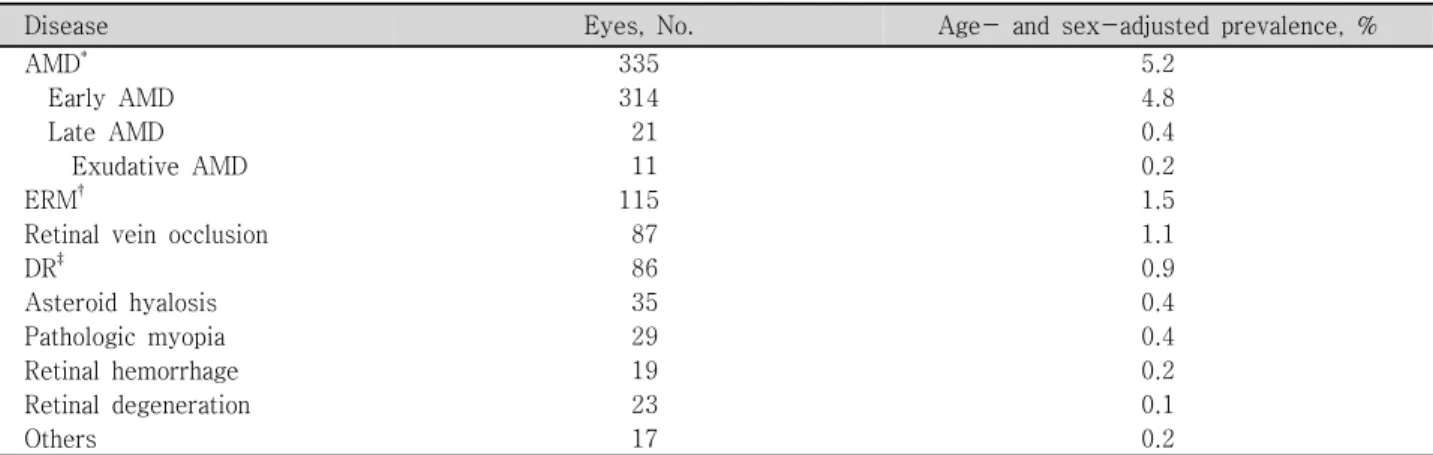
Table 2. Age- and sex-adjusted prevalence of vitreoretinal diseases in the study population
Disease Eyes, No. Age- and sex-adjusted prevalence, %
AMD* 335 5.2
Early AMD 314 4.8
Late AMD 21 0.4
Exudative AMD 11 0.2
ERM† 115 1.5
Retinal vein occlusion 87 1.1
DR‡ 86 0.9
Asteroid hyalosis 35 0.4
Pathologic myopia 29 0.4
Retinal hemorrhage 19 0.2
Retinal degeneration 23 0.1
Others 17 0.2
*AMD=age-related macular degeneration; †ERM=epiretinal membrane; ‡DR=diabetic retinopathy.
드루젠도 초기 나이관련황반변성에 포함하였다. 후기 나이 관련황반변성은 삼출성 나이관련황반변성이 있거나 지도모양 위축이 있는 경우로 정의하였다.
당뇨는 The new American Diabetes Association (ADA) criteria를 바탕으로 당뇨를 진단 받은 과거력이 있거나, 건 강검진 결과에서 공복혈당(fasting plasma glucose)이 7.0 mmol/l (126 mg/dl) 이상인 경우를 당뇨 환자로 정의하였 다.6당뇨 환자 중에 ETDRS에서 사용한 modified Airlie House classification을 참고하여 미세혈관류, 망막출혈, 경성삼출물, 면화반, 망막내미세혈관이상, 염주정맥, 정맥고리 등이 있는 경우를 당뇨망막병증으로 정의하였다.7망막이나 시신경 유두 에 신생혈관이 있거나, 범망막광응고의 흉터가 있는 증식당뇨 망막병증도 당뇨망막병증에 포함하였다.
망막앞막은 망막 앞 세포의 얇은 층 때문에 생기는 초기의 셀로판 황반 반사와 표면의 망막 주름과 당김을 동반하는 두꺼운 막 때문에 생기는 후기의 황반 앞 섬유증을 포함하 였다.8,9
망막정맥폐쇄는 망막중심정맥폐쇄와 망막분지정맥폐쇄를 모두 포함하였다. 망막중심정맥폐쇄는 망막부종, 시신경유 두의 충혈 또는 부종, 망막정맥의 확장, 망막 출혈 등을 특징
으로 하여 정의하였으며,10비슷한 양상을 나타내나, 폐쇄된 정맥의 정점에서 동정맥 교차에 의한 망막 구역에 한정되어 지는 경우는 망막분지정맥폐쇄로 생각하였다.
통계분석은 SPSS 12.0 (SPSS Inc., Chicago, IL, USA)를 사용하였으며, 유병률의 산출은 2006년 통계청에서 발표한 2005년 주민등록인구현황을 기준으로 직접표준화법을 사용 하여 연령 및 성별을 보정하였다. 각 질환의 연령 및 성별에 따른 차이는 카이제곱검정을 이용하여 비교하였으며, 유의 수준은 p<0.05로 정의하였다.
결 과
총 연구 대상자는 50세 이상의 성인 총 11,180명이었다.
이중 61.8%가 광역시 이상의 대도시에 거주하고 있었고, 28.1
%는 중소도시에, 10.1%는 농촌에 거주하고 있었다. 참여자 중 남자가 6276명, 여자가 4904명으로 56.1%가 남자였다.
평균 연령은 57.3±6.4세(50세~92세)이었으며, 남자의 평균 연령은 57.1±6.4세(50세~88세), 여자의 평균 연령은 57.6
±6.3세(50세~92세)이었다.
50세 이상의 한국 성인에서 유리체망막 질환의 연령 성별
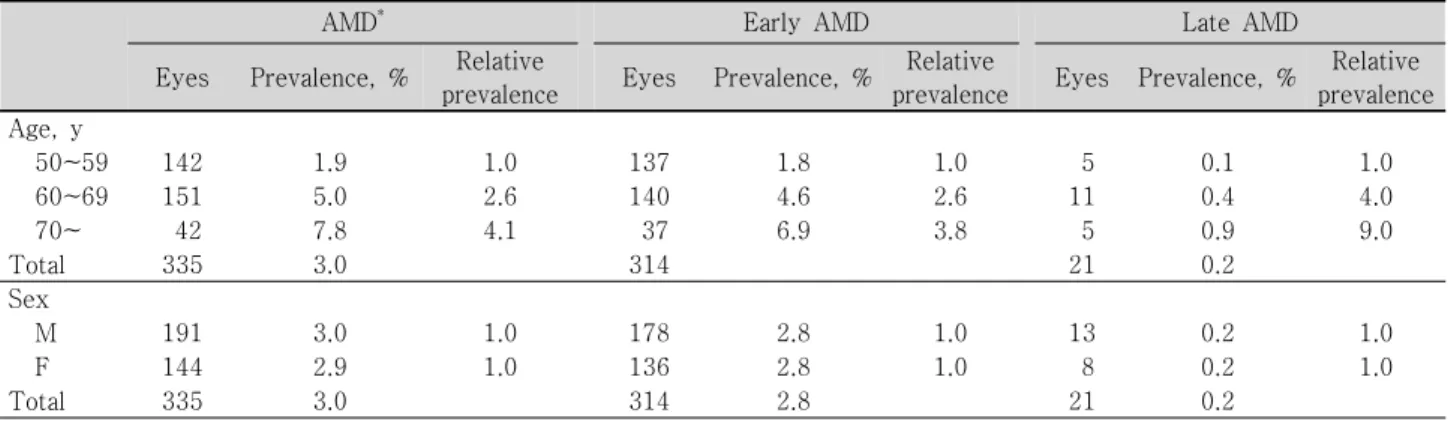
Table 3. Prevalence of age-related macular degeneration (AMD) in the study population by age and sex
AMD* Early AMD Late AMD
Eyes Prevalence, % Relative
prevalence Eyes Prevalence, % Relative
prevalence Eyes Prevalence, % Relative prevalence Age, y
50~59 142 1.9 1.0 137 1.8 1.0 5 0.1 1.0
60~69 151 5.0 2.6 140 4.6 2.6 11 0.4 4.0
70~ 42 7.8 4.1 37 6.9 3.8 5 0.9 9.0
Total 335 3.0 314 21 0.2
Sex
M 191 3.0 1.0 178 2.8 1.0 13 0.2 1.0
F 144 2.9 1.0 136 2.8 1.0 8 0.2 1.0
Total 335 3.0 314 2.8 21 0.2
*AMD=age-related macular degeneration.
Table 4. Prevalence of epiretinal membrane (ERM), retinal vein occlusion, and diabetic retinopathy (DR) in the study population by age and sex
ERM* Retinal vein occlusion DR†
Eyes Prevalence, % Relative
prevalence Eyes Prevalence, % Relative
prevalence Eyes Prevalence, % Relative prevalence Age, y
50~59 49 0.6 1.0 35 0.5 1.0 35 0.5 1.0
60~69 52 1.7 2.8 41 1.4. 2.8 41 1.4 2.8
70~ 14 2.6 4.3 11 2.0 4.0 10 1.9 3.8
Total 115 1.0 87 0.8 86 0.8
Sex
M 46 0.7 1.0 50 0.8 1.0 55 0.9 1.0
F 69 1.4 2.0 37 0.8 1.0 31 0.6 0.7
Total 115 1.0 87 0.8 86 0.8
*ERM=epiretinal membrane; †DR=diabetic retinopathy.
보정 후 유병률은 9.9%였다. 유리체망막 질환의 유병률은 통계학적으로 유의하게 나이가 많을수록 증가하였으나(p= 0.000), 성별에 따른 차이는 없었다(p=0.553)(Table 1).
유리체망막 질환의 질환별 유병률은 Table 2와 같았다.
가장 흔한 유리체망막 질환은 나이관련황반변성으로 연령 성별 보정 후 유병률은 5.2%였으며, 초기 나이관련황반변성 의 연령 성별 보정 후 유병률은 4.8%, 후기 나이관련황반변 성의 연령 성별 보정 후 유병률은 0.4%였다. 삼출성 나이관련 황반변성의 연령 성별 보정 후 유병률은 0.2%였다. 다음으로 흔한 질환은 망막앞막이었으며, 망막중심정맥폐쇄 및 망막 분지정맥폐쇄를 포함한 망막정맥폐쇄와 당뇨망막병증이 그 뒤를 이었다. 망막앞막, 망막정맥폐쇄, 당뇨망막병증의 연령 성별 보정 후 유병률은 각각 1.5%, 1.1%, 0.9%였다. 특히 당뇨 환자 중에서 당뇨망막병증을 가지는 경우는 9.0%였다.
기타 질환으로는 황반원공(5명), 망막변성(5명), 고혈압망막 병증(3명), 눈톡소플라스마의증(2명), 시신경위축(1명), 망막 색소변성(1명) 등이 있었다.
나이관련황반변성의 유병률과 초기 및 후기로 나누어 알아 본 유병률 모두 통계학적으로 유의하게 나이가 많을수록 증가
하였으나(각각, p=0.000, p=0.000, p=0.000), 성별에 따른 차이는 없었다(각각, p=0.742, p=0.842, p=0.594). 나이 관련황반변성의 유병률은 50~59세에서 1.9%, 60~69세에서 5.0%, 70세 이상에서 7.8%였으며, 초기 나이관련황반변성의 유병률은 50~59세에서 1.8%, 60~69세에서 4.6%, 70세 이 상에서 6.9%, 후기 나이관련황반변성의 유병률은 50~59세 에서 0.1%, 60~69세에서 0.4%, 70세 이상에서 0.9%였다 (Table 3). 성별에 따른 나이관련황반변성의 유병률은 Table 3과 같았다.
망막앞막 및 당뇨망막병증, 망막정맥폐쇄의 나이와 성별에 따른 유병률은 Table 4와 같았다. 망막앞막 및 망막정맥폐쇄, 당뇨망막병증, 모두 통계학적으로 유의하게 나이가 많을수 록 유병률이 높았다(각각, p=0.000, p=0.000, p=0.000).
망막정맥폐쇄 및 당뇨망막병증은 성별에 따른 차이는 없었 으나(각각, p=0.801 p=0.142), 망막앞막은 통계학적으로 유의하게 여성에서 많이 나타났다(p=0.000).
고 찰
본 연구에 따르면 건강검진을 위해 방문한 50세 이상의 한국 성인에서 유리체망막 질환의 연령 성별 보정 후 유병률 은 9.9%였다. 이는 40세 이상의 성인을 대상으로 한 아시아 의 The Aravind Comprehensive Eye Study나 The Tehran Eye Study보다는 낮았으나, 미국의 라틴아메리카 사람을 대상 으로 한 The Los Angeles Latino Eye Study 보다는 높았다.
The Aravind Comprehensive Eye Study에서 40세 이상의 성인의 유리체망막 질환 유병률은 10.8%였다.3The Tehran Eye Study에서 전 연령에 대한 유리체망막 질환의 유병률은 8.56%였으며, 40세 이상의 성인에서의 유리체망막 질환의 유병률은 21.02%였다.4The Los Angeles Latino Eye Study 에서 당뇨가 없는 40세 이상의 성인에서 망막병증의 유병률 은 6.6%였다.
앞선 연구들과 마찬가지로 본 연구에서도 유리체망막 질환 의 유병률은 연령이 증가할수록 증가하였으나, 성별에 따른 차이는 없었다.
50세 이상의 한국 성인에서 가장 흔한 유리체망막 질환은 다른 연구에서와 마찬가지로 나이관련황반변성으로 나타났다.
병원 자료를 바탕으로 한 Nwosu의 연구와 Oluleye et al의 연구에서 나이관련황반변성이 가장 흔한 유리체망막 질환이 었으며,2,11The Aravind Comprehensive Eye Study와 The Tehran Eye Study에서도 나이관련황반변성이 가장 흔한 유리 체망막 질환이었다.3,4
본 연구에서 밝혀진 50세 이상의 한국 성인에서 나이관련 황반변성의 연령 성별 보정 후 유병률은 5.2%로 앞선 아시아 에서의 연구와 비슷하였다. 40세 이상을 대상으로 한 Beijing Eye Study와 Singapore Malay Eye Study에서 유병률은 각각 5.4%, 5.7%였으며,12,1350세 이상을 대상으로 한 Funagata Study에서 유병률은 4.9%였다.14이전의 여러 연구와 마찬 가지로 본 연구에서도 나이관련황반변성은 연령과 밀접한 관계 가 있었으나, 성별에 따른 차이는 없었다.12,14-16
후기 나이관련황반변성의 성별 연령 보정 후 유병률은 0.4%
로, 아시아 국가에서의 유병률과 비슷하였으나, 서양에서의 유병률 보다는 낮았다. 서양에 비해 아시아에서 후기 나이관련 황반변성의 유병률이 낮은 이유로 유전 및 환경적인 요인이 있을 것으로 생각되며, 추후 더 자세한 연구가 필요할 것으로 사료된다.
Funagata Study에서는 서양의 연구와는 다르게 후기 나이 관련황반변성이 남성에서 더 호발하며, 그 원인으로 일본에서 남성의 흡연율이 높기 때문이라고 하였다. 하지만, 일본과 마찬가지로 남성의 흡연율이 높은 한국에서 이루어진 본 연구 에서 후기 나이관련황반변성의 유병률에 있어서 남녀 차이는
없었다. 이 결과는 본 연구에서의 흡연율이 자가 작성을 바탕 으로 해서 실제 흡연율 보다 낮게 측정되었기 때문이라고 생각 된다. 본 연구에서 흡연율은 13%로 제 3기 국민건강영양조사 에서 알려진 한국 성인의 실제 흡연율 34%와 차이가 있었다.
낮게 측정된 흡연율로 인해 나이관련황반변성에 대한 흡연의 효과가 저평가 되었을 가능성이 있다.
본 연구에서 알려진 망막앞막의 연령 성별 보정 후 유병률 은 1.5%로 앞선 연구에서 알려진 망막앞막의 유병률 보다 낮았다. 아시아인을 대상으로 한 연구에서 망막앞막의 유병 률은 서양인 및 라틴아메리카 사람을 대상으로 한 연구보다 낮았다. 서양인을 대상으로 한 Beaver Dam Eye Study에서 는 11.8%,17Blue Mountains Eye Study에서는 7%,9Visual Impairment Project Study에서는 6.0%였으며,18미국에 거 주하는 라틴아메리카 사람을 대상으로 한 Los Angeles Latino Eye Study에서는 이보다 높아 18.5%였다.19이에 반해, 아시 아인을 대상으로 한 Hisayama Study에서는 4.0%,20Beijing Eye Study에서는 2.2%였다.21하지만, 최근에 이루어진 Singa- pore Malay Eye Study와 Funagata Study에서는 유병률이 각각 7.9%와 5.44%로 서양의 연구와 큰 차이를 보이지 않
았다.22,23실제 망막앞막의 유병률이 인종간의 차이가 있는지
는 더 많은 연구가 필요할 것으로 사료된다.
이전의 대부분의 연구에서 망막앞막의 유병률은 나이와 유의한 관계가 있었으며,9,17-23본 연구에서도 나이가 증가 할수록 유의하게 증가하였다. 또한 본 연구에서 망막앞막이 남자보다 여자에서 통계학적으로 유의하게 더 많았는데, Los Angeles Latino Eye Study와 Singapore Malay Eye Study 에서도 여자에서 통계학적으로 유의하게 더 많았으며,19,22 다른 여러 연구에서도 비슷한 경향을 보였다.9,20,23
본 연구에서 중심망막정맥폐쇄와 분지망막정맥폐쇄를 포함 한 망막정맥폐쇄의 연령 성별 보정 후 유병률은 1.1%로 이전 의 연구들과 비슷하였다. 백인을 대상으로 한 Blue Mountains Eye Study에서는 1.6%,10 Beaver Dam Eye Study에서는 0.8%였으며,24아시아인을 대상으로 한 Beijing Eye Study 에서는 1.3%,25Singapore Malay Eye Study에서는 0.7%의 유병률을 보였다.26
이전의 연구들과 마찬가지로, 망막정맥폐쇄의 유병률은 연령 이 증가할수록 유의하게 증가하였으나, 성별에 따른 차이는
없었다.10,24-26망막정맥폐쇄는 많은 연구에서 양안의 침범이
드물다고 알려져 있으며,10,24본 연구에서는 양안을 침범한 경우는 한 경우도 없었다. 또한 대부분의 연구에서 분지망막 정맥폐쇄가 상이측에서 호발하는 것으로 밝혀졌으며,10,24본 연구에서도 상이측에 병변이 있는 경우가 67.1%로 비슷한 결과를 보였다.
본 연구에서 당뇨망막병증의 연령 성별 보정 후 유병률은
0.9%로 이전의 서양에서 이루어진 연구에서의 유병률 보다 낮았고, 아시아에서의 유병률과 비슷하였다. 서양에서 이루어 진 The Blue Mountains Eye Study에서 당뇨망막병증의 유병 률은 2.3%였고,27이탈리아의 Giuffrè et al의 연구에서 유병 률은 4.4%였으며,28미국의 The Eye Diseases Prevalence Research Group의 연구에서는 3.4%였다.29반면 아시아에서 시행된 The Aravind Comprehensive Eye Study에서의 당뇨 망막병증의 유병률은 0.5%였으며,3The Tehran Eye Study 에서는 0.84%였다.4
본 연구에서 당뇨병 환자 중에서 당뇨망막병증의 유병률 은 9.0%였으며, 이는 서양은 물론 아시아의 다른 연구 보다 도 낮았다. 서양의 The Blue Mountain Eye Study에서는 32.4%,27Giuffrè et al의 연구에서는 34.1%,28The Eye Di- seases Prevalence Research Group의 연구에서는 40.3%
였다.29아시아의 The CURES Eye Study에서는 17.6%,30The Aravind Comprehensive Eye Study에서는 11.4%,3Narendran et al의 연구에서는 26.8%,31 Dnadona et al의 연구에서는 22.4%,32The Singapore Malay Eye Study에서는 35.0%의 유병률을 보였다.33본 연구와 아시아의 연구를 바탕으로 볼 때 당뇨병 환자 중에서 산출한 당뇨망막병증 유병률이 아시 아에서 서양보다 대체적으로 낮은 것으로 나타났다. 당뇨병의 유병률이 아시아에서는 최근에 급격히 증가하고 있어 아시아 인에서도 당뇨망막병증의 유병률은 당뇨병의 유병률처럼 향후 수년 안에 증가할 가능성이 있다. 한국에서도 당뇨망막병증의 원인이 되는 당뇨병의 유병률이 빠른 속도로 증가하고 있다.
미국에서의 당뇨병 유병률은 1988~1994년에 8.2%, 1999~
2002년에 9.3%로 크게 변화가 없었으나,34한국인에서의 당뇨 병 유병률은 1997년 6.9%, 2003년에 11.7%로 급증하였다.35 당뇨병의 유병률이 급증하는 것을 고려하면, 향후 당뇨망막 병증의 유병률은 더 증가할 것으로 예상할 수 있다.
본 연구의 당뇨병 환자 중에서 당뇨망막병증의 유병률이 다른 아시아의 연구보다도 낮은 것은 본 연구가 건강 검진 자료 를 사용하였기 때문이라고 생각된다. 일반적으로 건강 검진은 건강한 사람을 대상으로 하기 때문에, 당뇨망막병증이 생길 가능성이 있는 중증의 당뇨 환자들은 제외되었을 가능성이 있다.
본 연구에 의하면 나이관련황반변성 및 망막앞막, 망막정맥 폐쇄, 당뇨망막병증 등의 유리체망막 질환은 고령의 환자 일수 록 유병률이 높았다. 이는 유리체망막 질환이 미래의 한국에 서 더 큰 안과적인 문제로 대두될 가능성을 의미한다. 통계청 에서 2006년에 발표한 장래인구추계에 따르면 2005년에는 65세 이상의 인구가 전체인구에 9.1%였으나, 2010년에는 11.0%, 2020년에는 15.6%로 증가하고, 2030년에는 24.3%
까지 증가할 것으로 예상되고 있다.36한국의 고령사회 진입
속도가 선진국에 비해 매우 빠르게 진행되고 있는 점을 생각 해 볼 때, 나이관련황반변성을 포함한 유리체망막 질환의 유병 률은 향후 더 증가할 것이다.
본 연구의 제한점으로서 첫째, 일개 대학병원 종합건강검진 센터 수진자를 대상으로 하였기 때문에 지역 대표성이 부족 하고 둘째, 종합건강검진을 하는 참여자들의 경제사정 및 교육 정도가 우월한 경향이 있어 선택 편견이 있을 수 있다는 것이 다. 이러한 제한점을 극복하고자 국내 연령대별 총인구수를 이용하여 직접법으로 연령보정을 하였고, 선택 편견을 최소화 하기 위해 참여자의 수를 10,000명 이상의 대규모로 포함시 켰다. 셋째, 안저사진을 황반을 중심으로 한 장만 촬영하였기 때문에 망막의 주변부를 확인할 수 없어 다른 연구에 비해 진단률이 낮았을 가능성이 있다. 특히, 당뇨망막병증의 경우 미세혈관류, 점상출혈 등의 초기 병변이 후극부에 제한되어 있지 않기 때문에, 유병률 산출에 더 큰 영향을 받았을 수 있다.
그럼에도 불구하고, 본 연구는 대규모의 한국인 성인을 대상 으로 한 유리체망막 질환의 유병률을 조사한 보고이며, 각 질환의 유병률의 연령 및 성별에 따른 경향을 이전의 다른 연구들과 비교하였다는 점에서 의미가 있다.
결론적으로, 한국의 성인에서 유리체망막 질환의 유병률은 다른 나라와 비슷하였다. 한국의 고령사회 진입 속도와, 당뇨 병 유병률의 증가 속도를 생각할 때, 나이관련황반변성과 당뇨 망막병증을 포함한 유리체망막 질환이 앞으로 시력 저하 및 실명에 중요한 원인이 될 것으로 예상할 수 있으며, 이에 대한 대비가 필요하겠다. 향후 전국적인 자료를 바탕으로 한 유리 체망막 질환의 유병률 및 위험요인에 대한 연구가 이루어져야 할 것이다.
참고문헌
1) Ahn JE, Seo KY, Kho HJ, et al. The change of blindness-causes in Korea. J Korean Ophthalmol Soc 1998;39:2761-7.
2) Nwosu SN. Prevalence and pattern of retinal diseases at the Guinness Eye Hospital, Onitsha, Nigeria. Ophthalmic Epidemiol 2000;7:41- 8.
3) Nirmalan PK, Katz J, Robin AL, et al. Prevalence of vitreoretinal disorders in a rural population of southern India: the Aravind Com- prehensive Eye Study. Arch Ophthalmol 2004;122:581-6.
4) Hatef E, Fotouhi A, Hashemi H, et al. Prevalence of retinal diseases and their pattern in Tehran; the Tehran Eye Study. Retina 2008;28:
755-62.
5) International ARM Epidemiological Study Group. An international classification and grading system for age-related maculopathy and age-related macular degeneration. Surv Ophthalmol 1995;39:367-74.
6) American Diabetes Association. Diagnosis and classification of dia- betes mellitus. Diabetes Care 2008;31:S55-60.
7) Early Treatment Diabetic Retinopathy Study Research Group. Gra- ding diabetic retinopathy from stereoscopic color fundus photographs-
an extension of the modified Airlie House Classification. ETDRS report number 10. Ophthalmology 1991;98:786-806.
8) Klein R, Klein BE, Wang Q, Moss SE. The epidemiology of epire- tinal membranes. Trans Am Ophthalmol Soc 1994;92:403-30.
9) Mitchell P, Smith W, Chey T, et al. Prevalence and associations of epiretinal membranes: the Blue Mountains Eye Study, Australia.
Ophthalmology 1997;104:1033-40.
10) Mitchell P, Smith W, Chang A. Prevalence and associations of retinal vein occlusion in Australia. The Blue Mountains Eye Study. Arch Ophthalmol 1996;114:1243-7.
11) Oluleye TS, Ajaiyeoba AI. Retinal diseases in Ibadan. Eye 2006;20:
1461-3.
12) Li Y, Xu L, Jonas JB, et al. Prevalence of age-related maculopathy in the adult population in China: the Beijing Eye Study. Am J Oph- thalmol 2006;142:788-93.
13) Cackett P, Tay WT, Aung T, et al. Education, socio-economic status and age-related macular degeneration in Asians: the Singapore Malay Eye Study. Br J Ophthalmol 2008;92:1312-5.
14) Kawasaki R, Wang JJ, Ji GJ, et al. Prevalence and risk factors for age-related macular degeneration in an adult Japanese population:
the Funagata study. Ophthalmology 2008;115:1376-81.
15) Chen SJ, Cheng CY, Peng KL, et al. Prevalence and associated risk factors of age-related macular degeneration in an elderly Chinese population in Taiwan: the Shihpai Eye Study. Invest Ophthalmol Vis Sci 2008;49:3126-33.
16) Vingerling JR, Dielemans I, Hofman A, et al. The prevalence of age-related maculopathy in the Rotterdam Study. Ophthalmology 1995;102:205-10.
17) Klein R, Klein BE, Wang Q, Moss SE. The epidemiology of epire- tinal membranes. Trans Am Ophthalmol Soc 1994;92:403-25.
18) McCarty DJ, Mukesh BN, Chikani V, et al. Prevalence and asso- ciations of epiretinal membranes in the visual impairment project.
Am J Ophthalmol 2005;140:288-94.
19) Fraser-Bell S, Ying-Lai M, Klein R, et al. Prevalence and associations of epiretinal membranes in latinos: the Los Angeles Latino Eye Study. Invest Ophthalmol Vis Sci 2004;45:1732-6.
20) Miyazaki M, Nakamura H, Kubo M, et al. Prevalence and risk factors for epiretinal membranes in a Japanese population: the Hisayama study. Graefes Arch Clin Exp Ophthalmol 2003;241:642-6.
21) You Q, Xu L, Jonas JB. Prevalence and associations of epiretinal membranes in adult Chinese: the Beijing eye study. Eye 2008;22:
874-9.
22) Kawasaki R, Wang JJ, Mitchell P, et al. Racial difference in the pre- valence of epiretinal membrane between Whites and Asians. Br J Ophthalmol 2008;92:1320-4.
23) Kawasaki R, Wang JJ, Sato H, et al. Prevalence and associations of epiretinal membranes in an adult Japanese population: the Funagata study. Eye 2009;23:1045-51.
24) Klein R, Klein BE, Moss SE, Meuer SM. The epidemiology of retinal vein occlusion: the Beaver Dam Eye Study. Trans Am Ophthalmol Soc 2000;98:133-41.
25) Liu W, Xu L, Jonas JB. Vein occlusion in Chinese subjects. Ophthal- mology 2007;114:1795-6.
26) Lim LL, Cheung N, Wang JJ, et al. Prevalence and Risk Factors of Retinal Vein Occlusion in an Asian Population. Br J Ophthalmol 2008;92:1316-9.
27) Mitchell P, Smith W, Wang JJ, Attebo K. Prevalence of diabetic retinopathy in an older community. The Blue Mountains Eye Study.
Ophthalmology 1998;105:406-11.
28) Giuffrè G, Lodato G, Dardanoni G. Prevalence and risk factors of diabetic retinopathy in adult and elderly subjects: The Casteldaccia Eye Study. Graefes Arch Clin Exp Ophthalmol 2004;242:535-40.
29) The Eye Diseases Prevalence Research Group. The prevalence of diabetic retinopathy among adults in the United States. Arch Oph- thalmol 2004;122:552-63.
30) Rema M, Premkumar S, Anitha B, et al. Prevalence of diabetic re- tinopathy in urban India: the Chennai Urban Rural Epidemiology Study (CURES) eye study, I. Invest Ophthalmol Vis Sci 2005;46:
2328-33.
31) Narendran V, John RK, Raghuram A, et al. Diabetic retinopathy among self reported diabetics in southern India: a population based assessment. Br J Ophthalmol 2002;86:1014-8.
32) Dandona L, Dandona R, Naduvilath TJ, et al. Population based assessment of diabetic retinopathy in an urban population in sou- thern India. Br J Ophthalmol 1999;83:937-40.
33) Wong TY, Cheung N, Tay WT, et al. Prevalence and Risk Factors for Diabetic Retinopathy The Singapore Malay Eye Study. Oph- thalmology 2008;115:1869-75.
34) Cowie CC, Rust KF, Byrd-Holt DD, et al. Prevalence of diabetes and impaired fasting glucose in adults in the U.S. population: Na- tional Health And Nutrition Examination Survey 1999-2002. Dia- betes Care 2006;29:1263-8.
35) Song KH, Nam-Goong IS, Han SM, et al. Change in prevalence and 6-year incidence of diabetes and impaired fasting glucose in Korean subjects living in a rural area. Diabetes Res Clin Pract 2007;
78:378-84.
36) Korea National Statistical Office. Population Projections for Korea:
2005~2050 (Based on the 2005 Census). Available at: http://www.
nso.go.kr/
=ABSTRACT=
The Prevalence of Vitreoretinal Diseases in a Screened Korean Population 50 Years and Older
Dong Ju Youm, MD1, Hyun-Sub Oh, MD1, Hyeong Gon Yu, MD, PhD2, Su Jeong Song, MD, PhD1
Department of Ophthalmology, Sungkyunkwan University School of Medicine, Kangbuk Samsung Hospital1, Seoul, Korea Depratment of Ophthalmology, Seoul National University College of Medicine2, Seoul, Korea
Purpose: To describe the prevalence of vitreoretinal diseases in the Korean population 50 years or older at a health screening center.
Methods: The participants of this study included 11,180 adults 50 years of age and older who visited the Health Promotion Center of Kangbuk Samsung Hospital from January to December 2006. Digital images of non-mydriatic fundus photographs were examined.
We calculated the sex- and age-adjusted prevalence of vitreoretinal diseases using the direct standardized method based on the number of resident registrations.
Results: The age- and sex-adjusted prevalence of vitreoretinal diseases in Korean adults 50 years of age and older was 9.9%.
The prevalence of vitreoretinal diseases significantly increased with age (P=0.000). There was no significant gender difference in the prevalence of vitreoretinal diseases (p=0.553). Age-related macular degeneration was the most common vitreoretinal disease, with an age- and sex-adjusted prevalence of 5.2%. Epiretinal membrane, retinal vein occlusion, and diabetic retinopathy were common vitreoretinal diseases in that order, and the age- and sex-adjusted prevalences were 1.5%, 1.1%, and 0.9%, respectively.
Conclusions: The prevalence of vitreoretinal diseases in a screened Korean population 50 years and older was 9.9%. Vitreoretinal diseases are a major ophthalmic problem in Korea. As the Korean population continues to age and the prevalence of diabetes increases, further investigations about the epidemiology and prevention of vitreoretinal diseases are needed.
J Korean Ophthalmol Soc 2009;50(11):1645-1651
Key Words: Age-related macular degeneration, Diabetic retinopathy, Prevalence, Retinal vein occlusion, Vitreoretinal diseases
Address reprint requests to Su Jeong Song, MD, PhD
Department of Ophthalmology, Kangbuk Samsung Hospital, Sungkyunkwan University School of medicine
#108 Pyeong-dong, Jongno-gu, Seoul 110-746, Korea
Tel: 82-2-2001-2250, Fax: 82-2-2001-2262, E-mail: [email protected]