pISSN: 0378-6471 eISSN: 2092-9374 http://dx.doi.org/10.3341/jkos.2012.53.6.849
= 증례보고 =
심바스타틴이 인체망막색소상피세포에서 생산된 혈관내피성장인자에 미치는 효과
김경진⋅김경실⋅김나래⋅진희승
인하대학교 의과대학 안과학교실, 인하 시과학연구소
목적: 심바스타틴(simvastatin)이 산화 스트레스하의 인체망막색소상피세포에서 생산된 혈관내피성장인자에 어떠한 영향을 미치는지 알아보고자 하였다.
대상과 방법: Simvastatin과 H2O2를 농도를 달리하여 인체망막색소상피세포에 처리한 뒤 MTT assay를 통해 세포 활성도를 측정하였 다. Simvastatin을 전 처치한 인체망막색소상피세포에 H2O2 100 μm을 처리하여 산화 스트레스를 준 후 ELISA를 이용하여 VEGF를 측정하였다.
결과: Simvastatin 10 μm까지는 세포 활성도에 영향이 없었으나 10 μm 이상의 농도에서 세포 활성도가 감소하는 양상을 보였다. 산화 스트레스 하의 인체망막색소상피세포를 5 μm 이하의 simvastatin에 전 처치 시 VEGF의 발현이 감소하였다. 노출시간에 따른 발현은 3시간 노출 시 가장 감소되었으며 24시간 노출 시까지 지속되었다.
결론: Simvastatin은 산화 스트레스 하의 인체망막색소상피세포에서 VEGF의 생성을 감소시키며 VEGF의 증가가 병인으로 작용하는 망막 질환을 예방하는데 도움이 될 수 있을 것으로 기대된다.
<대한안과학회지 2012;53(6):849-855>
■ 접 수 일: 2011년 7월 15일 ■ 심사통과일: 2011년 10월 10일
■ 게재허가일: 2012년 4월 24일
■ 책 임 저 자: 진 희 승
인천광역시 중구 인항로 27 인하대학교병원 안과
Tel: 032-890-3408, Fax: 032-890-2417 E-mail: [email protected]
* This study was supported by a National Research Foundation of Korea (NRF) grant funded by the Korean government (MEST) (No. 2010- 0012245) and by the Converging Research Center Program funded by the Ministry of Education, Science and Technology (2011K000697).
연령관련황반변성은 50세 이상에서 일어나는 황반의 변 성질환으로 서구에서는 55세 이상 노인 인구에서 심한 시 력저하를 유발하는 주된 원인이 되는 질환이며, 현재 우리 나라에서도 인구의 노령화와 함께 그 발생이 점점 증가하 고 있는 추세이다.1-3연령관련황반변성은 초기에 드루젠의 축적으로 특징지어지는 건성의 형태이지만 일부에서 진행 하여 맥락막 혈관신생으로 특징지어지는 삼출성의 형태로 나타나며, 심한 시력저하는 대부분 맥락막 혈관신생을 동반 하는 삼출성 연령관련황반변성에 의한다.4,5 망막이나 망막 색소상피세포 아래에서 발생한 맥락막 신생혈관은 누출된 삼출물이나 혈액에 의해 직접적인 망막 손상을 일으킬 수 있으며 이차적인 허혈 및 섬유성 혈관조직 등에 의해 시력 저하를 유발하게 된다.6이러한 연령관련황반변성에 동반된 맥락막 신생혈관 발생 원인에 대해서는 산화 스트레스, 면
역반응, 염증반응, 저산소 등의 다양한 원인이 제시되고 있 으며 혈관내피성장인자가 신생혈관 발생에 핵심 인자임이 밝혀진 이후 항 혈관내피성장인자를 이용한 치료가 임상에 서 널리 시행되고 있다.7-10
혈관신생을 촉진하는 인자로는 VEGF (vascular endo- thelial growth factor), bFGF (basic fibroblast growth factor), IGF-Ⅱ (insulin-like growth factor-Ⅱ) 등이 있고 억제하는 인자로는 PEDF (pigment epithelium-derived factor), TGF–β (transforming growth factor–ß), platelet factor-4, angiostatin, endostatin 등이 있다. 인체망막상 피세포는 혈관형성 유도 인자인 VEGF와 혈관형성 억제 인 자인 PEDF를 적절히 분비하여 외측으로는 맥락막 모세혈 관의 정상적인 기능상태를 유지하고 내측으로는 시세포 층 의 무혈관 상태를 유지시킨다. 정상적으로 인체 내에서는 이들 인자간의 균형이 유지되어 불필요한 혈관신생을 막고 있으나 산화 스트레스에 의한 인체망막상피세포에서의 VEGF의 분비 변화는 맥락막 신생혈관 발생을 유도하여 삼 출성 연령관련황반변성을 유발할 수 있다.11-15
Statins은 콜레스테롤 합성에 필수적인 효소인 히드록시 메틸글루타릴-코에이 환원효소(3-hydroxy-methyl glu- taryl-coenzyme A reductase)에 대한 경쟁적 억제제로 지 질 저하 약제로서 임상적으로 많이 사용되고 있다. Statins 은 심혈관 질환 사망률을 줄여주고 관상동맥질환 환자에서
죽상 경화증의 진행을 막아주는 것으로 알려졌다. 그러나 최근 statins의 콜레스테롤 저하 효과뿐만 아니라 콜레스테 롤 독립적인 효과, 즉 항산화효과나, 항염증효과, 항혈관생 성효과와 같은 pleiotropic effect가 주목받고 있으며 in vivo 연구에서 statins이 혈청 내 VEGF의 농도를 낮추는 효과가 보고되었다.16-21저자들은 본 연구를 통해 산화 스 트레스에 노출된 인체망막색소상피세포에서 simvastatin에 의한 VEGF의 발현 변화를 관찰하고자 하였다.
대상과 방법
세포주 배양
인체망막색소상피세포주(ARPE-19;ATCC No. CRL-2302) 를37℃, 95% air, 5% CO2가 공급되는 습윤화된 배양기에 서 배양하였다. 배양액은 56℃에서 30분간 열처리한 Fetal Bovine Serum (Gibco BRL, Grand Island, NY, USA) 10%
와 penicillin (100 U/ml), streptomycin (100 μm/ml)과 amphotericin B (1 μm/ml)를 포함한 항생제(antibiotic- antimycotic, Gibco BRL, Grand Island, NY, USA)를 함유 한 DMEM:F-12 배지(Gibco BRL, Grand Island, NY, USA)를 사용하였다. 배양액은 3일마다 새로 교환하였다.
세포활성도 검사
인체망막색소상피세포의 세포사를 유발하지 않는 sim- vastatin (Sigma-Aldrich, St. Louis, MO, USA)과 H2O2의 적정 농도를 알아보기 위하여 농도반응검사를 통해 세포 활성도를 측정하였다. 96 well microplate (Falcon, Lincoln Park, NJ, USA)에 망막색소상피세포를 5×10³ cells/well 로 희석시키고 세포 부유액 90 ul씩을 각각 분주하였다. 24 시간 배양 후에 H2O2 (0, 10, 20, 30, 50, 100, 200, 300, 400 μm)와, Simvastatin (0, 0.5, 1, 2, 5, 10, 20, 30 μm) 을 농도별로 10 ul씩 넣고 대조군으로 phosphate buffered saline (PBS) 10 ul를 넣은 후 2일간 37℃, 5% CO2배양기 에서 배양하였다. 모든 well에 3-(4,5-dimethylthiazol- 2-yl)-2,5-diphenyl tetrazolium bromide (MTT) 용액 (Sigma-Aldrich, St. Louis, MO, USA) 10 ul를 가해주고 다시 37℃, 5% CO2에서 4시간 더 배양하여 MTT가 환원 되도록 하였다. 각 well에서 media를 버린 다음 DMSO (Sigma-Aldrich, St. Louis, MO, USA)를 150 ul씩 넣고 10분 동안 흔들어서 생성된 MTT formazan 결정을 녹여서 microplate reader (Bio-Tek, Vermont, MA, USA)를 이 용하여 540 nm에서 흡광도를 측정하였다. 세포활성도의 정
도는 실험군과 대조군의 흡광도의 차이를 %로 나타내었다.
VEGF 생산의 측정
인체망막색소상피세포를 simvastatin에 농도별로(1, 2.5, 5, 10 μm) 전 처치한 후 3시간 뒤에 H2O2 100 μm을 처리 하여 산화 스트레스를 준 후 cell culture supernate sample 을 수확한 뒤 ELISA (Enzyme-linked immunosorbent assay)를 이용하여 VEGF를 측정하였다. 항체가 부착된 96 well microplate에 시료를 첨가하고 실온에서 2시간 배양 후 세척한 뒤 conjugate넣고 실온에서 2시간 배양한 뒤 세 척 후 substrate를 첨가하고 25분 후 stop solution을 첨가 하였다. 분광광도계로 540 nm에서 흡광도를 측정하였다.
VEGF의 측정은 ELISA kits (R&D system, Minneapolis, MN, USA)를 이용하였다. VEGF kit는 네 가지의 VEGF isoform 중 VEGF121와, VEGF165를 측정하였으며, 측정 최 소값은 VEGF는 15.6 pg/ml (intra-assay coefficient of variation 4.1%, inter-assay coefficient of variation 5.4%)이었다. 3회 반복 측정하여 평균값을 최종값으로 사 용하였다.
통계분석
실험 결과는 3회 이상의 독립된 실험 후 평균 ±표준편 차의 형태로 표시하였다. 통계분석은 SPSS version 19.0을 이용하였으며 Kruskal-wallis test를 이용하여 대조군과 실험군 간의 차이를 비교하였다. p<0.05인 경우 통계적인 유의성이 있는 것으로 간주하였다.
결 과
Simvastatin이 인체망막상피세포의 활성도에 미치는 영향
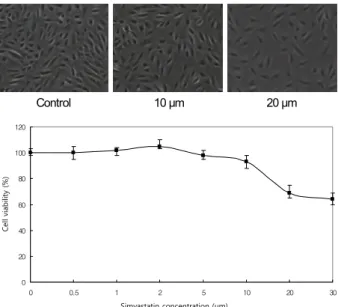
MTT assay를 통하여 세포사를 유발하지 않으면서 세포 변형을 일으킬 수 있는 simvastatin과 H2O2의 적정 농도를 조사하였다. Simvastatin 1 μm에 24시간 배양하였을 때 세 포생존율이 DMSO를 넣은 대조군에 비해 99.3 ±2.3% 수 준을 보였으며, 5 μm에서는 98.3 ± 2.8%를 보였다.
Simvastatin 10 μm에서는 93.0 ±1.5%였으나 20 μm 이 상의 농도에서는 농도 증가에 따라 세포생존율이 점차적으 로 감소하는 양상을 보여 simvastatin 10 μm 이상의 농도 에서는 독성이 나타나기 시작하는 것을 확인할 수 있었다 (Fig. 1). H2O2농도 100 μm에서는 99.3 ±4.3%를 보였으 나 H2O2 200 μm에서는 85.0 ±2.5%였으며 이후 H2O2농
Control 10 μm 20 μm
0 20 40 60 80 100 120
0 0.5 1 2 5 10 20 30
Simvas tatin Concentration (µM )
Cell viability (%)
Simvastatin concentration (μm)
Cell viability (%)
Figure 1. The viablity of ARPE-19 cells on exposure to
simvastatin. Human retinal pigment epithelial cells were cul- tured for 24 hours with various concentrations of simvastatin.Cell viability is fair at less than 10 μm simvastatin, but cell sur- vival showed progressive decrease with increasing concen- tration of simvastatin more than 10 μm and toxicity was ob- served at 20-μm simvastatin. The cell viability was determined using MTT assay. The experiment was performed three times independently.
0 20 40 60 80 100 120
0 10 20 30 50 100 200 300 400
H2O2 Concentration (µM )
Cell viability (%)
H2O2 Concentration (μm)
Cell viability (%)
Figure 2. The viablity of ARPE-19 cells on exposure to H
2O2. Human retinal pigment epithelial cells were cultured for 24 hours with various concentrations of H2O2. Cell viability is fair at less than 200 μm, but cell viability showed progressive decrease with increasing concentration of H2O2 more than 200 μm. The experiment was performed three times independently.*
*
*
*
0 50 100 150 200
C ON 50 100 200
H2O2( µM )
VEGF Concentration (pg/ml)
H2O2 (μm)
VEGF concentration (pg/ml)
Figure 3. Effect of H
2O2 on VEGF expression in ARPE-19.Human retinal pigment epithelial cells were cultured at vari- ous concentrations of H2O2. Concentration-dependent in- crease in VEGF expression occurred following treatment with 50, 100 and 200 μm simvastatin for 24 hours (*p < 0.05).
* *
*
*
*
0 50 100 150 200
C O N H 100 H 100
+ S i m1μM H 100 +S i m 2.5μM
H 100 +S i m 5μM
H 100 +S i m 10μM
VEGF concentration (pg/ml)VEGF concentration (pg/ml)
H 100 +
Sim 1 μm H 100 + Sim 2.5 μm H 100 +
Sim 5 μm H 100 + Sim 10 μm
Figure 4. Effect of simvastatin (Sim) on VEGF expression in
exogenous H2O2 (H 100) stimulated ARPE-19. (concentration dependent). Human retinal pigment epithelial cells were pre- treated at various concentration of simvastatin for 3 hours and further incubated with 100 μm H2O2. Simvastatin concen- tration of 1, 2.5 and 5 μm inhibited the VEGF expression of ARPE-19 under oxidative stress (*p < 0.05).도 증가에 따라 세포생존율이 점차적으로 감소하는 양상을 보였다(Fig. 2).
인체망막상피세포에서 산화스트레스에 의한 VEGF 발 현 증가
인체망막색소상피세포를 산화제인 H2O2에 농도를 달리
하여(0, 50, 100, 200 μm) 노출시킨 후 24시간 배양한 뒤 ELISA를 통해 VEGF의 발현을 측정하였다. H2O2에 노출되 기 전에 비하여 H2O2에 노출되었을 경우에 VEGF의 발현이 유의하게 증가하는 것을 확인할 수 있었으며 이는 200 μm 까지 H2O2 농도가 높아질수록 증가하는 경향을 보여 인체 망막상피세포에서 산화스트레스에 의해 VEGF의 발현이 증가하는 것을 확인할 수 있었다(Fig. 3).
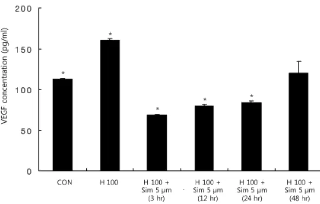
산화스트레스하의 인체망막상피세포에서 Simvastatin 전 처치 후 VEGF 발현 감소
인체망막색소상피세포를 다양한 농도의(0, 1, 2.5, 5, 10 μm) simvastatin에 3시간 동안 전 처치한 후 산화제인 H2O2 100 μm에 노출시킨 뒤 24시간 배양 후 ELISA를 통
* *
*
*
*
0 5 0 1 0 0 1 5 0 2 0 0
C ON H 100 H 100
+ S im 5 μ M ( 3 h ou r s )
H 100 + S i m 5 μM ( 12 h ou r s )
H 100 + S i m 5 μM ( 24 h ou rs )
H 100 + S i m 5 μ M ( 48 h ou r s )
VEGF concentration (pg/ml)VEGF concentration (pg/ml)
H 100 + Sim 5 μm (3 hr)
H 100 + Sim 5 μm (12 hr)
H 100 + Sim 5 μm (24 hr)
H 100 + Sim 5 μm (48 hr) H 100
CON
Figure 5. Effect of simvastatin (Sim) on VEGF expression in
exogenous H2O2 (H 100) stimulated ARPE-19. (time depend- ent). Human retinal pigment epithelial cells were pretreated with 5 μm simvastatin for various incubation time and further incubated with 100 μm H2O2. The decreased expression of VEGF was evident as early as 3 hours after incubation, and lasted for at least 24 hours (*p < 0.05).해 VEGF의 발현을 측정하였다. Simvastatin을 전 처치하 기 전에 비하여 1, 2.5, 5 μm의 simvastatin을 전 처치하였 을 경우 산화 스트레스하의 인체망막상피세포에서 VEGF 의 발현이 유의하게 감소하는 것을 확인할 수 있었으나 10 μm의 simvastatin을 전 처치하였을 경우 VEGF는 유의한 변화를 보이지 않았다(Fig. 4). 인체망막색소상피세포를 simvastatin 5 μm에 시간대별로(3, 12, 24, 48시간) 노출 시킨 뒤 ELISA를 통해 VEGF 발현을 측정하였다. 산화 스 트레스하의 인체망막상피세포에서 simvastatin에 3시간 노 출했을 때 VEGF의 발현이 가장 억제되었으며 VEGF 억제 효과는 노출 시간이증가함에 따라 감소하였으나 24시간까 지 지속되었다(Fig. 5).
고 찰
Simvastatin이 산화 스트레스하의 인체망막색소상피세 포에서 생산된 VEGF에 미치는 효과를 알아본 결과 산화 스트레스하의 인체망막색소상피세포를 5 μm 이하의 sim- vastatin에 전 처치 시 VEGF의 발현이 유의하게 감소하였 으며 노출시간에 따른 발현은 simvastatin에 3시간 노출 시 VEGF의 농도가 가장 감소되었고 24시간 노출 시까지 지속 되었다.
맥락막 모세혈관과 망막의 시세포층 사이에 위치한 망막 색소상피세포는 자체적으로 혈관형성 유도 인자인 VEGF 와 혈관형성 억제 인자인 PEDF를 생산한다. 이러한 신생혈 관 유도인자와 억제인자를 적절히 분비하여 외측으로는 맥 락막 모세혈관의 정상적인 기능상태를 유지하고 내측으로 는 시세포층의 무혈관 상태를 유지시킨다.22-25 그러나 망막
색소상피세포가 저산소나 산화 스트레스 같은 비정상적인 환경에 노출될 때 혈관형성 관련 인자인 VEGF나 PEDF의 생산과 분비는 변화되고 이러한 변화가 맥락막 신생혈관의 발생을 유도한다.11,12,26 또한 이러한 변화를 정상적인 상태 로 유도할 수 있는 약물이 투여된다면 신생혈관은 억제될 수 있다.27 VEGF는 40 kDa의 당단백질로 여러 가지 병적 상황에서 비정상적인 혈관 형성과 혈관의 투과성을 증가시 키는 데 중요한 역할을 하는 것으로 알려졌으며, 이에 대한 항체 요법이 현재 연령관련황반변성에 동반된 맥락막 신생 혈관의 치료에 활발히 이용되고 있다.14
Statins은 지질 수치를 조절하여 동맥 경화를 예방함으로 써 관상동맥 질환의 위험을 낮추며 뇌졸중과 알츠하이머 병의 예방에도 효과가 있는 것으로 알려졌다. 최근 연구에 따르면 statins의 동맥경화 예방효과는 연령관련황반변성 의 예방에도 효과가 있을 것으로 주목받고 있다. Freidman 의 혈관 모델(vascular model)에 따르면 맥락막 관류압의 감소는 망막상피세포의 지질 대사와 망막상피세포에서 분 비되는 지질 단백의 제거를 저하시키고 이는 드루젠의 형 성, 색소 변성, 지도형 위축뿐만 아니라 석회화와 브루크막 의 파괴도 유발한다. 이때 statins에 의한 혈관 내 지질의 감소는 정수압을 낮추고 맥락막 모세혈관 망에 혈류를 증 가시켜 안구 내 지질의 침착을 막는다.20이와 관련하여 망 막색소상피세포 차원에서 statins의 효과를 확인하였는데 in vitro 연구에서 인체망막상피세포(ARPE-19)가 apoB100 지질 단백을 합성하고 분비하는 것을 증명하였으며 statins 이 인체망막상피세포에 의해 형성된 콜레스테롤을 낮춤으 로써 인체망막상피세포에서 apoB100 지질단백의 분비를 억제하는 것을 증명하였다.28
최근 statins의 항산화작용이 연령관련황반변성의 예방 에 효과가 있다는 근거들이 보고되고 있다. Blue Mountain Eye Study에 따르면 지속적으로 statins을 복용하는 것이 연령관련황반변성 예방에 종합 비타민이나 항산화제를 복 용한 것과 대등한 효과가 있는 것으로 나타났으며, statins 이 protein kinase를 활성화시켜 산화 질소(nitric oxide)의 생성을 증가시키고 이는 맥락막의 혈류를 향상시켜 허혈 성 손상으로부터 혈관을 보호해준다는 연구가 보고되었다.
또한 연령관련황반변성은 만성 염증을 유발하는 염증세포 가 결집하게 되는데 statins은 직접적으로 C 반응성 단백 (C-reactive protein, CRP)의 내피세포에의 작용을 억제 할 뿐만 아니라 CRP 수치를 낮춤으로써 항염증작용을 매개 하는 것으로 알려졌다.17-20
최근 여러 연구에서 statins이 혈중 VEGF의 농도를 감소 시키고 VEGF의 형성에 관계된 전사인자도 하향 조절시킨 다는 것이 밝혀졌으며 statins이 VEGF 수용체의 인산화를
억제시킴으로써 인체 탯줄정맥세포 및 망막 내피세포에서 항혈관생성 효과를 나타낸다는 연구가 보고되었다.29-32또 한 staitins이 signal transducer and activator of tran- scription 3 (STAT3)와 Hypoxia-inducible factors-1 (HIF-1)을 억제함으로써 VEGF와 intercellular adhesion molecule-1 (ICAM-1)을 감소시켜 망막 신생혈관 형성을 억제한다는 연구 결과가 보고되었다.33안구 내 VEGF의 증 가는 연령관련황반변성의 맥락막 신생혈관 형성에 중요한 역할을 하므로 statins의 전신적 사용은 세포, 분자 단위의 작용으로 맥락막 신생혈관 발생과 진행을 줄여줄 것이다.
즉, statins은 지질 수치를 변화시킴으로써 연령관련황반변 성을 예방할 뿐만 아니라 콜레스테롤 독립적인 pleiotropic effect, 특히 항산화작용 및 항염증작용, 항혈관생성작용을 통해서 연령관련황반변성을 예방할 수 있을 것이다.34-36
본 연구에서는 산화 스트레스하에 있는 인체망막색소상 피세포에 simvastatin의 농도를 달리하여 가한 뒤 VEGF의 발현을 비교해 보았다. 적정농도의 simvastatin을 구하기 위해 세포활성도 검사를 시행한 결과 10 μm까지는 93.0 ± 1.5%의 높은 세포 생존율을 보였으나 10 μm 이상에서 simvastatin의 농도가 증가함에 따라 세포 생존율이 계속적 으로 감소하여 세포 독성을 나타내는 농도가 10 μm 이상임 을 알 수 있었다(Fig. 1). 산화 스트레스하에 있는 인체망막 색소상피세포에서 simvastatin에 의한 VEGF의 발현을 알 아보는 실험에서는 5 μm 이하의 simvastatin을 전 처치한 경우 H2O2만 단독으로 처리한 경우보다 VEGF의 발현이 유 의하게 감소하여 simvastatin이 항혈관내피성장인자로 작 용하여 VEGF의 발현을 감소시킨 것을 알 수 있었다.
Simvastatin의 노출 시간에 따른 VEGF의 발현 변화는 3시 간 노출 시부터 유의하게 감소하였으며 이는 24시간 노출 시까지 지속되어 비교적 짧은 시간의 노출만으로도 VEGF 억 제 효과가 나타나기 시작하는 것을 알 수 있었다(Fig. 4, 5).
여러 임상 연구에서 statins의 복용과 연령관련황반변성 의 유병률에 대하여 연구를 진행하고 있으나 그 결과에 대 해서는 아직 확립되어 있지 않으며 상대적으로 연령관련황 반변성 유병률이 높은 statins 복용 군의 특성상 장기간에 걸쳐 이루어진 대규모의 전향적 연구가 필요한 상황이 다.17-20,37-41
이번 연구는 statins이 산화 스트레스하의 인 체망막상피세포에서 VEGF의 생산을 억제 한다는 사실을 최초로 증명함으로써 statins의 연령관련황반변성 예방 효 과에 대한 이론적 근거를 마련해주었다는 점에서 의미가 있으며, 기존 연구가 statins의 콜레스테롤 저하 효과에 주 목했던 점에 비해 최근 주목 받고 있는 statin의 pleiotropic effect에 주목했다는 점에서 그 의의가 있다고 하겠다.
이 연구의 제한점은 simvastatin의 항혈관내피성장인자
효과가 실제로 인체 망막에서 혈관 신생 억제 효과를 나타 내는지 증명하지 못하였다는 것과 simvastatin에 의한 효과 가 다양한 statins의 일반적인 효과로 판단하기 어렵다는 점이다. 또한 simvastatin이 어떠한 경로(pathway)로 항혈 관내피성장인자 효과를 나타내는지 증명하지는 못하였다.
그러나 simvastatin의 항혈관신생 효과는 현재 다양한 실험 실 연구에서 진행되고 있으며 동물 실험에서 맥락막 혈관 신생을 억제해 주었다는 보고를 비추어 볼 때 statins이 실 제로 혈관 신생을 억제하는 효과가 있다고 생각할 수 있
다.42,43 또한 최초로 발견된 statins이며 천연 산물(natural
products)인 lovastatin의 합성아날로그(synthetic analog) 로서 다양한 실험실 연구에서 simvastatin이 대표적 약제로 사용되고 있다는 점에서 이번 연구에서는 simvastatin을 사 용하였으며 추후 simvastatin의 항혈관내피성장인자 효과 의 경로에 관하여서는 더 많은 연구가 필요할 것이다.
결론적으로 simvastatin은 산화 스트레스에 노출된 인체 망막색소상피에서 VEGF의 발현을 감소시키며 simvastatin 은 연령관련황반변성과 같이 VEGF의 증가가 병인으로 작 용하는 망막 질환을 예방하는 데 도움이 될 수 있을 것으로 기대된다.
참고문헌
1) Klein R, Klein BE, Linton KL. Prevalence of age-related maculopathy. The Beaver Dam Eye Study. Ophthalmology 1992;
99:933-43.
2) Klein R, Davis MD, Magli YL, et al. The Wisconsin age-related maculopathy grading system. Ophthalmology 1991;98:1128-34.
3) Ferris FL 3rd. Senile macular degeneration: review of epidemio- logic features. Am J Epidemiol 1983;118:132-51.
4) Ferris FL 3rd, Fine SL, Hyman L. Age-related macular degener- ation and blindness due to neovascular maculopathy. Arch Ophthalmol 1984;102:1640-2.
5) Cho SW, Bae JH, Song SJ. Anatomical non-responder to intravitreal bevacizumab for neovascular age-related macular degeneration. J Korean Ophthalmol Soc 2010;51:1464-70.
6) Hanahan D, Folkman J. Patterns and emerging mechanisms of the angiogenic switch during tumorigenesis. Cell 1996;86:353-64.
7) Ng EW, Adamis AP. Targeting angiogenesis, the underlying dis- order in neovascular age-related macular degeneration. Can J Ophthalmol 2005;40:352-68.
8) Kaiser PK, Brown DM, Zhang K, et al. Ranibizumab for predom- inantly classic neovascular age-related macular degeneration: sub- group analysis of first-year ANCHOR results. Am J Ophthalmol 2007;144:850-7.
9) Gragoudas ES, Adamis AP, Cunningham ET Jr, et al. Pegaptanib for neovascular age-related macular degeneration. N Engl J Med 2004;351:2805-16.
10) Cleary CA, Jungkim S, Ravikumar K, et al. Intravitreal bev- acizumab in the treatment of neovascular age-related macular de-
generation, 6- and 9- month results. Eye (Lond) 2008;22:82-6.
11) Kim YD, Park YC, Choi GJ. Expression of angiogeneis-related factors in retinal pigment epithelial cells under hypoxia. J Korean Ophthalmol Soc 2006;47:629-36.
12) Kim JM, Kim JY, Lee YH, Choi GJ. Angiogenesis according to ex- pressive change of angiogenic related factor in human RPE under oxidative stress. J Korean Ophthalmol Soc 2005;46:366-76.
13) Boyd SR, Zachary I, Chakravarthy U, et al. Correlation of in- creased vascular endothelial growth factor with neovascularization and permeability in ischemic central vein occlusion. Arch Ophthalmol 2002;120:1644-50.
14) Ogata N, Nishikawa M, Nishimura T, et al. Unbalanced vitreous levels of pigment epithelium-derived factor and vascular endothe- lial growth factor in diabetic retinopathy. Am J Ophthalmol 2002;134:348-53.
15) Lee YC, Yoon TJ, Choi GJ, Kim DH. Effect of triamcinolone on angiogenesis-related factors of cultured retinal pigment epithelial cells. J Korean Ophthalmol Soc 2009;50:594-602.
16) Bartoli M, Al-Shabrawey M, Labazi M, et al. HMG-CoA reductase inhibitors (statin) prevents retinal neovascularization in a model of oxygen-induced retinopathy. Invest Ophthalmol Vis Sci 2009;50:
4934-40.
17) García Layana A, Salinas Alamán A, Recalde Maestre S, Fernández Robredo P. [Antioxidants and ARMD]. Arch Soc Esp Oftalmol 2007;82:397-8.
18) Guymer RH, Dimitrov PN, Varsamidis M, et al. Can HMG Co-A reductase inhibitors ("statins") slow the progression of age-related macular degeneration? The age-related maculopathy statin study (ARMSS). Clin Interv Aging 2008;3:581-93.
19) Tan JS, Mitchell P, Rochtchina E, Wang JJ. Statins and the long-term risk of incident age-related macular degeneration: the Blue Mountains Eye Study. Am J Ophthalmol 2007;143:685-7.
20) Peponis V, Chalkiadakis SE, Bonovas S, Sitaras NM. The con- troversy over the association between statins use and progression of age-related macular degeneration: a mini review. Clin Ophthalmol 2010;4:865-9.
21) Khandhadia S, Lotery A. Oxidation and age-related macular de- generation: insights from molecular biology. Expert Rev Mol Med 2010;12:e34.
22) Dawson DW, Volpert OV, Gillis P, et al. Pigment epithelium-derived factor: a potent inhibitor of angiogenesis. Science 1999;285:245-8.
23) Bussolino F, Mantovani A, Persico G. Molecular mechanisms of blood vessel formation. Trends Biochem Sci 1997;22:251-6.
24) Raymond L, Jacobson B. Isolation and identification of stim- ulatory and inhibitory cell growth factors in bovine vitreous. Exp Eye Res 1982;34:267-86.
25) Schlingemann RO. Role of growth factors and the wound healing response in age-related macular degeneration. Graefes Arch Clin Exp Ophthalmol 2004;242:91-101.
26) Ohno-Matsui K, Morita I, Tombran-Tink J, et al. Novel mechanism for age-related macular degeneration: an equilibrium shift between the angiogenesis factors VEGF and PEDF. J Cell Physiol 2001;
189:323-33.
27) Penfold PL, Gyory JF, Hunyor AB, Billson FA. Exudative macular degeneration and intravitreal triamcinolone. A pilot study. Aust N Z J Ophthalmol 1995;23:293-8.
28) Wu T, Fujihara M, Tian J, et al. Apolipoprotein B100 secretion by cultured ARPE-19 cells is modulated by alteration of cholesterol levels. J Neurochem 2010;114:1734-44.
29) Jaumdally RJ, Goon PK, Varma C, et al. Effects of atorvastatin on circulating CD34+/CD133+/ CD45- progenitor cells and indices of angiogenesis (vascular endothelial growth factor and the angio- poietins 1 and 2) in atherosclerotic vascular disease and diabetes mellitus. J Intern Med 2010;267:385-93.
30) Vincent L, Soria C, Mirshahi F, et al. Cerivastatin, an inhibitor of 3-hydroxy-3-methylglutaryl coenzyme a reductase, inhibits endo- thelial cell proliferation induced by angiogenic factors in vitro and angiogenesis in in vivo models. Arterioscler Thromb Vasc Biol 2002;22:623-9.
31) Park HJ, Zhang Y, Georgescu SP, et al. Human umbilical vein en- dothelial cells and human dermal microvascular endothelial cells offer new insights into the relationship between lipid metabolism and angiogenesis. Stem Cell Rev 2006;2:93-102.
32) Hata Y, Miura M, Asato R, et al. Antiangiogenic mechanisms of simvastatin in retinal endothelial cells. Graefes Arch Clin Exp Ophthalmol 2010;248:667-73.
33) Bartoli M, Al-Shabrawey M, Labazi M, et al. HMG-CoA reductase inhibitors (statin) prevents retinal neovascularization in a model of oxygen-induced retinopathy. Invest Ophthalmol Vis Sci 2009;50:
4934-40.
34) Lutty G, Grunwald J, Majji AB, et al. Changes in choriocapillaris and retinal pigment epithelium in age-related macular degeneration.
Mol Vis 1999;5:35.
35) Le YZ, Bai Y, Zhu M, Zheng L. Temporal requirement of RPE-de- rived VEGF in the development of choroidal vasculature. J Neurochem 2010;112:1584-92.
36) Ferrara N. Vascular endothelial growth factor: basic science and clinical progress. Endocr Rev 2004;25:581-611.
37) Shalev V, Sror M, Goldshtein I, et al. Statin use and the risk of age related macular degeneration in a large health organization in Israel. Ophthalmic Epidemiol 2011;18:83-90.
38) Fong DS, Contreras R. Recent statin use and 1-year incidence of exudative age-related macular degeneration. Am J Ophthalmol 2010;149:955-8.
39) Maguire MG, Ying GS, McCannel CA, et al. Statin use and the in- cidence of advanced age-related macular degeneration in the Complications of Age-related Macular Degeneration Prevention Trial. Ophthalmology 2009;116:2381-5.
40) Kaiserman N, Vinker S, Kaiserman I. Statins do not decrease the risk for wet age-related macular degeneration. Curr Eye Res 2009;34:304-10.
41) Chuo JY, Wiens M, Etminan M, Maberley DA. Use of lipid-low- ering agents for the prevention of age-related macular degener- ation: a meta-analysis of observational studies. Ophthalmic Epidemiol 2007;14:367-74.
42) Sagara N, Kawaji T, Takano A, et al. Effect of pitavastatin on ex- perimental choroidal neovascularization in rats. Exp Eye Res 2007;84:1074-80.
43) Zambarakji HJ, Nakazawa T, Connolly E, et al. Dose-dependent ef- fect of pitavastatin on VEGF and angiogenesis in a mouse model of choroidal neovascularization. Invest Ophthalmol Vis Sci 2006;47:
2623-31.
=ABSTRACT=
Effects of Simvastatin on the Expression of VEGF in Human Retinal Pigment Epithelial Cells
Kyoung Jin Kim, MD, Kyong Sil Kim, Na Rae Kim, MD, Hee Seung Chin, MD, PhD
Department of Ophthalmology and Inha Vision Science Laboratory, Inha University School of Medicine, Incheon, Korea
Purpose: To examine the effect of simvastatin on vascular endothelial growth factor (VEGF) expression in cultured human retinal pigment epithelial (RPE) cells under oxidative stress.
Methods: RPE cell viability was measured using a 3-(4,5-dimethylthiazol-2-yl)-2,5-diphenyl tetrazolium bromide (MTT) as- say after 24 hours of incubation with various concentrations of simvastatin or H2O2. Cultured human RPE cells were pre- treated with various concentrations of simvastatin and then incubated with 100 μm H2O2. After 24 hours of incubation, an enzyme-linked immunosorbent assay (ELISA) was performed to evaluate the expression of VEGF.
Results: Simvastatin showed no toxicity up to 10 μm, but cell viability gradually decreased with increased concentration of simvastatin. Human RPE cells showed increased VEGF expression when exposed only to H2O2. When RPE cells were preincubated with simvastatin and later exposed to H2O2, VEGF expression was relatively lower.
Conclusions: Simvastatin downregulated the expression of VEGF in human RPE cells under oxidative stress. Simvastatin may have some clinical benefits in preventing retinal diseases associated with VEGF.
J Korean Ophthalmol Soc 2012;53(6):849-855
Key Words: Age-related macular degeneration, Oxidative stress, Retinal pigment epithelial cell, Simvastatin, Vascular en- dothelial growth factor
Address reprint requests to Hee Seung Chin, MD, PhD Department of Ophthalmology, Inha University Hospital
#27 Inhang-ro, Jung-gu, Incheon 400-711, Korea
Tel: 82-32-890-3408, Fax: 82-32-890-2417, E-mail: [email protected]