pISSN: 0378-6471 eISSN: 2092-9374
http://dx.doi.org/10.3341/jkos.2013.54.10.1520
= 증례보고 =
당뇨환자에서 당뇨망막병증과 베바시주맙 안내주입술이 맥락막 두께에 미치는 영향
박병준⋅정혜원⋅김형찬 건국대학교 의학전문대학원 안과학교실
목적: 당뇨환자에서 당뇨망막병증과 베바시주맙 안내주입술이 맥랙막두께에 미치는 영향을 알아보고자 하였다.
대상과 방법: 총 105명 105안을 대상으로 하였다. 당뇨환자를 no change, 비증식당뇨망막병증(non-proliferative diabetic retinopathy, NPDR), 증식당뇨망막병증(proliferative diabetic retinopathy, PDR)으로 분류하고, NPDR 환자들은 다시 mild, moderate, severe NPDR로 나누었다(총5군). 각 군에서 15명 15안 및 대조군 15명 15안의 맥락막두께를 비교하였다. 또한 당뇨망막병증으로 베바시주맙 안내주입술을 시행받은 15명 15안의 주입술 전과 주입술 한달 후 중심와아래 맥락막두께 변화를 분석하였다. 맥락막두께는 스펙트럼 도메인 빛간섭단층촬영의 Enhanced Depth Imaging (EDI-OCT)으로 측정하였다.
결과: 대조군을 환자군과 비교하였을때 맥락막두께는 moderate, severe NPDR, PDR군이 대조군에 비해 유의하게 얇았다. 당뇨망막병 증 정도에 따라 연속한 두 군을 비교 시 moderate군은 mild NPDR군에 비해, PDR군은 severe NPDR군에 비해 유의하게 얇았다. 또한 베바시주맙 안내주입술 전과 후를 비교하였을 때 맥락막두께는 187.3 μm에서 168.9 μm로 유의하게 감소하였다.
결론: 당뇨망막병증의 정도가 심해질수록 맥락막두께는 감소하였으며 베바시주맙 안내주입술 후 맥락막두께는 전에 비하여 얇아졌다.
<대한안과학회지 2013;54(10):1520-1525>
■Received: 2013. 3. 18. ■ Revised: 2013. 5. 9.
■Accepted: 2013. 8. 24.
■Address reprint requests to Hyung Chan Kim, MD, PhD Department of Ophthalmology, Konkuk University Medical Center, #120-1 Neungdong-ro, Gwangjin-gu, Seoul 143-729, Korea
Tel: 82-2-2030-8180, Fax: 82-2-2030-5273 E-mail: [email protected]
* This study was presented as a narration at the 108th Annual Meeting of the Korean Ophthalmology Society 2012.
당뇨망막병증은 시력 상실을 일으키는 가장 흔한 질환 중 하나로 진행 정도에 따라 mild, moderate, severe non proliferative diabetic retinopathy (NPDR), proliferative diabetic retinopathy (PDR)로 나눌 수 있다. 특히 심각한 중심 시력저하를 일으키는 원인인 당뇨황반부종은 미세혈 관의 내피세포 및 혈관기저막 손상 등으로 혈관의 지지구 조 약화 및 염증 반응이 생기고 이로 인한 혈관 투과성 변 화 등으로 발생된다고 알려졌다. 당뇨망막병증의 치료 방법 으로는 laser photocoagulation, 유리체강내 트리암시놀론 주입술, 항혈관내피성장인자(anti vascular endothelial growth factor, anti VEGF) 안내주입술, 유리체절제술 등 이 있다. 베바시주맙 안내주입술은 혈관내피성장인자와 연 관된 염증 반응을 막아주어 혈관 투과성을 감소시키고 당 뇨황반부종을 호전시켜, 시력 회복에 효과가 있다.1,2 또한
PDR 환자에서 베바시주맙 안내주입술은 신생혈관의 형성 및 혈관 투과성을 감소시키는 효과가 있다고 알려졌다.3,4
맥락막은 다수의 혈관을 포함하는 층으로서 시세포층 및 망막색소상피층과 같은 망막 외층에 혈액과 산소를 공급한 다. 하지만 심한 당뇨망막병증이 있거나 당 조절이 잘되지 않는 당뇨 환자의 경우 맥락막 모세혈관의 폐쇄, 혈관 퇴행, 신생혈관 등과 같은 맥락막 혈관병증이 호발하여 맥락막 혈류 및 두께에 영향을 미치는 것으로 알려졌다.5 실제로 당뇨망막병증 환자에게서 laser Doppler flowmetry를 사용 하여 중심와아래 맥락막 혈류를 측정한 결과 혈류의 감소 가 관찰되었다.6이러한 혈류 감소는 시세포층의 손상을 가 져와 시력 저하를 야기할 수 있으며 이전 연구에서 혈류 감 소의 영향으로 당뇨망막병증 환자에서 정상인에 비하여 맥 락막 두께가 더욱 얇은 것을 확인하였다.6,7또한 맥락막 혈 관은 혈관내피성장인자에 크게 영향을 받는데 그 예로, 실 험용 쥐에서 맥락막과 연접해있는 망막색소상피층의 혈관 내피성장인자 분비를 막을 경우 맥락막 혈관 및 모세혈관 이 형성되지 않았다.8베바시주맙 안내주입술시 베바시주맙 은 망막층을 통과하여 맥락막에 전달되고 맥락막 혈관벽에 축적된다.9 이러한 결과로 미루어 anti-VEGF 안내주입술 로 혈관내피성장인자의 효과를 막을 경우 맥락막 혈관의 투과성 및 크기와 맥락막두께에 영향을 미칠 것을 예상할 수 있다. 지금까지 조직학적 검사 외에 맥락막두께 및 구조
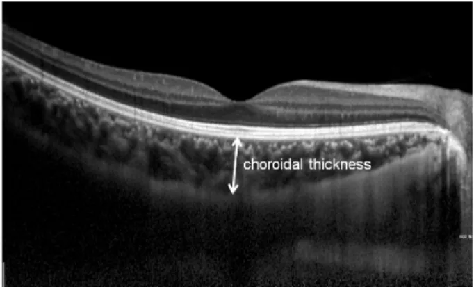
Figure 1. The choroid is seen in cross-section using EDI-
OCT. Subfoveal choroidal thickness was measured vertically from the outer border of the retinal pigment epithelium to the inner border of the sclera.를 관찰하는 것은 어려웠지만 최근 개발된 enhanced depth imaging spectral-domain optical coherence tomography (EDI OCT)를 통해 맥락막의 정량적 평가가 가능해졌고, 이를 통해 맥락막의 구조 및 두께 변화에 대한 연구가 활발 히 이루어지고 있다.10하지만 아직까지 당뇨환자의 맥락막 두께와 anti-VEGF 안내주입술이 맥락막에 주는 영향에 대 해서는 거의 알려진 것이 없다.
이에 본 연구에서는 당뇨환자에서 당뇨 망막병증의 정도 가 맥락막두께에 미치는 영향 및 베바시주맙 안내주입술 후 맥락막두께의 변화를 알아보고자 하였다.
대상과 방법
2011년 1월부터 2012년 12월까지 건국대학교병원 안과 외래를 방문한 총 105명 105안을 대상으로 하였다. 대조군 과 함께 당뇨 환자를 no change, mild, moderate, severe NPDR, PDR군 등 5군으로 나누고, 6군에서 각각 15명 15 안의 맥락막두께를 비교하였다. 또한 당뇨망막병증으로 베 바시주맙 안내주입술을 시행받은 15명 15안의 주입술 전 과 첫 주입술 한달 후의 중심와아래 맥락막두께 변화를 비 교하였다. 전체 대상의 의무기록과 검사기록은 후향적으로 분석하였다. 맥락막두께에 영향을 줄 수 있는 laser photo- coagulation, 유리체 절제술 등의 치료를 받았거나 고도근 시, 중심성 장액맥락망막병증, 나이관련황반변성 같은 안과 질환이 있는 환자는 대상에서 제외하였다. 또한 베바시주맙 안내주입술은 당뇨황반부종이 있거나 증식성 당뇨망막병증 으로 인한 신생혈관이 존재하는 환자에게 실행하였으며 유 리체강내 트리암시놀론 주입술 등과 같이 베바시주맙 외 다른 어떠한 당뇨망막병증치료를 받은 경우도 대상에서 제 외하였다.
모든 환자들을 대상으로 나이, 성별, 최대교정시력 및 구면대응치를 조사하였고, 양안의 세극등현미경검사, 안 저검사, Spectralis HRA+OCT (Heidelberg Engineering, Heidelberg, Germany)를 이용한 EDI-OCT를 시행하였다.
베바시주맙 안내주입술을 시행한 군은 주입술 전과 첫 주입술 4주 후에 앞에서 기술한 검사들을 시행하여 전후 차 이를 비교하였다. 베바시주맙 안내주입술은 환자를 시술 전 0.5% proparacaine hydrochloride (Alcaine, Alcon. Inc)를 점안하고 안내 감염을 예방하기 위해 0.05% povidone io- dine으로 안검 및 안구소독을 하였다. 이후 개검기로 눈꺼 풀을 벌리고 각막변연부로부터 수정체안의 경우 3.5 mm, 인공수정체안의 경우 3 mm 떨어진 하이측 공막에 30 gauge 주사기로 0.05 ml 베바시주맙을 유리체강 내로 투여 하였으며 점안 항생제(moxifloxacin, vigamox, Alcon. Inc) 를 시술 후 1주일간 하루 2시간 간격으로 점안하여 이차 감 염을 예방하였다.
맥락막두께는 EDI-OCT 촬영 후 중심와아래 바깥 망막 색소상피 경계에서 내측 공막 경계까지의 거리로 정의하였 으며, 자체 프로그램에서 제공되는 scale bar를 이용하여 측정하였다(Fig. 1).
통계적 분석은 SPSS (version 17.0)로 당뇨 정도에 따른 맥락막두께를 비교하기 위하여 Mann-Whitney U test를, 베바시주맙 안내주입술 전과 후의 맥락막두께를 비교하기 위하여 Wilcoxon signed rank test를 사용하였으며 p<0.05 일 경우 통계적으로 유의한 것으로 해석하였다.
결 과
각 군별 평균 나이(years, mean ±SD)와 구면대응치(D, mean ±SD)는 각각, 대조군 50.6 ±16.9, -0.32 ±1.06;
no change군 62.2 ± 14.1, 0.98 ± 2.80; mild NPDR군 61.0 ±11.3, -0.22 ±2.07; moderate NPDR군 61.2 ± 13.1, 0.40 ±1.55; severe NPDR군 57.9 ±10.1, -0.15
±1.39; PDR군 58.3 ±16.6, 0.25 ±1.37이었다(Table 1).
각 군간 평균 나이는 통계적으로 차이가 없었다(Mann- Whitney Utest, all p>0.05). 전체 환자에서 나이와 맥락 막두께를 비교한 결과 통계적으로 유의한 음의 상관 관계가 있었다(pearson correalation, r=-0.257, p=0.015).그러나 맥락막두께를 구면대응치와 비교한 결과 유의하지 않은 것 으로 나타났다(pearson correalation, p=0.378). 각 군의 평균 맥락막두께(μm, mean ± SD)는 대조군 277.9 ± 22.4, no change군 279.3 ±36.7, mild NPDR군 257.6 ± 25.9, moderate NPDR군 229.6 ±37.6, severe NPDR군 224.1 ±30.8, PDR군 192.0 ±36.7이었다(Table 1). 대
Table 1. Characteristics of control subjects and diabetic patients
Control No change Mild NPDR Moderate NPDR Severe NPDR PDR
Age (years) 50.6 ± 16.9 62.2 ± 14.1 61.0 ± 11.4 61.2 ± 13.0 57.9 ± 10.1 60.0 ± 9.5
Sex (M/F) 7/8 8/7 6/9 7/8 10/5 8/7
Spherical equivalent (D) -0.32 ± 1.06 0.98 ± 2.80 -0.22 ± 2.07 0.40 ± 1.55 -0.15 ± 1.39 0.43 ± 1.39 Choroidal thickness (μm) 277.9 ± 22.4 279.3 ± 36.7 257.6 ± 25.9 229.6 ± 37.6 224.1 ± 30.8 192.6 ± 47.2 Values are presented as mean ± SD.
NPDR = non proliferative diabetic retinopathy; PDR = proliferative diabetic retinopathy.
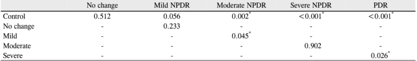
Table 2. Comparison of choroidal thickness among stages of diabetic retinopathy progression (p-value)
No change Mild NPDR Moderate NPDR Severe NPDR PDR
Control 0.512 0.056 0.002* <0.001* <0.001*
No change - 0.233 - - -
Mild - - 0.045* - -
Moderate - - - 0.902 -
Severe - - - - 0.026*
Dashes (-) indicate that the comparisons were not performed.
NPDR = non proliferative diabetic retinopathy; PDR = proliferative diabetic retinopathy.
*p < 0.05.
Figure 2. Mean subfoveal choroidal thickness according to se-
verity of diabetic retinopathy.Table 3. Baseline characteristics of diabetic patient with intra-
vitreal bevacizumab injectionPatients no. (M/F) 15 (9/6)
Age (years) 58.3 ± 8.6
Spherical equivalent (D) 0.27 ± 1.36
Injection no. 1.8 ± 0.8
BCVA (Snellen) Pre-IVB 0.6 ± 0.3
1 month after IVB 0.7 ± 0.3 Choroidal thickness (μm) Pre-IVB 187.3 ± 47.2 μm
1 month after IVB 168.9 ± 41.3 μm Values are presented as mean ± SD.
NPDR = non proliferative diabetic retinopathy; PDR = proliferative diabetic retinopathy; BCVA = best corrected visual acuity; IVB = intravitreal Bevacizumab injection.
조군과 환자군의 맥락막두께를 비교하였을 때 moderate, severe NPDR, PDR군의 맥락막두께가 대조군에 비해 유의 하게 얇았다(Mann-Whitney U test, moderate NPDR p=0.002; Severe NPDR p<0.001; PDR p<0.001, Table 2). 또한 연속된 당뇨망막병증 정도 별로 비교하였을 경우 moderate군은 mild NPDR군에 비하여, PDR군은 severe NPDR군에 비하여 맥락막두께가 유의하게 얇았다(Mann- Whitney Utest, Mild to moderate NPDR, p=0.045; Severe NPDR to PDR, p=0.026, Table 2).
당뇨망막병증 정도에 따른 평균 맥락막두께를 비교할 경 우, 비록 모든 연속된 당뇨망막병증 군 간에 통계학적으로 유의한 차이를 보이지는 않았지만 당뇨망막병증이 심해질 수록 맥락막두께가 감소하는 경향이 있는 것을 확인할 수 있다(Fig. 2).
베바시주맙 안내주입술을 시행받은 15명 15안의 평균나
이는 58.3 ±8.6세, 구면대응치 0.27 ±1.36 diopter, 평균 주사 횟수는 1.8회였다. Severe NPDR 4명 4안, PDR 11명 11안이 포함되었으며 severe NPDR 4안은 모두 당뇨황반 부종이 존재하였고 PDR 중 8안은 신생혈관, 3안은 당뇨황 반부종 및 신생혈관 모두 존재하였다. 최대교정 시력은 주 입술 전 0.6, 첫 주입술 1달 후 0.7로 호전되었다. 맥락막두 께는 주입술 전 187.3 ± 47.2 μm에서 주입술 1달 후 168.9 ±41.3 μm로 감소하였으며 이는 통계적으로 유의한 차이가 있었다(Wilcoxon signed rank test, p=0.001, Table 3, Fig. 3).
고 찰
맥락막은 혈관이 풍부한 조직으로 맥락막의 정상적인 구 조와 기능은 망막 기능에 필수적인 요소이다. 맥락막은 정
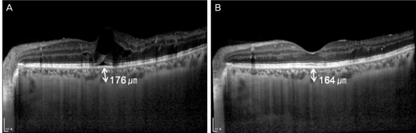
A B
Figure 3. Representative case of a 51-year-old man with diabetic macular edema was treated with IVB. (A) The VA was 20/30, and
the subfoveal choroidal thickness was 176 μm, at initial visit. (B) At one month after first IVB, the VA was improved to 20/25. The subfoveal choroidal thickness was 164 μm.상적으로 망막외층에 영양분 및 산소를 공급하지만 당뇨망 막병증이 진행된 경우 맥락막 모세혈관의 퇴화 등으로 인 하여 맥락막 혈류 감소가 일어나며 이로 인해 망막 시세포 층의 기능 상실 및 손상을 야기할 수 있다.11 또한 당뇨로 인한 맥락막 혈류 감소는 결국 맥락막두께를 감소시키는 것으로 보고되었다.6,7 이처럼 맥락막은 당뇨망막병증의 병 태생리에 중요한 역할을 하는 것으로 알려졌다. 또한 최근 혈관내피성장인자가 당뇨황반부종 및 증식당뇨망막병증의 정도와 서로 상관관계가 있다는 것이 밝혀지면서-anti VEGF 안내주입술이 당뇨황반부종과 당뇨망막병증 치료로 사용되고 있다.12-14 그러나 망막색소상피층에서 분비되는 혈관내피성장인자는 성인에서 혈관 조직의 생존 및 투과성 을 유지하는데 중요한 역할을 한다.15,16맥락막 혈관에는 혈 관내피성장인자 수용체들이 풍부하게 존재하며, 맥락막 혈 관의 생리학적 활동에 필수적인 혈관내피성장인자를 베 바시주맙 안내주입술을 통해 막게 되면 맥락막 혈관의 투 과성 감소를 야기하고, 결국 맥락막두께를 얇게 만들 수 있다. 최근까지 맥락막두께는 생체를 통한 조직학적 결과 로부터 얻은 정보에 의존할 수 밖에 없었다.17 근래에는 고해상도의 영상을 가능하게 한 스펙트럼 도메인 빛간섭단 층촬영(spectral-domain optical coherence tomography, SD-OCT) 장비가 도입되었으며, enhanced depth imaging spectral-domain optical coherence tomography (EDI-OCT) 를통해 맥락막의 영상화가 가능해졌다.18이후 이러한 장비 를 이용하여 많은 연구에서 정상안을 비롯하여 중심성 장 액맥락망막병증, 근시, 나이관련 황반변성 등 여러 망막질 환의 맥락막두께를 보고하였다.19-21
본 연구는 EDI-OCT를 이용하여 아직까지 국내에 발표 된바 없는 당뇨 환자와 베바시주맙 안내주입술을 시행한 환자의 맥락막두께를 측정하였다. 그 결과 moderate, se-
vere NPDR, PDR군이 대조군에 비하여 맥락막두께가 얇았 으며 특히 moderate NPDR군은 mild NPDR군에 비하여, PDR군은 severe NPDR군에 비하여 맥락막두께가 유의하 게 얇은 것을 확인할 수 있었다. 이는 맥락막두께의 감소를 야기하는 맥락막 혈관의 손상 정도와 혈류 감소가 당뇨망 막병증의 정도에 따라 다르다는 것을 시사한다. 또한 베바 시주맙 안내주입술 시행전과 첫 주입술 1달 후를 비교하였 을 때 맥락막두께가 유의하게 얇아진 것으로 미루어 베바 시주맙 안내주입술이 맥락막 순환 저하에 미치는 영향을 예측할 수 있었다. 베바시주맙 안내주입술 시행전의 맥락막 두께가 대조군에 비하여 얇은 이유는 안내주입술을 시행한 환자 모두가 황반부종 및 신생혈관이 발생할 정도로 당뇨 망막병증이 진행되어 이미 맥락막 혈관 및 구조에 영향을 끼쳤기 때문이라고 생각한다.
정상적인 노화과정으로 인하여 맥락막두께는 1년에 약 1.4-1.56 μm 정도 감소하는 것으로 알려졌다.18또한 근시 환자의 맥락막두께에 대한 연구에서 정상인이 고도근시 환 자보다 두께가 3배 정도 두꺼우며 구면대응치가 1 diopter 증가할때 약 9.3 μm 정도 맥락막두께가 증가한다는 결과가 발표 되었다.22,23본 연구에서는 이전에 발표된 연구와 마찬 가지로 나이에 따른 맥락막두께의 유의한 감소가 관찰되었 다. 하지만 분석 결과 이전 연구 결과와는 다르게 구면대응 치와 맥락막두께는 유의한 상관성을 보이지 않았다. 이는 대상 환자에서 고도근시 환자를 제외하였기 때문에 구면대 응치의 편차가 크지 않아 맥락막두께에 미치는 영향이 상 대적으로 적었기 때문인 것으로 추측된다.
본 연구는 기록 분석이 후향적으로 진행된 것과 상대적 으로 대상 환자가 적은 것을 제한점으로 꼽을 수 있다. EDI OCT 검사가 고해상도의 영상을 제공하지만 일부 검사에서 는 artifact가 발생할 수 있어 맥락막두께의 측정 시 오차를
유발할 수 있다는 점, 맥락막두께를 자동화된 프로그램에 의해 측정하지 않고 검사자에 의해 수동으로 측정되었다는 것 또한 제한점이다. 맥락막두께에 영향을 줄 수 있는 요소 의 상관분석에서 당뇨황반부종 유무는 제외되었다. 그 이유 는 당뇨황반부종이 맥락막두께에 미치는 영향이 아직 정확 히 밝혀지지 않았기 때문이다.7,24향후 대규모 환자를 대상 으로 장기적인 추적관찰을 시행하여 당뇨황반부종을 포함 한 맥락막두께에 영향을 줄 가능성이 있는 여러 요소를 분 석한 연구가 필요할 것이다.
결론적으로 맥락막두께는 당뇨망막병증의 정도가 진행 된 경우 얇았다. 또한 베바시주맙 안내주입술을 시행한 경 우 시행 전에 비해 시행 1달 후 맥락막두께는 얇아졌다.
EDI-OCT는 맥락막두께를 평가할 수 있는 비침습적 방법 으로써 당뇨 환자에서 맥락막의 구조적, 기능적 손상을 예 측하는데 도움이 될 수 있다.
REFERENCES
1) Seo JW, Park IW. Intravitreal bevacizumab for treatment of dia- betic macular edema. Korean J Ophthalmol 2009;23:17-22.
2) Velez-Montoya R, Fromow-Guerra J, Burgos O, et al. The effect of unilateral intravitreal bevacizumab (avastin), in the treatment of diffuse bilateral diabetic macular edema: a pilot study. Retina 2009;29:20-6.
3) Avery RL. Regression of retinal and iris neovascularization after in- travitreal bevacizumab (Avastin) treatment. Retina 2006;26:352-4.
4) Spaide RF, Fisher YL. Intravitreal bevacizumab (Avastin) treat- ment of proliferative diabetic retinopathy complicated by vitreous haemorrhage. Retina 2006;26:275-8.
5) Cao J, McLeod S, Merges CA, Lutty GA. Choriocapillaris degen- eration and related pathologic changes in human diabetic eyes.
Arch Ophthalmol 1998;116:589-97.
6) Regatieri CV, Branchini L, Carmody J, et al. Choroidal thickness in patients with diabetic retinopathy analyzed by spectral-domain op- tical coherence tomography. Retina 2012;32:563-8.
7) Querques G, Lattanzio R, Querques L, et al. Enhanced depth imag- ing optical coherence tomography in type 2 diabetes. Invest Ophthalmol Vis Sci 2012;53:6017-24.
8) Marneros AG, Fan J, Yokoyama Y, et al. Vascular endothelial growth factor expression in the retinal pigment epithelium is essen- tial for choriocapillaris development and visual function. Am J Pathol 2005;167:1451-9.
9) Heiduschka P, Fietz H, Hofmeister S, et al. Penetration of bev-
acizumab through the retina after intravitreal injection in the monkey. Invest Ophthalmol Vis Sci 2007;48:2814-23.
10) Spaide RF, Koizumi H, Pozzoni MC. Enhanced depth imaging spectral-domain optical coherence tomography. Am J Ophthalmol 2008;146:496-500.
11) McLeod DS, Lutty GA. High-resolution histologic analysis of the human choroidal vasculature. Invest Ophthalmol Vis Sci 1994;
35:3799-811.
12) Funatsu H, Yamashita H, Sakata K, et al. Vitreous levels of vas- cular endothelial growth factor and intercellular adhesion molecule 1 are related to diabetic macular edema. Ophthalmology 2005;112:
806-16.
13) Arevalo JF, Fromow-Guerra J, Quiroz-Mercado H, et al. Primary intravitreal bevacizumab (Avastin) for diabetic macular edema: re- sults from the Pan-American Collaborative Retina Study.
Ophthalmology 2007;114:743-50.
14) Aiello LP, Avery RL, Arrigg PG, et al. Vascular endothelial growth factor in ocular fluid of patients with diabetic retinopathy and other retinal disorders. N Engl J Med 1994;331:1480-7.
15) Maharaj AS, D’Amore PA. Roles for VEGF in the adult.
Microvasc Res 2007;74(2-3):100-13.
16) Saint-Geniez M, Maldonado AE, D’Amore PA. VEGF expression and receptoractivation in the choroid during development and in the adult. Invest Ophthalmol Vis Sci 2006;47:3135-42.
17) Chen TC, Cense B, Miller JW, et al. Histologic correlation of in vivo optical coherence tomography images of the human retina.
Am J Ophthalmol 2006;141:1165-8.
18) Margolis R, Spaide RF. A pilot study of enhanced depth imaging optical coherence tomography of the choroid in normal eyes. Am J Ophthalmol 2009;147:811-5.
19) Lee SH, Chung H, Kim HC. subfoveal choroidal thickness in fel- low eyes of patients with central serous chorioretinopathy. J Korean Ophthalmol Soc 2012;53:982-7.
20) Fujiwara T, Imamura Y, Margolis R, et al. Enhanced depth imaging optical coherence tomography of the choroid in highly myopic eyes. Am J Ophthalmol 2009;148:445-50.
21) Spaide RF. Age-related choroidal atrophy. Am J Ophthalmol 2009;147:801-10.
22) Ikuno Y, Kawaguchi K, Nouchi T, Yasuno Y. Choroidal thickness in healthy Japanese subjects. Invest Ophthalmol Vis Sci 2010;
51:2173-6.
23) Ikuno Y, Tano Y. Retinal and choroidal biometry in highly myopic eyes with spectral-domain optical coherence tomography. Invest Ophthalmol Vis Sci 2009;50:3876-80.
24) Esmaeelpour M, Považay B, Hermann B, et al. Mapping choroidal and retinal thickness variation in type 2 diabetes using three-di- mensional 1060-nm optical coherence tomography. Invest Ophthalmol Vis Sci 2011;52:5311-6.
=ABSTRACT=
Effects of Diabetic Retinopathy and Intravitreal Bevacizumab Injection on Choroidal Thickness in Diabetic Patients
Byeong Jun Park, MD, Hye Won Chung, MD, PhD, Hyung Chan Kim, MD, PhD
Department of Ophthalmology, Konkuk University Medical Center, Konkuk University School of Medicine, Seoul, Korea
Purpose: To evaluate the effect of diabetic retinopathy on choroidal thickness and the changes of choroidal thickness after intravitreal bevacizumab injection (IVB) in diabetic patients.
Methods: The present study included 105 patients (105 eyes). Patients were classified into 6 groups: control group (A); no change (B), mild (C), moderate (D), and severe (E) non proliferative diabetic retinopathy (NPDR); and proliferative diabetic retinopathy (PDR) (F), with 15 diabetic patients in each group. All patients underwent enhanced depth imaging spectral- domain optical coherence tomography (EDI OCT) to evaluate choroidal thickness. An additional 15 patients (15 eyes) with diabetic retinopathy treated with IVB were also included. These patients underwent EDI OCT before and 1 month after IVB.
Results: Mean choroidal thickness was significantly thinner in the moderate and severe NPDR, and PDR groups com- pared with the control group. Moreover, when comparing sequential stages of diabetic retinopathy progression, the choroi- dal thickness in the moderate NPDR stage and PDR stage was found to be significantly thinner than in the mild and severe NPDR, respectively. Additionally, choroidal thickness was 187.3 μm before IVB and significantly decreased to 168.9 μm 1 month after IVB (p < 0.05).
Conclusions: Choroidal thickness is related to the severity of diabetic retinopathy and is significantly decreased after IVB.
J Korean Ophthalmol Soc 2013;54(10):1520-1525
Key Words: Bevacizumab, Choroidal Thickness, Diabetic Retinopathy, Enhanced Depth Imaging Optical Coherence Tomography
Address reprint requests to Hyung Chan Kim, MD, PhD
Department of Ophthalmology, Konkuk University Medical Center
#120-1 Neungdong-ro, Gwangjin-gu, Seoul 143-729, Korea
Tel: 82-2-2030-8180, Fax: 82-2-2030-5273, E-mail: [email protected]