대한골절학회지 제17권, 제3호, 2004년 7월 Journal of the Korean Fracture Society
Vol. 17, No. 3, July, 2004
도수 정복 및 K-강선 내고정술을 이용한 Mason 제 2형 요골 두 골절의 치료
권병기·이 송·안동기·박준성·차상규
서울성심병원 정형외과
목 적: Mason 분류 제 2형 요골 두 골절에 대하여 C-arm guide 하에 K-강선을 이용한 도수 정복술 및 내고정술 시행 후 기능적 및 방사 선학적으로 임상적 결과를 분석하고자 하였다.
대상 및 방법: 2001년 3월부터 2003년 10월 까지 Mason 분류 제 2형 요골 두 골절로 내원한 환자 중 C-arm guide 하에 도수정복 후 K-강 선을 내고정술을 시행받은 7례를 대상으로 하였으며, 평균 연령은 38세 (5세~57세)였고, 평균 추시 기간은 20개월 (5개월~36개월)이었다. 이 들 7례를 대상으로 최종 추시시 방사선학적 평가와 기능적 평가로써 Broberg와 Morrey의 functional rating system에 따라 우수 (Excellent), 양호 (Good), 보통 (Fair) 및 불량 (Poor)으로 분류하여 결과를 판정하였다.
결 과: 최종 추시 결과 주관절의 운동 범위는 굴곡 구축 평균 1.4도 (범위: 0도~10도), 후속 굴곡 평균 146.4도 (140도~150도)였으며, 전완부 의 회외전 평균 74.2도 (70도~80도), 회내전 평균 75도 (70도~80도)였다. 기능적 평가 결과 우수 6례 및 양호 1례였으며, 불량에 해당하는 예는 없었다. 방사선학적 평가상 전례에서 골유합 소견을 관찰 되었으며, 유합까지의 기간은 평균 5주 (범위: 4주~6주)였으며, 전례에서 심각 한 합병증 등은 발생하지 않았다.
결 론: Mason 분류 제 2형 요골 두 골절의 치료에 있어서 C-arm guide하에 K-강선을 이용한 골절편 정복술 및 내고정술은 합병증 발생 없 이 만족할 만한 결과를 얻을 수 있는 유용한 방법이라 사료된다.
색인 단어: 요골 두 골절, 도수적 정복, K-강선, 내고정술
The Treatment of Mason Type II Radial Head Fractures using Closed Reduction and K-wire Fixation
Byung Ki Kwon, M.D., Song Lee, M.D., Dong Ki Ahn, M.D., Joon Seong Park, M.D., Sang Kyu Cha, M.D.
Department of Orthopedic Surgery, Seoul Sacred Heart General Hospital, Seoul, Korea
Purpose: To analyze the clinical results of the treatment of Mason type II radial head fractures using closed reduction and K-wire internal fixation under C-arm guide by radiologically and functionally.
Materials and Methods: Between March 2001 and October 2003, 7 patients with Mason type II radial head fracture were treated by closed reduction and internal fixation using K-wires under C-arm guide. The average age of the patients was 38 (5 to 57) years old , and average duration of follow up was 20 (5 to 36) months. At last follow up, we evaluated the radiological results and functional results by classifying excellent, good, fair and poor according to functional rating system of Broberg and Morrey.
Results: The range of motion of the elbow at last follow up, average flexion contracture was 1.4 (0 to 10) degrees, further average flexion was 146.4 (140 to 150) degrees, average supination was 74.2 (70 to 80) degrees and average pronation was 75 (70 to 80) degrees. In the functional results, 6 cases were excellent and 1 case was good. In the radiological evaluations, the average time of union was 5 (4 to 6) weeks after the operation and no serious complication was occurred in any cases.
Conclusion: In the treatment of Mason type II radial head fracures, closed reduction and K-wire internal fixation under C-arm guide was an effective method of treatment with satisfactory results and no complications.
Key Words: Radius head fracture, Closed reduction, K-wire, Internal fixation
277 통신저자 : 권 병 기
서울특별시 동대문구 청량리동 40-12 서울성심병원 정형외과
Tel : 02-966-1616·Fax : 02-968-2394 E-mail : [email protected]
Address reprint requests to : Byung Ki Kwon, M.D.
Department of Orthopedic Surgery, Seoul Sacred Heart General Hospital 40-12 Chungryangri-dong, Dongdaemoon-gu, Seoul 130-011, Korea Tel : 82-2-966-1616·Fax : 82-2-968-2394
E-mail : [email protected]
서 론
요골 두는 척골 근위부와 관절하여 주로 전완의 회내전 및 회외전 운동에 관여하면서 상완골의 소두와 관절하여 주 관절의 굴신운동에도 관여한다. 이러한 요골 두 골절의 치료 목적은 동통 없는 정상 관절운동과 주관절의 안정성을 회복 하는데 있다. 비전이성 요골 두 골절의 치료에 있어서 보존 적 치료에는 논란의 여지가 없으나, 전이성 요골 두 골절 치 료의 수술적 방법에 대하여는 여러 이견이 많다21). 요골 두 골절의 수술적 치료로는 대개 Mason 분류13) (Table 1) 제 2 형 이상에서 실시하며 방법으로는 골편절제술, 요골 두 제 거술, 관혈적 정복 및 내고정술과 요골 두의 인공삽입물 대 치술 등을 들 수 있다. 그 중 요골 두 제거술은 원위 요척 관 절의 불안정성, 수근 관절 및 주관절의 동통 및 퇴행성 관절 염, 관절의 불안정성, 이소성 골형성 등의 많은 문제점을 유 발할 수 있고11,12,17), 또한 관혈적 정복술 및 내고정술이나 인공삽입물 대치술은 골절부의 개방과 조작에 따른 연부 조직의 손상으로 인한 합병증이 발생할 수 있어 치료에 있 어서 고려해야 할 사항이다.
이에 저자들은 Mason 분류13) 제 2형 요골 두 골절로 내 원한 환자 7례에 대해서 경피적으로 K-강선을 삽입하여 골절정복 및 내고정술을 시행하고 최종 추시시 임상적 결 과를 문헌 고찰과 함께 보고하고자 한다.
대상 및 방법
2001년 3월부터 2003년 10월까지 Mason 분류13) 제 2형의 요골 두 골절로 내원한 환자 중 경피적으로 삽입한 K-강 선을 이용해 골절정복 및 내고정술을 시행한 7례를 대상으 로 하였다. 성별로는 남자 4례, 여자 3례였으며 수술시 연령 은 최소 5세, 최고 57세로 평균 36.1세였다. 수상의 원인으 로는 주관절 신전상태에서 추락 사고가 5례, 교통사고에 의 한 직접 손상이 2례였다. 수상 후 수술까지의 기간은 대개 1일에서 3일 이내였으며 수술 후 평균 추시기간은 20개월 (5개월~36개월)이었다.
수술은 환자를 앙와위에서 전신마취, 혹은 상완 신경총
차단술 시행 후 C-arm guide하에 골절부를 확인하고 주관절 혹은 전완부의 측면에서 경피적으로 K-강선 1개를 삽입하 여 K-강선을 요골 두의 골절선 사이로 전진 시켜서 골절 편 사이에 위치하게 한 후, 이 K-강선을 조작하여 골절정 복을 시도하였다. 골절부가 해부학적 정복이 되면 골절편 사이에 삽입한 K-강선을 드릴로 전진시켜서 반대쪽 피질골 까지 고정하고 필요한 경우 1개의 K-강선을 추가적으로 경 피적으로 삽입하여 골절편을 고정하였다. 수술직후 주관절 을 90도 굴곡 및 중립 회전위 상태에서 장상지 석고 고정을 하고 4주후 석고붕대와 내고정된 K-강선을 제거한 후 2주 간 착탈이 가능한 장상지 부목 (removable long arm splint)을 착용하게 하여 수동적 관절운동을 실시하였다. 수술 후 6주 부터 동통이 없는 한도 내에서 능동적 관절운동 및 근력운 동을 점차적으로 증가시켰다. 이들에 대해서 최종 추시시 방 사선학적으로 골유합 여부를 판단하였고, 기능적 평가로는 Broberg와 Morrey의 functional rating system3)을 따라 우수 (Excellent), 양호 (Good), 보통 (Fair) 및 불량 (Poor)으로 분류 하여 임상적 결과를 판정하였다 (Table 2).
Table 1. Mason classification
Type 1- Nondisplaced or minimally displaced fracture of the radial head
Type 2- Displaced fracture of the radial head Type 3- Comminuted fracture of the radial head
Type 4- Radial head fracture associated with elbow dislocation (with or without coronoid Fracture)
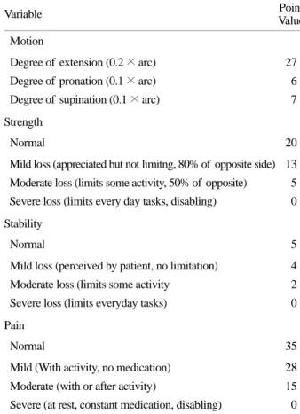
Table 2. Functional Rating Index (Modified After B.F. Morrey et al.)
Variable Point
Value
Motion Degree of extension (0.2 × arc)
Degree of pronation (0.1 × arc) Degree of supination (0.1 × arc)
27 6 7
Strength Normal 20
Mild loss (appreciated but not limitng, 80% of opposite side) Moderate loss (limits some activity, 50% of opposite) Severe loss (limits every day tasks, disabling)
13 5 0
Stability
Normal 5
Mild loss (perceived by patient, no limitation) Moderate loss (limits some activity
Severe loss (limits everyday tasks)
4 2 0
Pain Normal 35
Mild (With activity, no medication) Moderate (with or after activity)
Severe (at rest, constant medication, disabling)
28 15 0 (The translation of tital score to qualitiative groups was as follows:
95 to 100 points, excellant; 80 to 94 points, good; 60 to 79 points, fair; and zero to 59 points, poor.)
결 과
최종 추시 결과 주관절의 운동 범위는 굴곡 구축 평균 1.4도 (범위: 0도~10도), 후속 굴곡 평균 146.4도 (140도~150 도)였으며, 전완부의 회외전 평균 74.2도 (70도~80도), 회내 전 평균 75도 (70도~80도)였다 (Table 3, 4). 술후 관절의 기 능 평가를 위해 운동 범위, 근력, 관절 안정성, 동통에 근거 한 Broberg와 Morrey의 functional rating system3)을 적용한 결 과 6례가 우수, 1례가 양호였으며, 불량에 해당하는 예는 없 었다. 단순 방사선 검사상 골진 (callus)이 나타나고, 골절선 이 소실되기 시작하는 소견을 골유합으로 판정하였는데, 유 합까지의 기간은 평균 5주 (범위: 4주~6주)였다. 1례에서 경 도의 동통과 주관절 운동제한의 소견을 보였으나, 심한 동 통을 동반한 관절운동 장애, 불유합, 이소성 석회화 및 불안 정성등과 같은 심각한 합병증으로 요골 두 절제술을 시행한 례는 없었다.
증 례
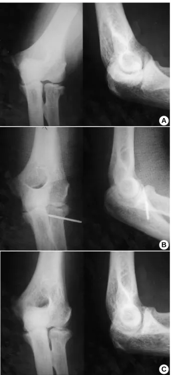
증례 1
35세 여자 환자로 낙상시 주관절 과신전 상태에서 땅을 짚 은 후 요골 두의 Mason분류13) 제 2형의 골절이 발생하였다 (Fig. 1A). 수상 1일 후 상완 신경총 차단술 하에서 C-arm guide하에 K-강선을 이용해서 골절편을 해부학적으로 정복 한 후 2개의 K-강선을 사용하여 내고정을 시행하였으며 (Fig. 2B), 술후 4주간 장상지 석고 붕대를 착용하였다. 수술 후 5개월 추시관찰에서 방사선 소견상 골유합은 견고하였고 (Fig. 1C), 통증은 없었다. 관절 운동 범위는 굴곡 구축 0도, 후속 굴곡 150도, 회내전 75도 및 회외전 80도로 정상이었 으며, 관절 불안정성 및 근력약화 소견 없는 상태로 Broberg 와 Morrey의 functional rating system3) 분류상 결과 평가는 우 수였다.
증례 2
57세 남자 환자로 낙상시 주관절 과신전 상태에서 땅을 짚은 후 부분적 분쇄 소견을 보이는 요골 두 골절로 내원 하였다 (Fig. 2A). 수상 1일 후 전신 마취하에 도수 정복을 한 후 1개의 K-강선을 이용하여 내고정을 시행하였다 (Fig.
2B). 수술 8개월 후 추시 관찰에서 방사선 소견상 견고한 골유합을 관찰 할 수 있었으나 (Fig. 2C), 심한 활동시 간헐 적인 동통을 호소하였다. 관절 운동 범위는 굴곡 구축 10도, 후속 굴곡 140도, 회내전 75도 및 회외전 70도였으며 Broberg 와 Morrey의 functional rating system3) 분류상 평가는 양호였다.
고 찰
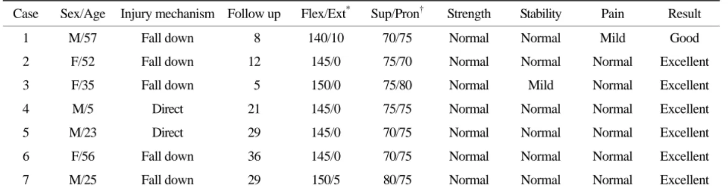
요골 두는 척골 절흔내에 위치하며 척골과 함께 전완부 의 회외전 및 회내전, 운동시에 상완골 소두와 관절하면서 주관절의 안정성에 매우 중요한 역할을 한다. Schwab 등18) Table 3. Patient data
Case Sex/Age Injury mechanism Follow up Flex/Ext* Sup/Pron† Strength Stability Pain Result 1 M/57 Fall down 8 140/10 70/75 Normal Normal Mild Good 2 F/52 Fall down 12 145/0 75/70 Normal Normal Normal Excellent 3 F/35 Fall down 5 150/0 75/80 Normal Mild Normal Excellent 4 M/5 Direct 21 145/0 75/75 Normal Normal Normal Excellent 5 M/23 Direct 29 145/0 70/75 Normal Normal Normal Excellent 6 F/56 Fall down 36 145/0 70/75 Normal Normal Normal Excellent 7 M/25 Fall down 29 150/5 80/75 Normal Normal Normal Excellent
*Flexion/Exention, †Supination/Pronation Motion, strength, stability, pain, and score are depicted in points
Table 4. Elbow evaluation result Mean motionz
Extension 1.4 degrees
Fexion 146.4 degrees
Pronation 75 degrees
Supination 74.2 degrees
Motion 39.7 Strength 20
Stability 4.8
Pain 33.2
Elbow score 97.5
Motion, strength, stability, pain, and score are depicted in points
은 외반 부하 (Valgus stress)에 저항하는 구조물로 요골 두의 중요성에 대해 언급한 바 있으며, Morrey 등16)은 요골 두를 외반력에 저항하는 중요 구조라 하여, 내측 측부인대에 이 어 secondary stabilizer라고 하였다. 종적인 안정성에 대하여
Halls와 Travill9)은 완관절에 가해진 종적 부하를 견디는 골 성 구조물의 역할에 대해 요상완 관절이 40%, 척상완 관절 이 60%의 부하를 담당한다고 하였다. 따라서 요골 두 골절 시에는 주관절의 관절운동과 안정성에 장애가 발생하여 심 Fig. 1. (A) Thirty-five year-old female patient sustained Mason
type II radial head fracture.
(B) Immediate postoperative radiograph shows reduced fracture fragment, which was fixed with 2 K-wires.
(C) Radiograph at postoperative 5 months shows anatomically healed radial head.
A
C B
Fig. 2. (A) graph of 57 year-old male patient shows comminuted radial head fracture.
(B) Fracture fragments were reduced and fixed with one K-wire.
(C) Radiograph of postoperative 8 months shows well heald radial head.
C B A
각한 관절 기능 장애를 초래할 수 있다. 성인의 요골 두 골 절은 모든 주관절 손상의 약 20%를 차지하며4), 수상 기전은 주관절을 신전한 상태에서 종적 하중이 요골 두에 가해질 때 가장 많이 발생하는 것으로 알려져 있다1,4). 전위가 있는 요골 두 골절의 경우 관절 불안정성 및 동통을 동반한 운동 장애 등의 합병증의 발생이 높으므로 수술적 치료를 요하며, 수술방법으로는 요골 두의 관혈적 정복 및 내고정술, 골편 제거술, 요골 두 절제술, 요골 두 인공성형치환술 등으로 나 눌 수 있다.
1897년 Helfrich가 요골 근위단의 골절을 처음으로 분류 하여 치료를 시작하였고, Schwartz와 Young19)은 요골 두 절 제술을 골절 치료의 한 방법으로 제안하였으며, Grossman8), Hammond10) 및 Sever20)은 회내전, 회외전 등의 운동 제한을 방지하기 위한 방편으로 골절편의 단순 제거술을 권장하기 도 하였다. 전위가 심하고 분쇄상인 요골 두 골절에서 요골 두 제거술은 주관절의 운동 강도나 운동 범위에는 많은 지 장을 초래하지만 널리 사용되어 왔다1).
1954년 Mason13)이 골절의 전이와 분쇄양상에 따라 분류 를 하였고, 치료에 있어서, 제 1형 골절은 보존적 치료를 시 행하고 조기 관절운동을 시행하고, Mason 제 2, 3형 골절은 요골 두 절제술과 다양한 내고정물을 이용한 관혈적 정복술 및 내고정술이 시행되어져 왔으나, 요골 두 절제술은 요골 단축, 관절 운동 장애 및 완관절 증상 등 술후 여러가지 합 병증을 유발할 수 있다. 이에 많은 저자들은 내고정술이 더 좋은 결과를 나타낸다고 보고하였고2,5,6,7,11)
, 대체적으로 Mason 제 3형 보다 제 2형에서 더 좋은 결과를 보고하고 있으며 이 때 사용되어진 내고정물로는 K-강선, 흡수성 나사못, Herbert 나사못, AO의 압박 나사못 및 금속판 등이다14,15). 흡수성 나 사못 사용시 골흡수, 내고정물에 의한 활액막염 등의 합병 증 발생이 보고 되었고2), 금속 고정물을 이용한 관혈적 정 복술 및 내고정술 또한 관절운동장애, 수술시 신경 손상, 이 소성 골형성, 부정유합, 불유합, 고정의 소실 및 무혈성 괴 사등의 합병증을 유발할 수 있다21).
이에 저자들은 관혈적 정복술에서 발생할 수 있는 합병증 을 최소화하면서, 가능한 요골 두를 해부학적 정복을 얻고자 하였으며, K-강선을 경피적으로 골절편 사이에 삽입하여 K-강선을 통해 골절편을 직접 조작함으로써 정확한 정복과 안정적인 고정이 가능하였고, 주관절을 개방해서 골절부를 조작함으로써 발생할 수 있는 연부조직의 합병증을 피할 수 있었으며, 전례에서 최종 추시시 만족 할만한 골유합 소견 과 기능적으로도 정상에 가까운 임상적 결과를 얻었다. 또 한 관혈적 정복술후 가장 심각한 합병증인 이소성 골형성 및 신경손상을 포함한 다른 중요 합병증 발생이 없었고, 연 부조직의 손상이 없었으므로 관절운동과 근력의 회복이 조 기에 가능하였으며, 관헐적 정복술에 비해 수술 시간이 짧
으며 수술부위를 개방하지 않기 때문에 마취 방법에 있어서 상완 신경총 차단술만으로도 충분히 가능하여 환자가 반드 시 입원할 필요가 없었다. 하지만 Mason 제 3형 이상의 분쇄 골절에서 시행한 예는 없으나 이러한 수기만으로는 좋은 결 과를 얻기 힘들 것으로 예상된다.
결 론
Mason 제 2형의 요골 두 골절의 치료에 있어서 C-arm guide하에 K-강선을 이용한 골절부 정복술 및 내고정술은 관혈적 정복술에 비해 간단하면서도 합병증 발생을 최소화 하면서 만족할 만한 결과를 얻을 수 있는 유용한 방법이라 사료된다.
참 고 문 헌
1) Adler JB and Shaftan GW: Radial head fractures. Is exci- sion necessary? J Trauma, 4: 115, 1964.
2) Baek GH, Sohn YJ, Lee CK and Chung MS: Bioabsor- bable Implant Fixation for the Treatment of Radial Head and Neck Fractures. J Korean Fracture Soc, 11: 70-77, 1998.
3) Broberg MA and Morrey BF: Results of delayed excision of the radial head after fractures. J Bone Joint Surg, 68-A:
669-674, 1986
4) Browner BD, Jupiter JB, Levine AM and Trafton PG:
Skeletal trauma, 2: 1125, 1992.
5) Bunker TD and Newman JH: The Herbert differential pitch bone screw in displaced radial head fractures. Injury, 16:
621-624, 1942.
6) Esser RD, Davis S and Taavao T: Fractures of radial head treated by intrnal fixation: late results in 26 cases. J Orthop Trauma, 9: 318-323, 1995.
7) Geel CW, Palmer AK and Ruedi T: Internal fixation of proximal radial head fractures. J Orthop Trauma, 4: 270-274, 1990.
8) Grossman J: Fracture of the head and neck of the radius. N Y J Med, 17: 472, 1923.
9) Halls AA and Travill A: Transmission of pressures across the elbow joint. Anat Rec, 150: 243-247, 1964.
10) Hammond R: Fracture of the head and neck of the radius.
Ann Surg, 53: 207, 1910.
11) Kang HJ, Park MS, Shin SJ, Kang HS and Park BM:
Operative Treatment of Radial Head Fracture of Mason type III. J Korean Fracture Soc, 12: 732-740, 1999.
12) King GJE, Evans DC and Kellam JF: Open reduction and
internal fixation of radial head fractures. J Orthop Trauma, 5:
21-28, 1991.
13) Mason ML: Some observation on the fracture of the radius.
J Bone Joint Surg, 42-A: 123-132, 1954.
14) McArthus RA: Herbert screw fixation of fracture of the head of radius. Clin Orthop, 224: 79-87, 1987.
15) Morrey BF: Current concepts in the treatment of fractures of the radial head, the olecranon, and the coronoid. Instructional course lectures. The Academy of American Orthopedic Su- rgeons, 22: 175-185, 1995.
16) Morrey BF, Tanaka S and An KN: Valgus stability of the elbow: A definition of primary and secondary constraints.
Clin Orthop, 265: 187-195, 1991.
17) Ring D, Quintero J and Jupiter JB: Open reduction and internal fixation of fractures of the radial head. J Bone Joint Surg, 84-A: 1811-1815, 2002.
18) Schwab GH, Bennett JB, Woods GW and Tullos HS:
Biomechanics of elbow instability: The role of the medial collateral ligament. Clin Orthop, 146: 42-52, 1980.
19) Schwartz RP and Young F: Treatment of fractures of the head and neck of the radial epiphysis in children. Surg Gynecol Obstet, 57: 528, 1933.
20) Server J: Fracture of the head and neck of the radius: A study of and results. JAMA, 84: 1551, 1925.
21) Shin DB, Lee YK, Ahn JY and Joo YK: Treatment Radial Head Fracture. J Korean Orthop Assoc, 29: 1835-1839, 1994.