The Antifungal Activity of Bee Venom against Dermatophytes
A-Reum Yu · Jum-Ji Kim · Gil-Sun Park · Su-Mi Oh · Chung Sub Han · Mi-Young Lee
Received: 27 September 2011 / Accepted: 21 February 2012 / Published Online: 31 March 2012
© The Korean Society for Applied Biological Chemistry 2012
Abstract The antifungal activities of the bee venom against Trichophyton mentagrophytes and Trichophyton rubrum were determined by using modified broth dilution assay. The most common dermatophytes, named T. mentagrophytes and T. rubrum, were known to cause a variety of cutaneous infections in humans and animals. The bee venom exhibited prominent antifungal activities against the two dermatophytes tested in this investigation.
Moreover, the antifungal activities of the bee venom were much stronger than that of fluconazole, one of the commercial antifungal drugs used in the treatment and prevention of superficial and systemic fungal infections. The result suggests that bee venom could be developed as a natural antifungal drug.
Keywords antifungal activity · bee venom · Trichophyton mentagrophytes · Trichophyton rubrum
Introduction
Dermatophytosis, mycotic infections caused by dermatophytes, is thought to be one of the most important public health problem yet unresolved (Chinelli et al., 2003). The most common dermatophytes, such as Trichophyton mentagrophytes and Trichophyton rubrum, were known to cause a variety of cutaneous infections in humans and animals. Especially T. rubrum causes the most common dermatophytic nail infections in humans (Summerbell, 1997). It is very difficult to treat onychomycosis, nail infection caused by dermatophytes or by nondermatophytic molds, due to its high probability of recurrence and the prolonged antifungal agent treatment time (Lee et al., 2010). Skin infections by dermatophytes
are often associated with relapses after cessation of the therapy, despite of the great advances in antifungal agent development (Mukherjee et al., 2003). Fluconazole, itraconazole, ketoconazole, terbinafine, and griseofulvin are commercial antifungal agents against dermatophytes (Goupta and Del Rosso, 2000; Hainer, 2003). However, some side effects including drug-resistance were observed in commercial antifungal agents (Lee et al., 2010). To overcome these problems of side-effects, natural products have been considered to be promising antifungal agents with less profound adverse effects. The antifungal activities of several phytochemicals including polyphenols, phenolics, terpenoids, and alkaloids have been reported (Chee et al., 2009).
Bee venom from honey bee (Apis mellifera L.) has been utilized for centuries as a pain reliever, anti-coagulant and anti- inflammatory agent for chronic diseases, such as Arthritis, Rheumatism, Tendonitis, Bursitis, Fibrosis and Multiple Sclerosis (Kwon et al., 2001; Kim et al., 2003; Peng et al., 2003; Han et al., 2007). Apitherapy which uses live honeybee stings has elucidated therapeutic value for piglets, calves and dairy cows with several respiratory diseases in Korea (Choi et al., 2003).
Bee venom has been reported to contain various bioactive substances including polypeptides (melittin, apamin, and mast cell degranulating peptide), amines (histamine, serotonin, dopamine, and norepinephrine), and enzymes (phospholipase, hyaluronidase, histidine decarboxylase) (Argiolas and Pisano, 1983). Two major components of bee venom, melittin and phospholipase A2, are generally thought to play an important role in the induction of irritation and allergic reaction associated with the bee stings (Kim et al., 2003). Melittin, a 26 amino acid polypeptide, has been known to have antibacterial effects (Eiseman et al., 1982; Akdis et al., 1996; Kwon et al., 2001). Recently, melittin-loaded perfluorocarbon nanoparticles possessed the ability to safely deliver significant payloads of melittin intravenously and to target and kill tumor cells (Pan et al., 2011). However, the information on the antifungal efficacy of the bee venom is not available.
In this investigation, we examined the antifungal activity of the bee venom against skin pathogens, named T. mentagrophytes and T. rubrum. We suggest the potential of bee venom as an antifungal ORIGINAL ARTICLE
A.-R. Yu · J.-J. Kim · G.-S. Park · M.-Y. Lee ( )
Department of Medical Biotechnology, SoonChunHyang University, Asan, Chungnam 336-600, Republic of Korea
E-mail: [email protected] S.-M. Oh · C. S. Han
DongSung Pharm. Co., Ltd, Asan, Chungnam 336-600, Republic of Korea
drug against various dermatophytes.
Materials and Methods
Collection and preparation of bee venom. The bee venom was collected by bee venom collector CJ201 (Chung Jin Biotech Co., Ltd., Ansan, Korea) that used electrical impulses to stimulate the bees to sting. The main components of bee venom were identified by liquid chromatography (LC) using Sephadex TM75 (Amersham Pharmacia Biotech, Piscataway, NJ) and source 15RPC ST column (Amersham Pharmacia Biotech). The contents of the main components of bee venom were calculated and compared with commercial melittin, apamin or phospholipase A2 (Sigma, St.
Louis, MO). Lyophilized whole bee venom was dissolved in distilled water at different concentrations, and then used in this experiment.
Preparation of test organisms. To examine the antifungal activity of bee venom, two strains of fungi (T. mentagrophytes (KCTC 6077) and T. rubrum (KCCM 60443)) were prepared as test organisms. One millimeter plugs of each fungus were inoculated on the Sabouraud dextrose Agar (SDA; Difco, Detroit, MI) plates, and then incubated at 28oC for 2 weeks.
Fluconazole preparation. The fluconazole was obtained from DongSung Pharm (Asan, Korea). Fluconazole was dissolved in distilled water as a stock solution, and then subsequently diluted.
The final concentrations of fluconazole used in this work were 15 and 30 ppm.
Evaluation of the antifungal activity of bee venom. The antifungal activity of bee venom was assayed using modified broth dilution methods (Kumar et al., 2010). We modified the antibacterial broth dilution method to apply it to the antifungal activity measurement. All of the fungi grown on the SDA plate were inoculated on the 20 mL of SDA media, and then incubated with shaking for 1 week. Each fungus solution was serially diluted with SDA medium, and then the diluted fungi were incubated
with various concentrations of bee venom at 28oC. After 1 h incubation, fungi solution with bee venom was immediately plated onto SDA agar plate for 5 days to examine the growth inhibition of the fungi. Colony forming units (CFUs) with bee venom were compared to those with fluconazole to compare the antifungal efficacy of bee venom with that of fluconazole.
Results
Composition of bee venom. The bee venom used this investigation consisted of melittin (50%), phospholipase A2 (12.8%) and apamine (2.8%) (data not shown), indicating no statistically significant differences from the contents of the main components compared to standard honeybee venom (Kim et al., 2005; Han et al., 2009). The structure of the main components of bee venom, named melittin and apamine were depicted in Fig. 1.
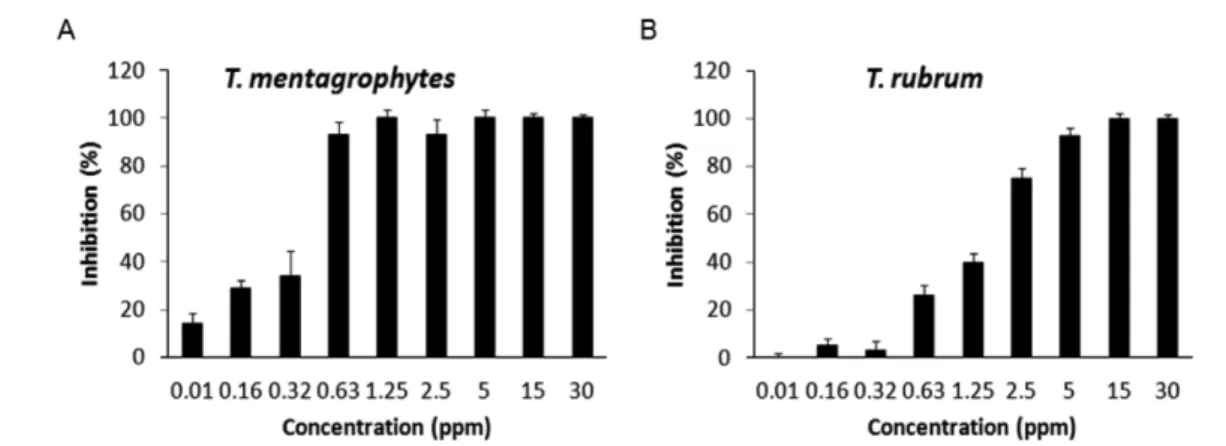
Antifungal efficacy of bee venom against dermatophytes. We evaluated the antifungal efficacy of the bee venom against T.
menatgrophytes and T. rubrum as shown in Fig. 2A and 2B by using modified broth dilution method. The broth dilution method was reported to be more reliable method for evaluating the susceptibility to antimicrobial agents than disc diffusion (Wiegand et al., 2008). At 0.63 ppm of bee venom, the growth of T.
menatgrophytes was approximately 92% inhibited, while only 26% of T. rubrum was inhibited for 1 h incubation of the same dose of bee venom. Bee venom showed much stronger antifungal effect against T. mentagraphytes than against T. rubrum.
Comparison of antifungal activity of bee venom with that of fluconazole. The antifungal efficacy of bee venom was was compared with that of fluconazole through colony formation on the SDA plates after broth dilution. The count for each colony of T. mentagrophytes and T. rubrum was compared with that of fluconazole, one of the commercial antifungal drugs. As shown in Fig. 3, the 15 and 30 ppm of the bee venom reduced all the populations of T. mentagrophytes within 5 min. In addition, the
Fig. 1 Chemical structure of (A) fluconazole and (B) principal constituents of the bee venom, named melittin and apamine.
same concentrations of the bee venom completely inhibited the growth of T. rubrum within 5 min (Fig. 4). On the other hand, however, fluconazole didn’t inhibit the growth of the same pathogens at all, although slight decrease in the counts was found as the incubation time increased. These results indicate that bee venom has much stronger antifungal activity than fluconazole.
Actually, the IC50 values of fluconazole against T. mentagrophytes at 28oC for 7 days was reported to be about 32 ppm (da Silva Barros et al., 2007). Approximately 50% growth inhibitions of T.
mentagrophytes and T. rubrum were found at 0.43 and 1.57 ppm
of the bee venom, respectively, for 1 h incubation when plotted on Fig. 2.
Discussion
Bee venom was known to contain several peptides like melittin, apamin, adolapin, mast cell degranulating peptide, enzymes, biologically active amines, and non-peptide components (Lariviere and Melzack, 1996; Kwon et al., 2002). Enzymes in the bee Fig. 2 Inhibitory effect of bee venom on the growth of (A) T. mentagrophytes and (B) T. rubrum. Each experiment was repeated three times and the results were designated as mean ± SD.
Fig. 3 Comparison of the antifungal activity of bee venom with fluconazole against T. mentagrophytes. 15 ppm and 30 ppm of bee venom and fluconazole were used. Each experiment was repeated more than three times.
venom included phospholipase A2, hyaluronidase, acid phosphomonoesterase, α-D-glucosidase, and lysophospholipase (Somerfield et al., 1984; Banks and Shipolini, 1986). Among them, melittin, a water-soluble cationic amphipathic 26 amino acid α-helical peptide, is a very nonspecific cytolytic peptide that attacks all lipid membranes leading to significant toxicity (Pan et al., 2011).
The frequent uses of antimicrobial and antifungal agents such as antibiotics enable many pathogens to acquire multiple drug resistance genes (Owens et al., 2001). The emergence of antibacterial resistant strains of animal pathogens and their potential health risk to humans have captured great attention (Pitkälä et al., 2004; Nair et al., 2005). Therefore, the development of a new antibacterial or antifungal agent with less adverse effects has been necessary for the successful treatment of dermatophytes infections (Komine et al., 2006).
Phytochemicals have been extensively studied to examine their antifungal effects against T. rubrum and T. mentagrophytes.
Turmeric oil from Curcuma longa (Zingiberaceae) had minimum inhibitory concentrations (MICs) in a range of 229.8–919.2 ppm (Apisariyakul et al., 1995). Furthermore, four phenolic amides, dihydro-N-caffeoyltyramine, trans-N-feruloyloctopamine, trans-N- caffeoyltyramine, and cis-N-caffeoyltyramine isolated from Lycium chinense were reported to have anti-fungal activity in a range of 5–10 ppm (Lee et al., 2004). 6α-O-(β-D-xylopyranosyl- (1→3)-β-D-quinovopyranosyl)-(25,S)-5α-spirostan-3β-ol had IC50
values of 25µg/mL against T. mentagrophytes and T. rubrum (Arif et al., 2011). From Solanum species, 6-α-O-β-D-xylopyranosyl- (1→3)-β-D-quinovopyranosyl-(25R)-5α-spirostan-3β,23α-ol was reported to be active in a rage of 12.5 to 200µg/mL against T.
mentagrophytes and T. rubrum. Limonene was also shown to exert a potent antifungal effect against T. rubrum with MIC value of 0.5% (Chee et al., 2009).
The antibacterial properties of bee venom as a natural antibacterial agent have been extensively studied, and bee venom therapy has been suggested to be used as an alternative to antibiotic therapy (Fennell et al., 1968; Somerfield et al., 1984; Saini et al., 1997).
A strong antibacterial activity of bee venom against both Gram- negative and Gram positive bacteria had been reported (Stocker and Traynor, 1986; Perumal Samy et al., 2007). Nakatuji et al.
(2009) also reported that bee venom could control the growth of S. aureus, which plays an important role in the pathogenesis of inflamed lesions in the case of acne vulgaris. Moreover, bee venom also exhibited antibacterial activities against skin bacteria such as P. acnes, S. epidermidis and S. pyrogenes (Han et al., 2010), while information on the antifungal activity of bee venom against dermatophytes is not available.
We demonstrated for the first time that bee venom has very strong antifungal efficacy, much stronger than that of fluconazole.
Moreover, anti-fungal activity of bee venom was much higher than that of various phytochemicals judging by their effective antifungal concentration ranges. This study raises the possibility Fig. 4 Comparison of the antifungal activity of bee venom with fluconazole against T. rubrum. 15 ppm and 30 ppm of bee venom and fluconazole were used. Each experiment was repeated more than three times.
that the bee venom could be used as an alternative strategy for treating fungal pathogens that would reduce antibiotic usage.
Further experiments might be necessary to evaluate the in vivo efficacy of bee venom and to determine their potential effects on the skin tissue as well as its mechanism of action on fungi.
Acknowledgment This work was supported by the Soonchunhyang University Research Fund (No. 2012-19920013).
References
Akdis CA, Akdis M, Blesken T, Wymann D, Alkan SS, Muller U, and Blaser K (1996) Epitope-specific T cell tolerance to phospholipase A2 in bee venom immunotherapy and recovery by IL-2 and IL-15 in vitro. J Clin Invest 98, 1676–1683.
Apisariyakul A, Vanittanakom N, and Buddhasukh D (1995) Antifungal activity of turmeric oil extracted from Curcuma longa (Zingiberaceae). J Ethnopharmacol 49, 163–169.
Argiolas A and Pisano JJ (1983) Facilitation of phospholipase A2 activity by mastoparans, a new class of mast cell degranulating peptides from wasp venom. J Biol Chem 258, 13697–13702.
Arif T, Mandal TK, and Dabur R (2011) Natural products: Antifungal agents derived from plants. In Opportunity, Challenge and Scope of Natural Products in Medicinal Chemistry. Tiwari VK (ed.), pp. 283–311, Research Signpost, Kerala, India.
Banks BEC and Shipolini RA (1986) Chemistry and pharmacology of honey- bee venom. In Venoms of the Hymenoptera. Piek T (ed.), pp. 329–416, Academic Press, London, UK.
Chee HY, Kim H, and Lee MH (2009) In vitro antifungal activity of limonene against Trichophyton rubrum. Microbiology 37, 243–246.
Chinelli PA, Sofiatti Ade A, Nunes RS, and Martin JE (2003) Dermatophyte agents in the city of São Plaulo, from 1992 to 2002. Rev Inst Med Trop Sao Paulo 45, 259–263.
Choi SH, Cho SK, Kang SS, Bae CS, Bai YH, Lee SH, and Pak SC (2003) Effect of apitherapy in piglets with preweaning diarrhea. Am J Chin Med 31, 321–326.
da Silva Barros ME, de Assis Santos D, and Hamdan JS (2007) Evaluation of susceptibility of Trichophyton mentagrophytes and Trichophyton rubrum clinical isolates to antifungal drugs using a modified CLSI microdilution method (M38-A). J Med Microbiol 56, 514–518.
Eiseman JL, Von Bredow J, and Alvares AP (1982) Effect of honeybee (Apis mellifera) venom on the course of adjuvant-induced arthritis and depression of drug metabolism in the rat. Biochem Pharmacol 31, 1139–
1146.
Fennell JE, Shipman WH, and Cole LJ (1968) Antibacterial action of melittin, a polypeptide from the venom. Proc Soc Exp Biol Med 127, 707–710.
Gupta AK and Del Rosso JQ (2000) An evaluation of intermittent therapies used to treat onychomycosis and other dermatomycoses with the oral antifungal agents. Int J Dermatol 39, 401–411.
Hainer BL (2003) Dermatophyte infections. Am Fam Physician 67, 101–108.
Han SM, Lee KG, Yeo JH, Kweon HY, Kim BS, Kim JM, Baek HJ, and Kim ST (2007) Antibacterial activity of the honey bee venom against bacterial mastitis pathogens infecting dairy cows. Int J Indust Entomol 14, 137–142.
Han SM, Lee KG, Yeo JH, Baek HJ, and Park KK (2009) Determination of major constituents of honeybee venom from Korea. Korean J Apiculture 24, 175–178.
Han SM, Lee KG, Yeo JH, Baek HJ, and Park KK (2010) Antibacterial and anti-inflammatory effects of honeybee (Apis mellifera) venom against acne-inducing bacteria. J Med Plants Res 4, 459–464.
Kim HW, Kwon YB, Ham TW, Rho DH, Yoon SY, Lee HJ, Han HJ, Yang IS, Beitz AJ, and Lee JH (2003) Acupoint stimulation using bee venom attenuates formalin induced pain behavior and spinal cord fos expression in rats. J Vet Med Sci 65, 349–355.
Kim KS, Choi US, Lee SD, Kim KH, Chung KH, Chang YC, Park KK, Lee
YC, and Kim CH (2005) Effect of bee venom on aromatase expression and activity in leukaemic FLG 29.1 and primary osteoblastic cells. J Ethnopharmacol 99, 245–252.
Komine Y, Komine K, Kai K, Itagaki M, Kuroishi T, Aso H, Obara Y, and Kumagai K (2006) Effect of combination therapy with lactoferrin and antibiotics against staphylococcal mastitis on drying cows. J Vet Med Sci 68, 205–211.
Kumar R, Shrivastava SK, and Chakraborti A (2010) Comparison of broth dilution and disc diffusion method for the antifungal susceptibility of Aspergillus flavus. Am J Biomed Sci 2, 202–208.
Kwon YB, Lee JD, Lee HJ, Han HJ, Mar WC, Kang SK, Beitz AJ, and Lee JH (2001) Bee venom injection into an acupuncture point reduces arthritis associated edema and nociceptive responses. Pain 90, 271–280.
Kwon YB, Lee HJ, Han HJ, Mar WC, Kang SK, Yoon OB, Beitz AJ, and Lee JH (2002) The water-soluble fraction of bee venom produces antinociceptive and anti-inflammatory effects on rheumatoid arthritis in rats. Life Sci 71, 191–204.
Lariviere WR and Melzack R (1996) The bee venom test: A new tonic-pain test. Pain 66, 271–277.
Lee MH, Lee KB, Oh SM, Lee BH, and Chee HY (2010) Antifungal activities of dieckol isolated from the marine brown alga Ecklonia cava against Trichophyton rubrum. J Korean Soc Appl Biol Chem 53, 504–
507.
Lee DG, Park Y, Kim MR, Jung HJ, Seu YB, Hahm KS, and Woo ER (2004) Anti-fungal effects of phenolic amides isolated from the root bark of Lycium chinense. Biotechnol Lett 26, 1125–1130.
Mukherjee PK, Leidich SD, Isham N, Leitner I, Ryder NS, and Ghannoum MA (2003) Clinical Trichophyton rubrum strain exhibiting primary resistance to terbinafine. Antimicrob Agents Chemother 47, 82–86.
Nair MK, Joy J, Vasudevan P, Hinckley L, Hoagland TA, and Venkitanarayanan KS (2005) Antibacterial effect of caprylic acid and monocaprylin on major bacterial mastitis pathogens. J Dairy Sci 88, 3488–3495.
Nakatuji T, Kao MC, Fang JY, Zouboulis CC, Zhang L, Gallo RL, and Huang CM (2009) Antimicrobial property of lauric acid against Propionibacterium acnes: Its therapeutic potential for inflammatory acne vulgaris. J Invest Dermatol 129, 2480–2488.
Owens WE, Nickerson SC, Boddie RL, Tomita GM, and Ray CH (2001) Prevalence of mastitis in dairy heifers and effectiveness of antibiotic therapy. J Dairy Sci 84, 814–817.
Pan H, Soman NR, Schlesinger PH, Lanza GM, and Wickline SA (2011) Cytolytic peptide nanoparticles ('NanoBees') for cancer therapy. Wiley Interdiscip Rev Nanomed Nanobiotechnol 3, 318–327.
Peng XL, Gao XL, Chen J, Huang X, and Chen HS (2003) Effects of intravenous Injections Paederiae and Stauntonia on spontaneous pain, hyperalgesia and inflammation induced by cutaneous chemical tissue injury in the rat. Sheng Li Xue Bao 55, 516–524.
Perumal Samy R, Gopalakrishnakone P, Thwin MM, Chow TK, Bow H, Yap EH, and Thong TWJ (2007) Antibacterial activity of snake, scorpion and bee venoms: A comparison with purified venom phospholipase A2 enzymes. J Appl Bacteriol 102, 650–659.
Pitkälä A, Haveri M, Pyörälä S, Myllys V, and Honkanen-Buzalski T (2004) Bovine mastitis in Finland 2001-prevalence, distribution of bacteria, and antimicrobial resistance. J Dairy Sci 87, 2433–2441.
Saini SS, Peterson JW, and Chopra AK (1997) Melittin binds to secretory phospholipase A2 and inhibits its enzymatic activity. Biochem Biophys Res Commun 238, 436–442.
Somerfield SD, Stach JL, Mraz C, Gervais F, and Skamene E (1984) Bee venom inhibits superoxide production by human neutrophils.
Inflammation 8, 385–391.
Stocker JF and Traynor JR (1986) The action of various venoms on Escherichia coli. J Appl Bacteriol 61, 383–388.
Summerbell RC (1997) Epidemiology and ecology of onychomycosis.
Dermatology 194, 32–36.
Wiegand I, Hilpert K, and Hancock RE (2008) Agar and broth dilution methods to determine the minimal inhibitory concentration (MIC) of antimicrobial substances. Nat Protoc 3, 163–175.