Arthroscopic Stabilization for Displaced Lateral Clavicular Fractures:
Can It Restore Anatomy?
Prince Shanavas Khan, Yon-Sik Yoo1 , Byung-Su Kim1, Seong-Jin Lee1, Jong Mun Ha1
Department of Orthopaedic Surgery, Pariyaram Medical College, Kerala, India, 1Department of Orthopaedic Surgery, Hallym University Dongtan Sacred Heart Hospital, Hwaseong, Korea
Background: The purpose of our study was to evaluate the accuracy of reduction based on postoperative computed tomography (CT) images after arthroscopic stabilization using tightrope system for unstable distal clavicle fracture.
Methods: Twelve patients with distal clavicle fracture combined with coracoclavicular (CC) ligament injury (type II, V) who received ar- throscopically assisted fixation using a flip button device were evaluated for accuracy of reduction using 3-dimensional postoperative CT scan by measuring the degree of distal clavicular angulation and clavicular shortening.
Results: Immediate postoperative plain radiograph confirmed restoration of the CC distance (CCD) in 10 patients. At final follow-up, the CCD remained reduced anatomically on plain radiographs in these patients. All patients showed excessive posterior angulation and shortening compared to the opposite side. The average Constant score recovered to 94.8 at final follow-up.
Conclusions: Indirect reduction and arthroscopic subacromial approach with flip button fixation of unstable distal clavicle fractures demonstrated favorable clinical results despite unavoidable posterior angulation of distal clavicle and shortening the total length of clavicle.
(Clin Shoulder Elbow 2016;19(3):143-148)
Key Words: Distal clavicle; Indirect fixation; Arthroscopy
Clinics in Shoulder and Elbow
Copyright © 2016 Korean Shoulder and Elbow Society. All Rights Reserved. pISSN 2383-8337
Clinics in Shoulder and Elbow Vol. 19, No. 3, September, 2016 http://dx.doi.org/10.5397/cise.2016.19.3.143
Received November 24, 2015. Revised February 1, 2016. Accepted February 10, 2016.
Correspondence to: Yon-Sik Yoo
Department of Orthopaedic Surgery, Hallym University Dongtan Sacred Heart Hospital, 7 Keunjaebong-gil, Hwaseong 18450, Korea Tel: +82-31-8086-2413, Fax: +82-31-8086-2029, E-mail: [email protected]
IRB approval (No. 2016-397-I).
Financial support: None. Conflict of interests: None.
Introduction
Distal-third clavicle fractures with disruption of the coraco- clavicular (CC) ligament are potentially unstable1) and are com- plicated by a high rate of non-union (33.3%) when treated con- servatively.2) Internal fixation in these fractures is often difficult, because of inability to attain screw purchase due to inadequate distal fragment size, comminution, and instability caused by torn CC ligament. Numerous techniques such as CC screws, tension bands, hook and locked plates, intramedullary screws, transacro- mial K-wire or Knowles pin fixation, and CC ligament repair2-6) alone or in combination have been described, but no gold standard has been established. Many of them were associated with favorable outcomes but potential complications (nonunion, symptomatic hardware, peri-implant fracture, infection, rotator
cuff tear, malunion, and acromioclavicular [AC] joint arthrosis) and morbidities were unavoidable.7-10)
Attention has recently shifted towards minimally invasive surgery and arthroscopic fixation of these unstable fractures.11-16) Midterm results of arthroscopic flip button fixation showing fa- vorable results in distal clavicle fractures have recently been pub- lished.13,14) Arthroscopic fixation has many advantages; however, postoperative X-rays can be deceiving in that it cannot identify occult mal-reduction evident on 3-dimensional computed to- mography (3D CT), which might affect the long term results of this novel technique. The purpose of our study was to evaluate the accuracy of reduction based on postoperative CT images after arthroscopic stabilization using a tightrope system for un- stable distal clavicle fracture.
Methods
This study was conducted retrospectively. Twelve patients with distal clavicle fracture combined with CC ligament injury who received arthroscopically assisted fixation using a flip button device (tightrope; Arthrex Inc., Naples, FL, USA) between Janu- ary 2013 and December 2013 in Hallym University Dongtan Sa- cred Heart Hospital were included in the study. This technique was intended for use in acute cases (within the first week of injury) for Craig type II and type V fractures. The study group in- cluded 6 men and 6 women with a mean age of 40 years (range, 26–50 years). Radiological imaging using anteroposterior (AP) and axillary view demonstrated Craig type IIB fractures in 7 pa- tients and type V fractures in 5 patients. All patients underwent surgery within the first week of injury. Patients with other fracture types (I, III, and IV) and those diagnosed with associated injuries of rotator cuff, labrum, or biceps tendon during arthroscopic ex- amination were excluded. All Patients had a minimum follow-up period of 2 years (range, 2.0–2.5 years).
Surgical Technique
Under general anesthesia with the patient in lateral decubitus position, diagnostic arthroscopy of the glenohumeral joint was performed through the posterior portal. Next, the anterolateral portal is made approximately 1.5 cm lateral to and in line with anterior border of acromion to perform a bursectomy. The anterior portal is established with direct visualization from the anterolateral portal. This portal allows access to the subcoracoid space for dissection around the inferior coracoid. Next, a 1.5-cm skin incision was made just medial to the fracture site along the midline of the distal clavicle (Fig. 1A).
An AC tightrope drill guide (Arthrex Inc.) was centered on the undersurface of the base of the coracoid through the anterior portal while its other end (the drill sleeve) was placed 1cm medi- al to the fracture in the center of the clavicle. With the drill guide
held in this position and under clear arthroscopic view, a 2.4- mm guide pin was drilled from the clavicle to the undersurface of the coracoid process. Next, a 4.0-mm tunnel was made using a tightrope reamer. The tightrope reamer was left in situ and the guide pin was removed. A guide wire was inserted through the tightrope reamer, passed out of the hole drilled in the coracoid and retrieved with a grasper placed through the anterior portal and the reamer was removed. The flip button device (2 metal buttons, one oblong and the other rounded, connected by a loop of #5 FiberWire; Arthrex Inc.) was inserted into the loop and pulled through the clavicle and the base of the coracoid.
The button was flipped and positioned horizontally under the coracoid using a suture retriever. The medial clavicular fragment was pushed inferiorly with a bone tamp and superior force ap- plied on elbow and arm to indirectly reduce the fracture (Fig.
1B, C). The flip button was tied down over the round button on the superior surface of the clavicle after adequate reduction was obtained under an image intensifier.
The affected shoulder was immobilized in a shoulder brace in a neutral position for 4 weeks postoperatively. Gentle pas- sive mobilization started at 4 weeks and active mobilization at 6 weeks after the surgery.
Outcome Measures
All patients were checked by 3D CT on postoperative 1 day.
Postoperative 3D CT scan was used for assessing the 3D quality of reduction by measuring the degree of distal clavicular angula- tion and clavicular shortening as described by Bachoura et al.
(Fig. 2).17) The restoration of relative clavicular length and degree of angulation on the distal clavicle were measured in the axial plane. Three midlines passing through the shaft of the medial segment, middle segment, and lateral segments of the clavicle were created. The angle between the lines from middle and lat- eral segments was measured in axial planes and compared with the contralateral side of the clavicle. The k statistic was used to
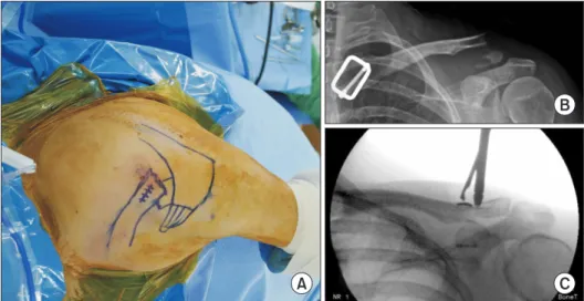
Fig. 1. Showing a reduction procedure under C-arm guidance before final fixation using the flip button device. (A) Surface anatomy and incision around the fracture site. (B) Pre- operative X-ray indicating a type IIB distal clavicle fracture. (C) Downward pushing of the medial fragment before the tie.
A
B
C
assess interobserver agreement for the measurement of angle.
The k value for interobserver agreement was 0.85 (p<0.001).
Clavicle length is measured as the straight distance between the medial and lateral ends of the clavicle, rather than the true length of the clavicle along its curvilinear surface.
Follow-up evaluation included the Constant score and the pain level based on the visual analogue scale (VAS; 0=no pain, 10=maximum pain). Maintenance of reduction was determined by measuring the vertical distance between the upper border of the coracoid process and the inferior cortex of the clavicle (CC distance, CCD) in standard AP view X-ray and comparing it to the contralateral side. Successful union was defined by oblitera- tion of the fracture gap on plain radiographs and no tenderness
or pain at the fracture site during shoulder exercise.
The Wilcoxon signed-rank test was used to compare the degree of posterior angulation and total clavicular length with those of the contralateral clavicle. Clinical scores were accessed using the independent t-test using IBM SPSS ver. 19.0 software (IBM Co., Armonk, NY, USA). The level of significance was set at p<0.05.
Results
An overview of patients’ demographics, including age and gender, type of fracture, time for union, and radiologic results, as well as the Constant score at the last follow-up is shown in
Fig. 2. Angle between the lateral and middle segments of the clavicle (black arrow), posterior angulation in the axial plane (yellow arrow) (A) and length of the clavicle (B) in the 3-dimensional computed tomography scans.
A B
Table 1. Baseline Patient Characteristics and Radiologic Results after Indirect Reduction for Distal Clavicle Fracture Patient No. Gender Age
(yr) Craig type
Angle between the lateral and
middle segments of clavicle (°) Clavicle length (cm) Time of
union (d) Constant score Affected side Contralateral side Affected side Contralateral side
1 Female 26 IIB 142.9 147.6 128.6 136.2 70 100
2 Female 36 V 120.4 128.8 129.8 136.3 55 92
3 Female 42 IIB 119.8 122.6 124.6 127.6 102 100
4 Male 38 V 130.7 135.4 152.3 156.5 62 92
5 Male 50 IIB 125.3 143.9 144.8 159.4 64 94
6 Male 48 V 118.7 140,3 142,3 147.7 69 100
7 Female 41 V 118.6 122.4 142.5 146.1 Resection 89
8 Female 33 IIB 122.7 144.5 131.1 133.9 73 95
9 Male 43 IIB 115.1 146.6 151.6 145.7 69 98
10 Male 39 V 150.8 152.7 151.7 151.2 83 92
11 Female 45 IIB 120.2 131.2 126.2 131.4 91 96
12 Male 40 IIB 116.3 131.5 141.3 146.7 Plate 89
Table 1. Pain, as assessed by the VAS, was significantly improved following surgery (p<0.02), from an average of 8.1 preopera- tively to 0.4 at final follow-up. The average Constant score was recovered to 94.8 at final follow-up. Immediate postoperative plain radiograph confirmed restoration of the CCD in 10 pa- tients. At final follow-up, the CCD remained reduced anatomi- cally on plain radiographs in these patients.
Two patients demonstrated incomplete reduction, resulting in failure to restore CCD. One patient with type IIB was over- reduced to 25% of the contralateral CCD, and another patient with type IIB fracture was incompletely reduced. Both patients underwent revision surgery, using plate and screw designed for distal clavicle fracture for first and excision of the distal fragment for the latter.
All patients showed excessive posterior angulation and short- ening compared to opposite side. The mean posterior angulation of distal clavicle and total clavicular length in 10 patients at final follow-up were 32.4 degree and 136.9 cm respectively, while those of the contralateral side were 23.4 degree and 143.8 cm, which were both statistically significant (p<0.03 and p<0.01).
Mean time to union was 13.7 weeks (range, 7.8–14.5 weeks) after surgery. All patients had returned to full overhead pre-injury level of activity at last follow-up.
Discussion
Our study, arthroscopic stabilization with tightrope through a subacromial approach, demonstrated favorable clinical results and union despite unavoidable posterior angulation of the distal clavicle as evident on 3D CT images.
Despite our results, we believe that more anatomical restora- tion will be better in clinical results on long term follow-up, and no existing data describes biomechanical and clinical ill-effect caused by posterior angulation, special attention must be paid to its long term influence.
As a minimal invasive technique, arthroscopic stabilization of distal third clavicle fractures has been introduced using sutures or double-button devices for Neer type II fractures. Minimal invasive procedures are theoretically more advantageous over open technique in terms of fracture healing as well as ligament healing as a consequence of less soft tissue stripping and de- vascularization around the fracture site.12,16) Arthroscopy allows for diagnosis and treatment of any intra-articular pathology.
Arthroscopic preparation of the coracoid base also provides a better view of the bone tunnel than an open surgical technique.
Flinkkilä et al.,18) in their comparative study, favored arthroscopic tight rope fixation over hookplate, but reduction was achieved under direct vision, exposing the fracture hematoma.
Pujol et al.16) treated 4 patients with indirect reduction and double button device fixation through the arthroscopic rota- tor interval approach claiming union and a full recovery of the
shoulder function in 6 months. Checchia et al.,12) using an ar- throscopic rotator interval approach and figure of 8 circlage loop technique around coracoid, treated 7 patients with a mean age of 46 years. Mean union time was 7 weeks. At mean follow-up of 15 months, there were 7 excellent to good results using Uni- versity of California, Los Angeles (UCLA and American Academy of Orthopaedic Surgeons (AAOS) scores. One patient had de- veloped stiffness of the shoulder probably due to use of a rotator interval approach. Loriaut et al.13) recently reported satisfactory functional outcome in 21 patients with type 2B displaced frac- ture of the clavicle who received an arthroscopically assisted fixation using a double button device through a rotator interval approach. Although the importance of an arthroscopic approach was highlighted, all of them opened the rotator interval for pre- paring the coracoid base.
Baumgarten11) treated a case with type II fracture using a flip button system through an arthroscopic subacromial approach, with fracture consolidation at 3 months, and return to pain- less, unrestricted activity at 5 months. Motta et al.14) treated 14 male patients, with type IIA, IIB, and 5 lateral clavicular fractures through an arthroscopic subacromial approach using TightRope.
All fractures healed without limitations in range of motion or loss of reduction. We are in agreement with Baumgarten11) and Motta et al.,14) who used a subacromial approach, because we believe that opening the rotator interval may lead to post-opera- tive instability or stiffness of the shoulder.19,20)
Arthroscopic indirect reduction techniques may not result in adequate apposition in all fracture patterns, and there may ex- ist residual instability between the fracture fragments,12,15) which was evident in one of our patients. Numerous complications in relation to arthroscopic fixation have been mentioned in the lit- erature, including implant failure with nonunion, transient adhe- sive capsulitis, symptomatic AC joint arthritis, coracoid fracture, and clavicular erosion.12,13,16,21) However, we did not encounter any of them except instability and over reduction.
Even though Loriaut et al.13) and Motta et al.14) reported satis- factory radiologic outcomes after arthroscopic stabilization based on plane radiographs, malreduction/posterior angulation, an occult complication and its effect on clinical outcome has never been mentioned previously as it is not evident on routine X- rays. Rieser et al.,22) in a biomechanical evaluation, showed that combination of a locking plate and tightrope was superior to either method alone as a result of greater construct strength and stiffness. Though fracture consolidation can be achieved without inter fragmentary fixation, arthroscopic stabilization using a dou- ble button device may not be sufficient to maintain anatomical reduction especially in the horizontal plane due to deforming forces exerted by muscle pull and gravity on fracture fragments as evidenced by shortening and increased posterior angulation of the distal clavicle in our cohort.
Although this case series constitutes level IV evidence (Oxford
grading), some general conclusions can be drawn due to the rar- ity of type II and type V fractures and few shoulder surgeons per- forming arthroscopic fixation. The quality of reduction does not appear to be related to the severity of injury. Despite a significant difference in the angulation and length of clavicle (p<0.03 and p<0.01), evaluation of CCD on the plain AP view X-rays did not reveal these abnormalities. Malreduction appears to be related to indirect reduction, improper placement of the clavicular tun- nel, or improper tensioning of the flip button device. Proper positioning of the coracoid tunnel at the center of the base,23) clavicular tunnel within the footprint of the CC ligament, results in anatomic restoration of distal clavicle fractures.
There were several limitations of this study. First, the relatively small number of patients proved to be a limiting factor in deter- mining the relevant outcome of the procedure. Second, because this was a retrospective comparative study after surgery, we had no control group. The third limitation was the absence of bio- mechanical theory regarding posterior angulation and shortening after arthroscopic fixation. We assumed that this problem might be concerned with muscle force or gravity.
Concerns have been raised regarding whether shortening and posterior angulation of the distal clavicle will result in altered scapular biomechanics and AC joint problems. Andermahr et al.,24) in their anatomical study, showed that malunion of the clavicle may cause glenoid malpositioning, which, in turn, can lead to functional deficits during abduction. Hence, further long term studies are necessary to address the untoward events due to altered angulation and radius of curvature in the transverse plane, and to refine the technique further in order to restore normal anatomy by placement of the bony tunnels or number of tunnels over the anatomic foot print of the CC ligament.
Conclusion
Indirect reduction arthroscopic subacromial approach with flip button fixation of unstable distal clavicle fractures demon- strated favorable clinical results despite unavoidable posterior angulation of the distal clavicle and shortening of the total length of the clavicle. Further long term studies are needed to evaluate the influence of a posteriorly angulated distal clavicle and result- ing in a shortened clavicle after arthroscopic flip button fixation of these fractures.
References
1. Neer CS 2nd. Nonunion of the clavicle. J Am Med Assoc.
1960;172:1006-11.
2. Oh JH, Kim SH, Lee JH, Shin SH, Gong HS. Treatment of distal clavicle fracture: a systematic review of treatment modalities in 425 fractures. Arch Orthop Trauma Surg. 2011;131(4):525- 33.
3. Ballmer FT, Gerber C. Coracoclavicular screw fixation for un- stable fractures of the distal clavicle. A report of five cases. J Bone Joint Surg Br. 1991;73(2):291-4.
4. Bezer M, Aydin N, Guven O. The treatment of distal clavicle fractures with coracoclavicular ligament disruption: a report of 10 cases. J Orthop Trauma. 2005;19(8):524-8.
5. Kao FC, Chao EK, Chen CH, Yu SW, Chen CY, Yen CY. Treat- ment of distal clavicle fracture using Kirschner wires and tension-band wires. J Trauma. 2001;51(3):522-5.
6. Kashii M, Inui H, Yamamoto K. Surgical treatment of distal clavicle fractures using the clavicular hook plate. Clin Orthop Relat Res. 2006;447:158-64.
7. Ding M, Ni J, Hu J, Song D. Rare complication of clavicular hook plate: clavicle fracture at the medial end of the plate. J Shoulder Elbow Surg. 2011;20(7):e18-20.
8. Flinkkilä T, Heikkilä A, Sirniö K, Pakarinen H. TightRope versus clavicular hook plate fixation for unstable distal clavicular frac- tures. Eur J Orthop Surg Traumatol. 2015;25(3):465-9.
9. Lee KW, Lee SK, Kim KJ, Kim YI, Kwon WC, Choy WS. Ar- throscopic-assisted locking compression plate clavicular hook fixation for unstable fractures of the lateral end of the clavicle:
a prospective study. Int Orthop. 2010;34(6):839-45.
10. Wijdicks FJ, Van der Meijden OA, Millett PJ, Verleisdonk EJ, Houwert RM. Systematic review of the complications of plate fixation of clavicle fractures. Arch Orthop Trauma Surg.
2012;132(5):617-25.
11. Baumgarten KM. Arthroscopic fixation of a type II-variant, un- stable distal clavicle fracture. Orthopedics. 2008;31(12). doi:
10.3928/01477447-20081201-02.
12. Checchia SL, Doneux PS, Miyazaki AN, Fregoneze M, Silva LA. Treatment of distal clavicle fractures using an arthroscopic technique. J Shoulder Elbow Surg. 2008;17(3):395-8.
13. Loriaut P, Moreau PE, Dallaudière B, et al. Outcome of arthro- scopic treatment for displaced lateral clavicle fractures using a double button device. Knee Surg Sports Traumatol Arthrosc.
2015;23(5):1429-33.
14. Motta P, Bruno L, Maderni A, Tosco P, Mariotti U. Acute lateral dislocated clavicular fractures: arthroscopic stabilization with TightRope. J Shoulder Elbow Surg. 2014;23(3):e47-52.
15. Nourissat G, Kakuda C, Dumontier C, Sautet A, Doursounian L.
Arthroscopic stabilization of Neer type 2 fracture of the distal part of the clavicle. Arthroscopy. 2007;23(6):674.
16. Pujol N, Desmoineaux P, Boisrenoult P, Beaufils P. Arthroscopic treatment of comminuted distal clavicle fractures (latarjet fractures) using 2 double-button devices. Arthrosc Tech.
2013;2(1):e61-3.
17. Bachoura A, Deane AS, Wise JN, Kamineni S. Clavicle mor- phometry revisited: a 3-dimensional study with relevance to operative fixation. J Shoulder Elbow Surg. 2013;22(1):e15-21.
18. Flinkkilä T, Ristiniemi J, Hyvönen P, Hämäläinen M. Surgical treatment of unstable fractures of the distal clavicle: a compar-
ative study of Kirschner wire and clavicular hook plate fixation.
Acta Orthop Scand. 2002;73(1):50-3.
19. Harryman DT 2nd, Sidles JA, Harris SL, Matsen FA 3rd. The role of the rotator interval capsule in passive motion and stabil- ity of the shoulder. J Bone Joint Surg Am. 1992;74(1):53-66.
20. Gaskill TR, Braun S, Millett PJ. Multimedia article. The ro- tator interval: pathology and management. Arthroscopy.
2011;27(4):556-67.
21. Shin SJ, Roh KJ, Kim JO, Sohn HS. Treatment of unstable distal clavicle fractures using two suture anchors and suture tension bands. Injury. 2009;40(12):1308-12.
22. Rieser GR, Edwards K, Gould GC, Markert RJ, Goswami T, Ru- bino LJ. Distal-third clavicle fracture fixation: a biomechanical evaluation of fixation. J Shoulder Elbow Surg. 2013;22(6):848- 55.
23. Campbell ST, Heckmann ND, Shin SJ, et al. Biomechanical evaluation of coracoid tunnel size and location for coracocla- vicular ligament reconstruction. Arthroscopy. 2015;31(5):825- 30.
24. Andermahr J, Jubel A, Elsner A, et al. Malunion of the clavicle causes significant glenoid malposition: a quantitative anatomic investigation. Surg Radiol Anat. 2006;28(5):447-56.