■Jin-Hee Yoon, BS; Ji-Won Park, PT, PhD1; Jong-In Youn, PhD
■Department of Biomedical Engineering, College of Medical Science, Catholic University of Daegu; 1Department of Physical Therapy, College of Medical Science, Catholic University of Daegu;
Purpose: The aim of this study was to do numerical analysis of the wavelength dependence in low level laser therapy (LLLT) using a finite element method (FEM).
Methods: Numerical analysis of heat transfer based on a Pennes' bioheat equation was performed to assess the wavelength dependence of effects of LLLT in a single layer and in multilayered tissue that consists of skin, fat and muscle.
The three different wavelengths selected, 660 nm, 830 nm and 980 nm, were ones that are frequently used in clinic settings for the therapy of musculoskeletal disorders. Laser parameters were set to the power density of 35.7 W/cm2, a spot diameter of 0.06 cm, and a laser exposure time of 50 seconds for all wavelengths.
Results: Temperature changes in tissue based on a heat transfer equation using a finite element method were simulated and were dominantly dependent upon the absorption coefficient of each tissue layer. In the analysis of a single tissue layer, heat generation by fixed laser exposure at each wavelength had a similar pattern for increasing temperature in both skin and fat (980 nm > 660 nm > 830 nm), but in the muscle layer 660nm generated the most heat (660 nm >> 980 nm > 830 nm).
The heat generation in multilayered tissue versus penetration depth was shown that the temperature of 660 nm wavelength was higher than those of 830 nm and 980 nm
Conclusion: Numerical analysis of heat transfer versus penetration depth using a finite element method showed that the greatest amount of heat generation is seen in multilayered tissue at = 660 nm. Numerical analysis of heat transfer may help lend insight into thermal events occurring inside tissue layers during low level laser therapy.
Keywords: Heat transfer, Finite element method, Low‐level laser therapy Received: October 3, 2010
Accepted: December 8, 2010
Corresponding author: Jong-In Youn, [email protected]
Laser Therapy (LLLT) Using a Finite Element Method
The Journal Korean Society of Physical Therapy
I. Introduction
Light-based treatment of tissue can be used for a variety of applications including biostimulation, sealing or welding blood vessels, and low-level laser therapy.1-7 It has been claimed that biostimulation is a result of heating of minute areas of tissue in a way that stimulates nerves and accelerates wound healing.1-3 Light-induced heating is typically performed with laser light of different wavelengths (from ultraviolet to infrared), power densities (i.e., the ratio between beam power and irradiated area), and duration of exposure.4,5 The amount of energy imparted to the tissue and hence the temperature rise can be changed by either varying the power density or the duration of
the time pulse of the laser.1 Low-level laser therapy (LLLT), which is used by physiotherapists, is the application of laser light in the range of 1 to 500 mW for the treatment of a wide variety of acute and chronic musculoskeletal aches and pains.5-9 The applied laser light typically has a narrow spectral bandwidth in the red or near infrared spectrum (600~1000 nm), with a power density or irradiance (power output divided by laser spot area) between 1 and 5 W/cm2.2,3
In clinical settings, LLLT has been used inthe treatment of muscle pain, and some positive findings have been discovered for neck muscle pain, and conditions like fibromyalgia, which may be associated with skeletal muscle fatigue.4-7 Many different models of treatment such as exercise, massage, drugs, TENS,
surgery and laser therapies have been used to treat musculoskeletal disorders. Low and medium energy lasers such as GaAs or He:Ne that emit light at wavelengths 600 to 980 nm have been used for various methods of physical therapy.8,9 Researchers found that low level lasers can affect many cellular and sub-cellular processes, and that specific doses of LLLT reduced muscle creatine kinase activity levels, thus indicating a decrease in muscle damage when compared with non-irradiated tissues.8-10 In wavelength dependence studies, researchers found that near-infrared wavelengths penetrate better through the human skin than red wavelengths for the treatment of muscle pains, and for this reason, lasers with near-infrared wavelengths are much more commonly used in the clinical practice of physiotherapists.4,5
Musculoskeletal treatment is a relatively new area of LLLT research for physiotherapists, but the optimal parameters for laser wavelengths and dosimetries of this application are not fully established. One possible mechanism behind the therapeutic effects of LLLT is the interaction of photons from laser irradiation at optimal doses with specific receptors in the mitochondria, and this mechanism increases mitochondrial functioning, ATP, RNA, and protein synthesis.2-11 These interactions lead to increased oxygen consumption and increased membrane potential and enhanced synthesis of NADH and ATP. Consequently, it increases cellular metabolism, possibly increasing wound healing and accelerating inflammatory processes.3 LLLT can also decrease oxidative stress and production of reactive oxygen species,2,11 improve mitochondrial function,12 and stimulate the mitochondrial respiratory chain, ATP synthesis,13 and the microcirculation.14 Although LLLT can have positive effects on hemoglobin and blood cells,15 hemoglobin absorbs laser light and may hamper even distribution of the LLLT dose.16 In larger human skeletal muscles, it may be difficult to distribute the light energy to the interested area due to spatial inhomogeneity of the tissue.
Numerical analysis of heat transfer plays an important role in biomedicine by either solving existing equations or assisting in determining unknown constitutive equations.16-20 A finite element method (FEM) is a simple tool for modeling heat diffusion and bioheat equations to describe light propagation into a tissue and the induced thermal effects over the depth of the tissue. In this study, FEM numerical analysis was performed to compare photothermal effects of different wavelengths in
order to evaluate the therapeutic effects of LLLT.
II. Methods
1. The heat distribution equation in tissue
The bioheat transfer equation (Eq. 1) can be used to estimate the laser-light-induced tissue temperature distribution and the resultant thermal damage.
( , ) [ ( , )] b b b[ art( , ) ( , )] ( , )
T r t
c k T r t C T r t T r t S r t
ρ ∂ t = ∇ ∇ +ρ ω − +
∂ (1)
where T is the temperature [°C], ρ is the density of the tissue [kg/cm3], c is the specific heat of the tissue [J/kg・°C], k is the thermal conductivity of tissue [W/cm・°C], r is the position vector [cm], t is the time [s], Tart is the temperature of arterial blood [°C], S is the deposited light power [W/cm3], and ωb
[mL/(g・min)] is the tissue average volumetric blood perfusion rate (since the density of blood ρb is to be considered as a constant value, it is possible to call ρb・ωb[kg/s・m3]). The coefficients ρ, k and ωb are functions of temperature T.19
Most theoretical analyses of heat transfer in living tissue are based on the Pennes' equation, which describes the influence of blood flow on temperature distribution in the tissue in terms of volumetrically distributed heat sinks or sources. The standard Pennes' bioheat equation is used to determine the temperature distribution within a multilayered skin model based on the physical structure of human skin. The numerical solution of the mathematical model provides a quantitative description of the thermal response of living tissues when exposed to a heating source such as laser light.20,21
The Pennes bioheat equation describes the thermal behavior based on the classical Fourier's law. (Eq. 2)
( , ) ( ) b b b( b ) met ext
T r t
c k T C T T Q Q
ρ ∂ t − ∇ ⋅ ∇ =ρ ω − + +
∂ (2)
where Qmet is the metabolic heat source and Qext is the external (Spatial) heat source.
2. The geometry of the 2D axisymmetric model The geometry in the simulation was based on a 2D
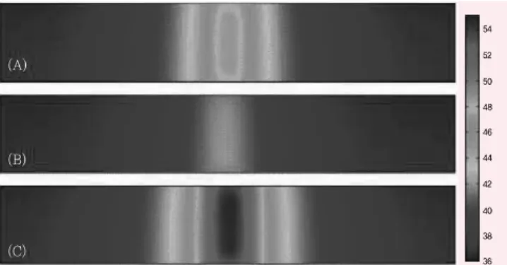
Figure 1. 2D axisymmetric FEM modeling of heat transfer in the skin layer (2 cm × 3 mm) at 660 nm (A), 830 nm (B), and 980 nm (C) wavelengths
axisymmetric model. The modeled tissue consisted of skin, fat, and muscle with depths of 3 mm, 3 mm, and 3 cm, respectively.
The tissue surrounding the vessel was treated as a homogenous tissue. A laser device sitting above the skin put out a circular beam with a radius of 2 cm that irradiated and heated the tissue.
3. Spatial heat source (Laser)
The laser energy deposition profile in the sample was modeled using a standard exponential decay based on the Beer-Lambert Law. The equation of the effects of the laser beam is described in Eq. 3.
(3)
where I0 is the irradiation intensity (W/m2) at the skin surface, is the absorptivity (1/m), and the parameter determines the width of the irradiated region (the standard deviation of the Gaussian function which describes the beam profile [m]). r and z are the 2D axial coordinates of the radius and depth, respectively [in meters].
4. Boundary conditions
(1) All boundaries except the top laser incident surface are insulated.
(2) At the top surface, convective heat exchange (convective heat transfer coefficient = 10 W/m2K) with surrounding ambient air (25°C) were considered.
(3) The temperature profile was symmetric about the z-axis.
(4) In the initial condition with a time of zero, the temperature was equal to the ambient temperature.
(5) Arterial blood temperature was constant at 37°C22
5. Induced laser parameters for the simulation (1) Laser wavelengths: 660 nm, 830 nm, and 980 nm (2) Temporal features of laser input: continuous wave (3) Spot diameter of the lasers: 600 microns
(4) Laser output: 100 mW (5) Irradiation time: 50 seconds
6. Physiological and optical properties of human tissue for the simulation input.
(1) Physiological properties of human tissue23
Density, ρ [kg/m3]: skin (1,116), fat (971), and muscle
(1,041)
Specific heat capacity, C [J/(kg․)]: skin (3,150), fat (2,250), and muscle (3,430)
Heat conductivity, k [W/mk]: skin (0.5), fat (0.28), and muscle (0.4975)
Blood perfusion rate, ωb [mL/kg・min]: skin (120), fat (28), and muscle (38)
(2) Absorption coefficients (1/cm) of different layers of human tissue at the three wavelengths24
Skin (thickness: 3 mm): 0.26 (λ= 660 nm), 0.12 (λ= 830 nm), and 0.38 (λ= 980 nm)
Fat (thickness: 3 mm): 0.1 (λ= 660 nm), 0.09 (λ= 830 nm), and 0.13 (λ= 980 nm)
Muscle (thickness: 30 mm): 0.85 (λ= 660 nm), 0.3 (λ=
830 nm), and 0.55 (λ= 980 nm)
III. Results
For the wavelength dependence studies, heat generation due to laser irradiation was simulated by 2D axisymmetric FEM modeling at each single tissue layer (skin, fat, and muscle). The 2D temperature profile in skin showed that heat transfer from 980 nm laser irradiation is significantly higher than for 830 nm and 660 nm wavelengths (Figure 1). The maximum temperature occurred at 980 nm (54.8°C), which may be due to the highest absorption coefficient of skin at that wavelength when compared to the other two wavelengths, 660 nm and 830 nm. Figure 2 shows the heat transfer of temperature profile in the fat layer.
Again, the maximum temperature was seen at 980 nm, but was lower than the skin layer (50°C). Heat generation from both 980
Figure 2. 2D axisymmetric FEM modeling of heat transfer in the fat layer (2 cm × 3 mm) at 660 nm (A), 830 nm (B), and 980 nm (C) wavelengths
Figure 3. 2D axisymmetric FEM modeling of heat transfer in the muscle layer (2 cm × 3 cm) at 660 nm (A), 830 nm (B), and 980nm (C) wavelengths
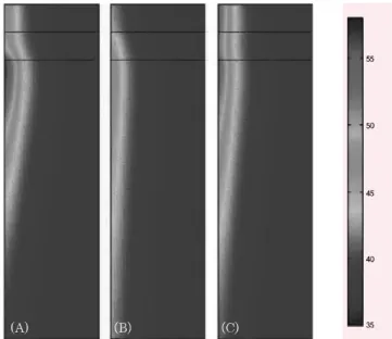
Figure 4. 2D axisymmetric FEM modeling of heat transfer in multi-layered tissue (1 cm × 3.3 cm) at 660 nm (A), 830 nm (B), and 980 nm (C) wavelengths.
Figure 5. Temperature distributions versus depth (A) and the radius (transverse) temperature distributions (B) in the multi-layered tissue after the laser irradiations of 660 nm, 830 nm, and 980 nm.
nm and 660 nm light in the fat layer was decreased, but the temperature for 830 nm irradiation increased about 3°C when compared to temperature profiles for the skin layer. Figure 3 shows the temperature profile in the muscle layer after 2D axisymmetric FEM modeling. Since the absorption coefficient of muscle at 660 nm is higher than that for skin or fat, an excessive temperature rise (up to 100°C) occurs at that wavelength. However, the maximum temperatures for 830 nm and 980 nm were 42.4°C and 57.7°C, respectively.
Figure 4 shows heat transfer distributions in multi-layered tissue. We evaluated the effects of light on each tissue layer. Heat generation in the multi-layered condition had a similar tendency in each tissue layer, but the excessive temperature rise in the
muscle layer at 660 nm wavelength was substantially reduced due to the presence of the skin and fat layers.
Figure 5(A) shows the temperature distributions versus depth and the transverse temperature profiles at each wavelength. Although λ=980 nm showed the highest temperature increase in the skin and fat layers, the highest
amount of heat transfer was achieved at λ=660 nm, which may indicate that this is the most effective wavelength in LLLT treatment of musculoskeletal disorders. The temperature increase at the top surface (5(B)) showed that λ=980 nm was highest when compared to the other two wavelengths.
IV. Discussion
Low-level laser therapy (LLLT) was introduced in a clinical randomized controlled trial on musculoskeletal pain in early 1980s.25 A broad range of doses (0.0001~38 J/cm2) have been reported to produce significant positive effects on musculoskeletal disorders in about one third of the LLLT
trials.26 Biostimulation, or laser-catalyzed reactions, refers to the application of electromagnetic energy by low-level laser to tissues, which is supposed to lead to the stimulation of all kinds of cell functions. The effects have been considered to stimulate and/or inhibit biochemical, physiological, and proliferative activities.25-30 The magnitude of this effect seems to be dependent upon the wavelength and dosage of the laser light because it has long been known that cells are sensitive to specific wavelengths.9
In recent studies, considerable pain reduction after LLLT has been reported in acute and chronic painful conditions such as rheumatoid arthritis (λ=830nm Ga-As-Al laser),27 knee osteoarthritis (λ=904 nm Ga-As laser),28 fibromyalgia (λ=904 nm Ga-As laser)7 and low back pain (λ=904 nm Ga-As laser).29 There were also a few studies of LLLT for skeletal muscle fatigue with the use of different wavelengths such as λ=655 nm Ga-Al-As laser,30,31 and λ=830 nm Ga-As-Al laser.10 Ernesto et al. performed clinical studies with single-laser diode probes to test if LLLT could delay the development of skeletal muscle fatigue and increase muscle recovery when applied before exercise. In these studies, LLLT decreased muscle fatigue and improved biochemical markers related to muscle recovery.10,31,32 Methodological differences in the application parameters of LLLT (wavelength, intensity, duration) may affect the final improvement in pain or the decrease in functional limitation.
Wavelength, power output, energy intensity, and application duration of LLLT are important parameters that affect the success of musculoskeletal therapy.33
After the appearance of Pennes' bioheat equation in 1948, a variety of models of heat transfer in different tissues of human body have been proposed.24-26 The standard Pennes' bioheat equation is used to determine the temperature distribution within a multilayered skin model based on the physical structure of human skin. The numerical solution of the mathematical model provides a quantitative representation of the thermal response of living tissues when exposed to a heating source.24-26 Therefore, the numerical analysis of the heat transfer and accurate estimation of the spatial and temporal distribution of the temperature in the skin, fat, and muscle is of great importance in the actual thermal therapies based on new technologies and devices in LLLT.20
Chukuka et al4 compared two wavelengths, near infrared (904 nm) and red (632 nm) and found that red light was
attenuated more by muscle than by any of the other tissues.
Thus, their data indicated that near infrared light penetrates muscle tissue more than 632.8 nm light. However, they did not consider other optical absorption effects above the muscle tissue such as skin and fat. Our findings indicate that red (660 nm) light is absorbed more by muscle than are near infrared lights (830 and 980 nm). Until now, many clinical settings have been used when performing LLLT therapy for a variety of musculoskeletal disorders. Our findings of temperature profiles for different wavelengths may help generate effective photo- therapy and lend insight into the thermal events occurring inside tissue layers during low level laser therapy.
V. Conclusion
The aim of this work was to investigate the numerical solutions of the bioheat transfer equation in order to derive the temperature distribution inside of the tissue during low-level laser therapy for musculoskeletal disorders. The 2D axisym- metric FEM modeling of each layer (skin, fat, and muscle) and of multi-layered tissues successfully demonstrates how the temperature profile changes at different laser wavelengths over the depth and surface transverse direction. The results show that the highest amount of heat transfer is achieved at = 660 nm. This local rise in temperature may cause conformational changes and trigger biochemical activities that may indicate that it is the most effective wavelength in LLLT treatment of musculoskeletal disorders.
Author Contributions
Research design: Youn JI, Park JW Acquisition of data: Yoon JH, Youn JI
Analysis and interpretation of data: Yoon JH, Youn JI, Park JW Drafting of the manuscript: Yoon JH, Youn JI, Park JW Research supervision: Youn JI
Acknowledgements
This research was supported by Basic Science Research Program through the National Research Foundation of Korea (NRF) funded by the Ministry of Education, Science and Technology (2009-0065758)
Reference
1. John E, Susan B, Joseph B. Introduction to biomedical engineering. In: Gerard LC, LiHong VW, Sohi R, eds, Biomedical optics and lasers: Physical interaction of light and physical sensing, USA, Elsevier Academic Press, 2005:
997-1000.
2. Avni D, Levkovitz S, Maltz L et al. Protection of skeletal muscles from ischemic injury: low-level laser therapy increases antioxidant activity. Photomed Laser Surg. 2005;23(3):273-7.
3. Huang YY, Chen AC, Carroll JD et al. Biphasic dose response in low level light therapy. Dose Response. 2009;7(4):358-83.
4. Chukuka S, Enwemeka CS. Attenuation and penetration of visible 632.8 nm and invisible infra-red 904 nm light in soft tissues. Laser Therapy. 2001;13:95-101.
5. Enwemeka CS. Intricacies of dose in laser phototherapy for tissue repair and pain relief. Photomed Laser Surg. 2009;
27(3):387-93.
6. Chow RT, Heller GZ, Barnsley L. The effect of 300 mW, 830 nm laser on chronic neck pain: a double-blind, radomized, placebo-controlled study. Pain. 2006;124(1-2):201-10.
7. Gür A, Karakoc M, Nas K et al. Efficacy of low power laser therapy in fibromyalgia: a single-blind, placebo-controlled trial. Lasers Med Sci. 2002;17(1):57-61.
8. Chukuka S, Kesavar G. The biological effects of laser therapy and other physical modalities on connective tissue repair processes. Laser Therapy. 2000;12 Special millennium Edition.
9. Beckerman H, de Bie RA, Boter LM et al. The efficacy of laser therapy for musculoskeletal and skin disorders: a criteria-based meta-analysis of randomized clinical trials. Phys Ther.
1992;72(7):483-91.
10. Leal Junior EC, Lopes-Martins RA, Vanin AA et al. Effect of 830 nm low-level laser therapy in exercise-induced skeletal muscle fatigue in humans. Lasers Med Sci. 2009;24(3):425-31.
11. Rizzi CF, Mauriz JL, Freitas Correa DS. Effects of low-level laser therapy (LLLT) on the nuclear factor (NF)-kappaB signaling pathway in traumatized muscle. Lasers Surg Med. 2006;
38(7):704-13.
12. Xu X, Zhao X, Liu TC et al. Low-intensity laser irradiation improves the mitochondrial dysfunction of C2C12 induced by electrical stimulation. Photomed Laser Surg. 2008;26(3):
197-202.
13. Silveira PC, Silva LA, Fraga DB et al. Evaluation of mitochondrial respiratory chain activity in muscle healing by low-level laser therapy. J Photochem Photobiol B. 2009;
95(2):89-92.
14. Tullberg M, Alstergren PJ, Ernberg MM. Effects of low-power laser exposure on masseter muscle pain and microcirculation.
Pain. 2003;105(1-2):89-96.
15. Stadler I, Evans R, Kolb B et al. In vitro effects of low-level laser irradiation at 660 nm on peripheral blood lymphocytes. Lasers Surg Med. 2000;27:255-61.
16. Torricelli A, Pifferi A, Taroni P et al. In vivo optical characterization of human tissues from 610 to 1,010 nm by time-resolved reflectance spectroscopy. Phys Med Biol.
2001;46(8):2227-37.
17. Craig JA, Barlas P, Baxter GD et al. Delayed-onset muscle soreness: lack of effect of combined phototherapy/
low-intensity laser therapy at low pulse repetition rates. J Clin Laser Med Surg. 1996;14(6):375-80.
18. Craig JA, Barron J, Walsh DM et al. Lack of effect of combined low intensity laser therapy/phototherapy (CLILT) on delayed onset muscle soreness in humans. Lasers Surg Med.
1999;24(3):223-30.
19. Ashley J, Welch M, Martinv JC. van Gemert: Optical-thermal response of laser-irradiated tissue. 1st ed. New York, Plenum Press, 1995:379-84.
20. Florin F, Andrei IB, Iulia MC. Computer-aided analysis of the heat transfer in skin tissue. Proceedings of the 3rd WSEAS Int.
2010.
21. Xu F, Lu TJ, Seffen KA. Biothermomechanics of skin tissues.
Journal of the Mechanics and Physics of Solids. 2008;
56(5):1852-84.
22. Jaunicha M, Rajea S, Kim K et al. Bio-heat transfer analysis during short pulse laser irradiation of tissues. International Journal of Heat and Mass Transfer. 2008;51(23-24):5511-21.
23. Francis AD. Physical properties of tissue a comprehensive reference book. London, Acadamic Press, 1990:10-42.
24. Simpson CR, Kohl M, Essenpreis M et al. Near-infrared optical properties of ex vivo human skin and subcutaneous tissues measured using the Monte Carlo inversion technique. Phys Med Biol. 1998;43(9):2465-78.
25. Goldman JA, Chiapella J, Bass N et al. Laser therapy of rheumatoid arthritis. Laser Surg Med. 1980;1(1):93-101.
26. Basford JR. Low intensity laser therapy: still not an established clinical tool. Laser Surg Med. 1995;16(4):331-42.
27. Johanssen B, Hauschild B, Remvig L et al. Laser therapy of rheumatoid arthritis. Scand J Rheumatol. 1994;23:145-7.
28. Gur A, Cosut A, Sarac AJ et al. Efficacy of different therapy
regimes of low-power laser in painful osteoarthritis of the knee:
A double-blind and randomized-controlled trial. Laser Surg Med. 2003;33(5):330-8.
29. Gur A, karakog M, Gevik R et al. Efficacy of low power laser therapy and exercise on pain and functions in chronic low back pain. Laser Surg Med. 2003;32(3):233-8.
30. Lopes-Martins RA, Marcos RL, Leonardo PS et al. Effect of low-level laser (Ga-Al-As 655 nm) on skeletal muscle fatigue induced by electrical stimulation in rats. J Appl Physiol.
2006;101(1):283-8.
31. Leal Junior EC, Lopes-Martins RA, Dalan F et al. Effect of 655 nm low-level laser therapy on exercise-induced skeletal muscle fatigue in humans. Photomed Laser Surg. 2008;26(5):419-24.
32. Leal Junior EC, Lopes-Martins RA, Baroni BM et al. Effect of 830 nm low-level lasertherapy applied before high-intensity exercises on skeletal muscle recovery in athletes. Lasers Med Sci.
2009;24(6):857-63.
33. Aral H, Murat B, Suleyman G et al. Efficacy of low level laser therapy in myofascial pain syndrome: An algometric and thermographic evaluation. Lasers Surg Med. 2003;33(5):
339-43.