• 검색 결과가 없습니다.

Knee Flexion Angles Influence Hip Extensor ActivityDuring Prone Heel Squeeze

N/A
N/A
Protected

Academic year: 2021

Share "Knee Flexion Angles Influence Hip Extensor ActivityDuring Prone Heel Squeeze"

Copied!
8
0
0

로드 중.... (전체 텍스트 보기)

전체 글

(1)

Knee Flexion Angles Influence Hip Extensor Activity During Prone Heel Squeeze

Hyo-jung Jeong

1,2

, BPT, PT, Ji-hyun Lee

1,2

, MSc, PT, Woo-jeong Choi

1,2

, BPT, PT, Heon-seock Cynn

1,3,4

, PhD, PT

1Applied Kinesiology and Ergonomic Technology Laboratory

2Dept. of Physical Therapy, The Graduate School, Yonsei University

3Dept. of Physical Therapy, College of Health Science, Yonsei University

4Dept. of Ergonomic Therapy, The Graduate School of Health and Environment, Yonsei University

Abstract 1)

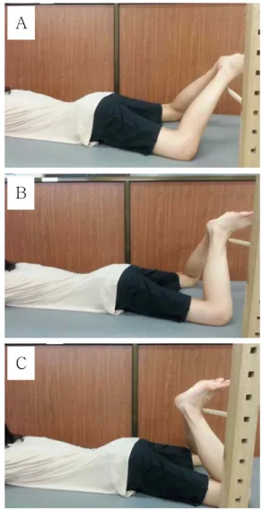
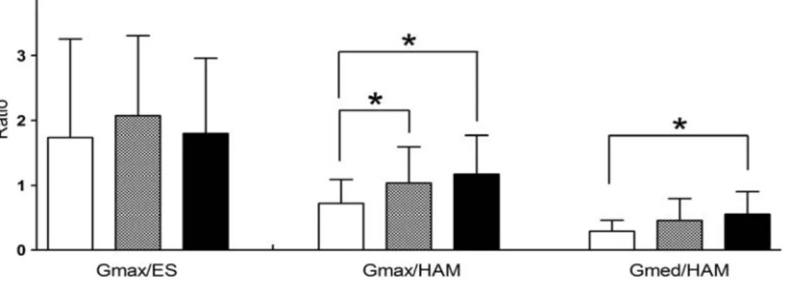
The purpose of this study was to determine the muscle activities of the erector spinae (ES), gluteus maximus (Gmax), gluteus medius (Gmed), and the hamstring (HAM) and the ratios of Gmax/ES, Gmax/HAM, and Gmed/HAM during the prone heel squeeze (PHS) with different knee flexion angles (45°, 90°, and 135°). Fifteen young and healthy subjects (8 men, 7 women) were recruited for the study.

Surface electromyography signals were collected on ES, Gmax, Gmed, and HAM during PHS. A separate one-way analysis of variance with repeated measures was used to determine the significance of the muscle activities of ES, Gmax, Gmed, and HAM and the ratios of Gmax/ES, Gmax/HAM, and Gmed/HAM with different knee flexion angles during PHS. There was a significant increase in the Gmax activity at the knee flexion of 90° in comparison with that of the 45° (p=.016). There were significant increases in the Gmed activity at the knee flexion of 90° (p=.008) and 135° (p=.006) in comparison with that of the 45°. There were significant decreases in the HAM activity at the knee flexion of 90° (p=.009) and 135° (p=.004) in comparison with that of the 45°. There were significant increases in the Gmax/HAM muscle activity ratio at the knee flexion of 90° (p=.007) and 135° (p=.012) in comparison with that of the 45°. There were significant increase in the Gmed/HAM muscle activity ratio at the knee flexion of 135°

in comparison with that of the 45° (p=.008). The knee flexion of 90° during PHS can induce decreasing activity of HAM and increasing activity of Gmax, and the knee flexion of 135° during PHS can induce decreasing activity of HAM and increasing activity of Gmed. Hence, PHS with different knee flexion positions could be considered for the different target muscle.

Key Words: Electromyography; Hip extensor muscles; Prone heel squeeze.

Introduction

Clinically, the weakness of the hip musculature has been reported to cause changes in gait bio- mechanics (Kennedy et al, 2009), increase pain (Fukuda et al, 2012; Souza and Powers, 2009; Tyler et al, 2006), and limit functional activities (Neumann, 2010). Weakness in the gluteus maximus (Gmax) and gluteus medius (Gmed) is theoretically thought to in- crease patellofemoral joint stress by resulting in femoral internal rotation, which causes patellofemoral

pain syndrome (Powers, 2003). Gmax weakness cre- ates slouched posture, causing gait difficulties (Kisner and Colby, 2007). Gmed weakness increases the excessive rotation of the pelvic and femur bones that leads to pain and injury (Philippon et al, 2011;

Zeller et al, 2003). Also, the weakness of Gmed con- tributes to the subtalar inversion resulting in the an- kle instability (Friel et al, 2006).

Due to the fact that a muscle imbalance in strength and activity patterns causes movement im- pairments, the dominance of the hamstring (HAM)

Corresponding author: Ji-hyun Lee [email protected]

(2)

Variables Mean±SD

a

Age (year) 21.7±.4

Height (㎝) 167.0±2.4

Weight (㎏) 60.5±3.1

BMI

b

(㎏/㎡) 21.5±.7

a

mean±standard deviation,

b

body mass index.

Table 1. General characteristics of subjects as the synergist muscle of the Gmax has been sug-

gested to contribute to compensatory motions, espe- cially of the spine (Chance-Larsen et al, 2010;

Sahrmann, 2002). An early pelvic anterior pelvic tilt- ing and rotation with increased erector spinae (ES) activity in individuals with lumbar rotation syndrome during prone knee flexion (Park et al, 2011) is the result of a dominant HAM with an imbalance due to a weak Gmax. Moreover, a dominant HAM, often developed by distance runners, causes a high in- cidence of running injuries (James et al, 1978; Wang et al, 1993).

Previous studies have demonstrated the optimal exercise position and compensatory lumbopelvic con- trol methods for proper exercise implementation by investigating the activities of ES, Gmax, Gmed, and HAM during the exercises (Chance-Larsen et al, 2010; Cynn et al, 2006; Kang et al, 2013; Park et al, 2011). Prone hip extension with knee flexion in dif- ferent hip abduction angles were investigated to ex- amine the influence of different hip abduction angles, which showed the most effective way of facilitating the Gmax muscle (Kang et al, 2013). More, an ab- dominal drawing-in maneuver during the prone hip extension showed lower the compensatory lumbopel- vic motion by reducing the contraction delay of the Gmax in relation to the biceps femoris (Chance-Larsen et al, 2010). Another research has also shown the use of a pressure biofeedback unit during side-lying hip abduction that provided lumbar stabilization which in result increased muscle activities of the Gmed (Cynn et al, 2006).

The prone heel squeeze (PHS) is one of the re- habilitation exercises performed to activate and strengthen the hip musculature. A previous study in- vestigated the muscle activities of the pectineus and piriformis during 13 hip rehabilitation exercises.

Among the exercises performed, PHS showed the highest muscle activity in the piriformis, which was performed at the knee flexion angle 45° (Giphart et al, 2012). Moreover, the activities of the Gmed and iliopsoas during the 13 hip rehabilitation exercises

including PHS were performed to identify the Gmed strengthening exercise while minimizing the iliopsoas activation. PHS was performed at the knee flexion of approximately 70° and had the highest peak of Gmed activation among the other hip exercises (Philippon et al, 2011). Despite previous studies of the PHS, no study has suggested standard knee flexion degrees when performing PHS. Moreover, no study has in- vestigated how different knee flexion angles would influence the back and hip extensor muscles during the PHS.

Thus, the aim of this study was to determine the muscle activities of the ES, Gmax, Gmed, and HAM and the ratios of Gmax/ES, Gmax/HAM, and Gmed/HAM with knee flexion angles of 45°, 90°, and 135° during PHS in healthy subjects. We hypothe- sized that the different knee angles would show dif- ferences in the muscle activities of the ES, Gmax, Gmed, and HAM, and the muscle activity ratios of the Gmax/ES, Gmax/HAM, and Gmed/HAM during the PHS in healthy subjects.

Methods

Subjects

G*power 3.1.6 software (Franz Faul, Kiel

University, Kiel, Germany) was used to estimate the

sample size in this study. A sample size of 7 sub-

jects was calculated from an effect size of .58 and

power of .80 collected from a pilot study of 4

subjects. Fifteen young and healthy subjects (8 men,

7 women) were recruited and participated in the

study (Table 1). The exclusion criteria were a his-

(3)

tory of lumbar, sacroiliac, or lower limb injury within the past year; past or present neurological, muscu- loskeletal, or cardiopulmonary diseases (Kang et al, 2013); hip flexor shortness assessed by the Thomas test (Vogt and Banzer, 1997); tensor fasciae latae shortness assessed by a modified Ober’s test; adduc- tor muscle shortness assessed by the adduction con- tracture test (Magee, 2002); and lumbar or hip pain during PHS. Prior to participation, all subjects signed an informed consent form. The study was approved by the Yonsei University Wonju Institutional Review Board (IRB 1041849-201409-BM-039-02).

Instrumentation

Surface electromyography (EMG) signals were collected by a Noraxon TeleMyo 2400T transmitter (Noraxon, Scottsdale, AZ, USA). EMG data were collected by MyoResearch XP Master Edition 1.06 software (Noraxon, Scottsdale, AZ, USA) at the sampling rate of 1000 ㎐ with the band-pass filter between 20 and 450 ㎐. The root-mean-square was calculated with a moving window of 50 ㎳.

Electrodes

The muscle activities were measured on each par- ticipant’s dominant and non-dominant side by calcu- lating the average of both sides. All of the subjects had a dominant side on the right. Prior to the ex- periment, the skin was first shaved and cleaned with alcohol to reduce skin impedance and for better elec- trical conductance. Disposable Ag/AgCl surface elec- trodes were positioned parallel to the muscle fibers with center-to-center spacing of 2 ㎝. All electrodes were placed bilaterally over the following muscles:

(1) ES: parallel to the spine, approximately 2 ㎝ lat- eral to the L3 vertebra determined by the iliac crest.

(2) Gmax: half the distance between the greater tro- chanter and S2 vertebra in the middle of the muscle in an oblique angle. (3) Gmed: parallel to the muscle fibers, a third of the distance between the iliac crest and the greater trochanter. (4) HAM: the center of the back of the thigh, approximately half of the dis-

tance from the gluteal fold to the back of the knee (Criswell, 2010).

Normalization

After attaching the electrodes, EMG data were collected to obtain the maximum voluntary isometric contraction (MVIC). The subjects’ testing positions were determined according to Kendall and McCreary (2005). The subjects’ positions were as follows: (1) ES: a prone position with the hands clasped behind the head. The subjects extended the trunk with the examiner holding the pelvis in the direction of the posterior pelvic tilt. (2) Gmax: a prone position with the knee flexed. Resistance was applied against the lower part of the posterior thigh, in the direction of the hip flexion. (3) Gmed: a side-lying position with the underneath leg slightly flexed at the hip and knee. The subjects abducted the hip with slight ex- tension and external rotation. Resistance was applied against the leg near the ankle in the direction of the hip abduction and flexion. (4) HAM: a prone position with the knee flexed between 50° and 70°. Resistance was applied against the leg proximal to the ankle.

Each trial was repeated two times with 30 s rest period between the two trials. To collect the MVIC data, resistance was applied for 5 s. The middle 3 s were used for the analysis.

Procedures

The subjects were asked to choose one number from 1 to 15 to select the random order of the knee flexion angle (available from: http://www.randomization.com;

accessed 10 July 2014). Once the number was se- lected, the subjects performed modified knee flexion based on the randomized plan.

The PHS starting position was as follows: the subjects were in the prone position with their hands overlapped in front of their forehead facing the table.

The subjects’ hips were abducted 30° according to

the line between the anterior superior iliac spine and

the midpoint of the patella (Kang et al, 2013). The

subjects’ knees were flexed using a 12-inch goni-

(4)

A

B

Figure 1. Prone heel squeeze exercise performed with the different knee flexion angles (A: knee flexion 45°, B:

knee flexion 90°, C: knee flexion 135°).

ometer (Baseline Plastic 360 Degree ISOM Goniometer 12" Length, Baseline

TM

, OR, USA) for knee flexion modified to 45°, 90°, and 135°. To begin the exercise, a wooden target bar was placed at the height of the subjects’ distal part of the tibia when the subjects’ heels touched (Figure 1). Prior to the exercise, 10 minutes were spent for familiarizing subjects with the PHS. During the familiarization, subjects were educated how to correctly perform the PHS. With the verbal instruction “start”, the subjects pressed their heels into one another for 6 s (Giphart et al, 2012). They were instructed to engage in maximum effort when pressing their heels. With the

verbal instruction “relax”, the subjects stopped the performance. The trials were done 3 times for every knee flexion angle. The middle 4 s of the perform- ance were used for analysis.

Statistical analysis

A Kolmogorov-Smirnov Z test was used to exam- ine whether the continuous data approximated a nor- mal distribution. A separate one-way analysis of variance with repeated measures was used to de- termine the significance of the ES, Gmax, Gmed, and HAM muscle activities and the Gmax/ES, Gmax/HAM, and Gmed/HAM muscle activity ratios at the different knee flexion angles during PHS. All alpha levels were set at .05. If a significant differ- ence was found, the Bonferroni correction was per- formed with alpha=.05/3=.017 for the post-hoc analysis. The analysis was undertaken using SPSS ver. 21.0 software (SPSS Inc., Chicago, IL, USA).

Results

All the continuous variables approximated a nor- mal distribution (p>.05). There were significant dif- ferences found in the Gmax (F=3.701, p=.050), Gmed (F=5.077, p=.013), and HAM (F=7.449, p=.003) muscle activity among the different knee angles during PHS.

The post-hoc test results were as follows: 1) there was a significant increase in the Gmax activity at the knee flexion of 90° in comparison with that of 45° (p=.016), 2) there were significant increases in the Gmed activity at the knee flexion of 90° (p=.008) and 135° (p=.006) in comparison with that of 45°, 3) there was a significant decrease in the HAM activity at the knee flexion of 90° (p=.009) and 135° (p=.004) in comparison with that of 45° (Figure 2).

There were significant differences found in the Gmax/HAM muscle activity ratio (F=3.918, p=.032) and Gmed/HAM muscle activity ratio (F=6.301, p=.006) among the different knee angles during PHS.

The post-hoc test results were as follows: 1) there

C

(5)

Figure 2. Comparisons of the muscle activities with knee flexion angles at 45°, 90°, and 135° (MVIC: maximal voluntary isometric contraction, ES: erector spinae, Gmax: gluteus maximus, Gmed: gluteus medius, HAM: hamstring, error bar:

standard deviation, *p<.017).

Figure 3. Comparisons of the muscle activity ratios with knee flexion angles at 45°, 90°, and 135° (ES: erector spinae, Gmax: gluteus maximus, HAM: hamstring, error bar: standard deviation, *p<.017).

was a significant increase in the Gmax/HAM muscle activity ratios at the knee flexion of 90° (p=.007) and 135° (p=.012) in comparison with that of 45°, 2) there was a significant increase in the Gmed/HAM muscle activity ratio at the knee flexion of 135° in compar- ison with that of 45° (p=.008) (Figure 3).

Discussion

The purpose of this study was to determine

whether different knee flexion angles can influence

the ES, Gmax, Gmed, and HAM muscle activities

and the ratios of Gmax/ES, Gmax/HAM, and

Gmed/HAM during PHS. The results of this study

concur with our hypothesis that there would be dif-

ferences in the muscle activity and muscle activity

ratios among the different knee angles. Because no

previous studies have investigated the muscle activ-

ities of ES, Gmax, Gmed, and HAM and the muscle

activity ratios of Gmax/ES, Gmax/HAM, and

Gmed/HAM with different knee flexion angles during

(6)

PHS, to our knowledge, this study is novel and can provide a viable base for selecting the optimal knee flexion angle for specific muscle activation during PHS in healthy subjects.

The Gmax muscle activity increased significantly 32% at the knee flexion of 90° in comparison with that of 45° during PHS. Previous study also reported results similar to the current study that the HAM muscle activity decreased as the knee flexion in- creased during the hip extension with different knee flexion angles (Kwon and Lee, 2013). The result of the current study can be explained by the possible occurrence of the posterior pelvic tilt at the knee flexion of 45° during PHS. Even though the kine- matic data of the pelvic movement was not meas- ured in this study, the increased activity of the HAM at the knee flexion of 45° may induce posterior pelvic tilt during PHS. Because the HAM is attached to the ischial tuberosity of the pelvis, the augmented activity of the HAM might have caused posterior pelvic tilt at the knee flexion of 45°. Once the pelvis was posteriorly tilted at the knee flexion of 45°, the gluteal muscle is placed into a shortened range, and due to the length-tension relationship, the isometric muscle tension in the shortened range is diminished compared with that of the mid-range at the knee flexion of 90°. Therefore, PHS at the knee flexion of 90° can be recommended to selectively activate Gmax.

The Gmed muscle activity increased significantly 44% and 50% at the knee flexion of 90° and 135° in comparison with that of 45°, whereas the HAM muscle activity decreased significantly 19% and 22%

at the knee flexion of 90° and 135° in comparison with that of 45° during PHS. The reduced muscle activity of the HAM and the increased muscle activ- ity of the Gmed at the knee flexion of 90° compared with that of 45° may be explained by the decreased knee extension moment arm at the knee flexion of 90° compared with that of 45°. While PHS is being performed at the knee flexion of 45°, clockwise knee extension torque is increased from the lengthened

moment arm and gravity, thus, to sustain PHS at the knee flexion of 45° or to counterbalance the in- creased clockwise knee extension torque, counter- clockwise knee flexion torque might be required.

This required counterbalance could be accomplished by increasing the HAM activity that occurred with the decreasing muscle activity of the Gmed at the knee flexion of 45°. The HAM muscle activity was also significantly reduced when the Gmed muscle activity increased at the knee flexion of 135° com- pared with that of 45°. This rise of the Gmed mus- cle activity can possibly be caused by the reduction of the HAM muscle activity due to the shortened length of the HAM at the knee flexion of 135° and the limited tension generation capability at the short- ened length due to active insufficiency. This finding is in agreement with the previous study, which also reported that the HAM muscle activity decreased as the knee flexion increased during hip extension with different knee flexion angles (Kwon and Lee, 2013).

Kennedy and Cresswell (2001) reported that when one joint is fixed as the muscle is shortened by the other joint during the movement, the number of muscle fibers used for the movement decreases and as a result the muscle activity decreases. Therefore, the knee flexion of 90° or 135° can be recommended to decrease the HAM muscle activity in comparison with that of 45° during PHS.

The Gmax/HAM muscle activity ratio significantly increased at the knee flexion of 90° and 135° in comparison with that of 45° during PHS. The greater Gmax/HAM muscle activity ratio suggests increased Gmax activity and decreased HAM activity. Our re- sults have shown a significant increase in the Gmax activity at the knee flexion of 90°, however, there was no significant increase in the Gmax activity at the knee flexion of 135°. Moreover, the HAM activity has shown a significant decrease at the knee flexion of 90° and 135° in comparison with that of 45°.

Therefore, we find that the knee flexion of 90° dur-

ing PHS is the better PHS position for increasing

the Gmax/HAM ratio and selectively increasing the

(7)

activation of the Gmax relative to the HAM activity among the different knee flexion angles.

The Gmed/HAM muscle activity ratio significantly increased at the knee flexion of 135° in comparison with that of 45° during PHS. The greater Gmed/HAM muscle activity ratio suggests increased Gmed activity and decreased HAM activity. The re- sults of our study have shown significant increases in the Gmed activity and decreases in the HAM ac- tivity at the knee flexion of 90° and 135° in compar- ison with that of 45°. Although the selective muscle has shown an increase in the Gmed activity in rela- tion to the HAM activity, the ratio of Gmed/HAM muscle activity has shown a significant increase only at the knee flexion of 135° in comparison with that of 45°. Therefore, we find that the knee flexion of 135° during PHS is the better PHS position for in- creasing the Gmed/HAM ratio and selectively in- creasing the activation of the Gmed relative to the HAM activity among the different knee flexion angles.

There were several limitations in this study. First, the subjects were recruited as a young healthy group. This limits the generalizability of the results, which cannot be generalized to patients performing PHS. Second, the muscle activities were only col- lected to analyze the PHS. Thus, future studies is needed to incorporate lumbopelvic kinematics on the muscle activities of ES, Gmax, Gmed, and HAM to analyze the compensatory movements during the PHS. Third, the study was designed as a cross-sec- tional study, thus, training effects of PHS with dif- ferent knee flexion angles could be examined. Future studies should investigate the longitudinal effects of PHS to confirm the findings of this study.

Conclusion

The purpose of this study was to determine whether different knee flexion angles can influence the muscle activities of ES, Gmax, Gmed, and HAM

and the ratios of Gmax/ES, Gmax/HAM, and Gmed/HAM during PHS. The findings of this study showed a significant increase in the Gmax activity at the knee flexion of 90° and Gmed activity of 90°

and 135° in comparison with that of 45°. Moreover, there was a significant increase in the Gmax/HAM muscle activity ratios at the knee flexion of 90° and 135° and the Gmed/HAM muscle activity ratio of 135° in comparison with that of 45°. The PHS dur- ing knee flexion of 90° induces decrease in the HAM muscle activity and increase in the Gmax muscle ac- tivity, and the knee flexion of 135° induces decrease in the HAM muscle activity and increase in the Gmed muscle activity. Therefore, we suggest PHS with different knee flexion positions for the different target muscles.

References

Chance-Larsen K, Littlewood C, Garth A. Prone hip extension with lower abdominal hollowing im- proves the relative timing of gluteus maximus activation in relation to biceps femoris. Man Ther. 2010;15(1):61-65. http://dx.doi.org/10.1016/

j.math.2009.07.001

Criswell E. Cram’s introduction to surface electromyography. 2nd ed. Sudbury, MA, Jones and Bartlett Publishers, 2011:342-369.

Cynn HS, Oh JS, Kwon OY, et al. Effects of lumbar stabilization using a pressure biofeedback unit on muscle activity and lateral pelvic tilt during hip abduction in sidelying. Arch Phys Med Rehabil. 2006;87(11):1454-1458.

Friel K, McLean N, Myers C, et al. Ipsilateral hip abductor weakness after inversion ankle sprain.

J Athl Train. 2006;41(1):74-78.

Fukuda TY, Melo WP, Zaffalon BM, et al. Hip post-

erolateral musculature strengthening in sedentary

women with patellofemoral pain syndrome: A

randomized controlled clinical trial with 1-year

follow-up. J Orthop Sports Phys Ther. 2012;42

(8)

This article was received September 24, 2014, was reviewed September 24, 2014, and was accepted October 31, 2014.

(10):823-830. http://dx.doi.org/10.2519/jospt.2012.4184 Giphart JE, Stull JD, Laprade RF, et al. Recruitment and activity of the pectineus and piriformis muscles during hip rehabilitation exercises: An electromyography study. Am J Sports Med.

2012;40(7):1654-1663. http://dx.doi.org/10.1177/036 3546512443812

James SL, Bates BT, Osternig LR. Injuries to runners. Am I Sports Med. 1978;6(2):40-50.

Kang SY, Jeon HS, Kwon O, et al. Activation of the gluteus maximus and hamstring muscles during prone hip extension with knee flexion in three hip abduction positions. Man Ther. 2013;18(4):

303-307. http://dx.doi.org/10.1016/j.math.2012.11.006 Kendall FP, McCreary EK, Provance PG, et al.

Muscles: Testing and function with posture and pain. 5th ed. Baltimore, MD, Lippincott Williams

& Wilkins, 2005:181-436.

Kennedy P, Cresswell A. The effect of muscle length on motor-unit recruitment during isometric plantar flexion in humans. Exp Brain Res. 2001;

137(1): 58-64.

Kennedy MJ, Lamontagne M, Beaulé PE.

Femoroacetabular impingement alters hip and pelvic biomechanics during gait. Walking bio- mechanics of FAI. Gait Posture. 2009;30(1):41-44.

http://dx.doi.org/10.1016/j.gaitpost.2009.02.008 Kisner C, Colby LA. Therapeutic Exercise:

Foundations and techniques. 5th ed. Philadelphia, PA, F.A. Davis Company, 2007:692-693.

Kwon YJ, Lee HO. How different knee flexion angles influence the hip extensor in the prone position.

J Phys Ther Sci. 2013;25(10):1295-1297. http://

dx.doi.org/10.1589/jpts.25.1295

Magee DJ. Orthopedic Physical Assessment. 4th ed.

Philadelphia, Elsevier Science Health Science Division, 2002:692-697.

Neumann DA. Kinesiology of the hip: A focus on mus- cular actions. J Orthop Sports Phys Ther. 2010;40 (2):82-94. http://dx.doi.org/10.2519/jospt.2010. 3025 Park KN, Cynn HS, Kwon OY, et al. Effects of the

abdominal drawing-in maneuver on muscle ac-

tivity, pelvic motions, and knee flexion during active prone knee flexion in patients with lum- bar extension rotation syndrome. Arch Phys Mede Rehabil. 2011;92(9):1477-1483. http://dx.doi.

org/10.1016/j.apmr.2011.03.020

Philippon MJ, Decker MJ, Giphart JE, et al.

Rehabilitation exercise progression for the glu- teus medius muscle with consideration for iliop- soas tendinitis: An in vivo electromyography study. Am J Sports Med. 2011;39(8):1777-1785.

http://dx.doi.org/10.1177/0363546511406848

Powers CM. The influence of altered lower-extremity kinematics on patellofemoral joint dysfunction: A theoretical perspective. J Ortho Sports Phys Ther. 2003;33(11):639-646.

Sahrmann S. Diagnosis and Treatment of Movement Impairment Syndromes. St Louis, Mosby, 2002:

30-31.

Souza RB, Powers CM. Differences in hip kinematics, muscle strength, and muscle activation between subjects with and without patellofemoral pain. J Ortho Sports Phys Ther. 2009;39(1):12-19. http://

dx.doi.org/10.2519/jospt.2009.2885

Tyler TF, Nicholas SJ, Mullaney MJ, et al. The role of hip muscle function in the treatment of pa- tellofemoral pain syndrome. Am J Sports Med.

2006;34(4):630-636.

Vogt L, Banzer W. Dynamic testing of the motor stereotype in prone hip extension from neutral position. Clin Biomech (Bristol, Avon). 1997;12 (2):122-127.

Wang SS, Whitney SL, Burdett RG, et al. Lower extremity muscular flexibility in long distance runners. J Ortho Sports Phys Ther. 1993;17(2):

102-107.

Zeller BL, McCrory JL, Kibler WB, et al. Differences

in kinematics and electromyographic activity be-

tween men and women during the single-legged

squat. Am J Sports Med. 2003;31(3):449-456.

수치

Table  1.  General characteristics of subjectsas the synergist muscle of the Gmax has been
Figure  1.  Prone heel squeeze exercise performed with the different knee flexion angles (A: knee flexion 45°, B:
Figure  3.  Comparisons of the muscle activity ratios with knee flexion angles at 45°, 90°, and 135° (ES: erector spinae, Gmax: gluteus maximus, HAM: hamstring, error bar: standard deviation, *p&lt;.017).

참조

관련 문서

③ A student who attended Korean course at KNU Korean Language Program and holds TOPIK Level 3 or a student who completed Korean course Level 4 at the KNU Korean Language

· 50% exemption from tuition fee Ⅱ for the student with a TOPIK score of level 3 or higher or completion of level 4 or higher class of the Korean language program at the

웹 표준을 지원하는 플랫폼에서 큰 수정없이 실행 가능함 패키징을 통해 다양한 기기를 위한 앱을 작성할 수 있음 네이티브 앱과

_____ culture appears to be attractive (도시의) to the

※ Screw home motion ⇒ 경골이 외측 회전 Open kinetic chain. http://moon.ouhsc.edu/dthompso/namics/screw3.gif.. Test

- quadriceps tendon 이 슬개골 하연에서 tibial tuberocity에 부착.

고관절 : 관골의 관골구(acetabulum)과 femur head sacroiliac joint: sacrum + ilium.. pubic symphysis : pubic + pubic

The “Asset Allocation” portfolio assumes the following weights: 25% in the S&amp;P 500, 10% in the Russell 2000, 15% in the MSCI EAFE, 5% in the MSCI EME, 25% in the