J. Exp. Biomed. Sci. 2012, 18(2): 175~179 pISSN : 1738-3226
A Hydrogel Film Containing Propolis Nanoparticles as a Wound Healing Membrane
Jin Kim1¶, Yong Moon Kim1¶, Dong-Woon Kim2 and Ki-Young Lee3,4,†
1Department of Advanced Chemicals and Engineering, Chonnam National University, Gwangju 500-757, Korea
2Department of Clinical Pathology, Gwangyang Health College, Gwangyang 545-703, Korea
3Faculty of Applied Chemical Engineering, 4The Research Institute for Catalysis, Chonnam National University, Gwangju 500-757, Korea
It is desirable that a wound healing membrane acts as a barrier for coverage of a damaged skin and has the biological activities such as anti-inflammatory effects. In this study, we prepared the hydrogel film containing the propolis nanoparticles as a wound healing membrane. The propolis nanoparticles were prepared by incorporation of propolis into the hydrophobic core of γ-cyclodextrin. The incorporation efficiency of propolis in the nanoparticles was 50 ± 2.3%.
Propolis nanoparticles observed by a scanning electron microscope (SEM) were spherical with the size of 30~40 nm.
The swelling behaviors of the hydrogel film containing propolis nanoparticles showed a similar pattern with the hydrogel film without propolis nanoparticles. The cumulative amount of propolis released from the hydrogel film containing propolis nanoparticles in the buffer of pH 7.4 and 5.5 was 86.0 ± 2.0% and 64.6 ± 1.0% of total propolis loaded in the hydrogel film within 9 h, respectively. These results provide a rationale for studying wound healing application of the hydrogel film containing propolis nanoparticles in a clinical setting.
Key Words: Propolis nanoparticles, Hydrogel film, Wound healing membrane
피부는 외부 환경으로부터 인체를 보호하는 기능을 갖 는 기관으로 피부가 손상을 입게 되면 위험한 상황에 노 출된다. 외부에서 가해진 힘에 의한 외상, 피부질환에 의 한 피부 손상, 화상 등과 같은 피부의 결손은 일상 중 흔하게 일어난다. 피부의 결손이 심한 경우 일차봉합이 불가능하여 이차적으로 각종 감염 등에 노출되기 쉽다.
따라서 손상된 피부 조직이 부작용 없이 복구되도록 신 속한 치료가 필요하며 적절한 창상피복재를 이용한 상처 치료가 필수적이다 (Sun et al., 2011). 기존의 창상피복재 는 물리적인 얇은 막을 상처 부위에 덮음으로써 외부로 부터 결손 부위를 보호하고 상처 부위가 마르지 않게 하 여 육아 조직의 형성을 촉진하는 기능을 한다. 창상피복
재로는 하이드로젤 (hydrogel) 필름, 나노섬유 필름 등이 사용되고 있고 피부 손상 부위를 외부와 격리시키는 막 (barrier)의 역할을 한다 (Grinstaff, 2007). 보다 바람직한 창상피복재는 피부 결손 부위에 물리적인 막으로 작용할 뿐만 아니라 항염증 (anti-inflammation) 등과 같은 생리활 성을 갖는 치료제를 포함한다면 효과적으로 피부 결손 부위를 치료하는데 도움을 줄 수 있을 것이다 (Ayvazyan et al., 2011).
Propolis는 식물들이 자신을 보호하기 위해 분비하는 수지성 화합물을 꿀벌들이 채취하여 타액과 혼합하여 만 든 황갈색 또는 암갈색 수지상 물질이며, 벌집 내부를 미 생물 등으로부터 보호해 주는 역할을 한다. Propolis는 주 로 resin, balsam, 그리고 phenol 화합물로 구성되어 있고, phenol 화합물의 주된 구성성분은 flavonoids이다. Propolis 는 항균 (Velikova et al., 2000), 살균 (Madhubala et al., 2011), 항염증 (Sartori et al., 2011), 항바이러스 (Amoros et al., 1992), 항암 (Watanabe et al., 2011), 항산화 (Isla et al., 2001) 및 항궤양 (Barros et al., 2008) 작용을 가지고 있
*Received: 27 February, 2012 / Revised: 4 June, 2012 Accepted: 4 June, 2012
¶J. Kim and Y. M. Kim equally contributed in this study.
†Corresponding author: Ki-Young Lee. Faculty of Applied Chemical Engineering, Chonnam National University, Gwangju 500-757, Korea.
Tel: +82-62-530-1843, Fax: +82-62-530-1676 e-mail: [email protected]
○CThe Korean Society for Biomedical Laboratory Sciences. All rights reserved.
Brief Communication
으며, 면역조절 (Dimov et al., 1991)에도 관여한다고 알려 져 있다. 이러한 생리활성들은 페놀화합물과 수지상 물질 들의 복합작용에 의해 상승효과를 갖는다 (Burdock, 1998;
Markham et al., 1996). Propolis가 다양한 생리활성을 갖고 있음에도 불구하고 식품, 의약품, 화장품 등과 같은 산 업에 응용하기 위해서는 해결해야 할 문제점들이 있다.
Propolis는 물에 용해되지 않는 난용성 성질, 강한 맛과 독특한 향을 가지고 있다. 이를 개선하기 위해, alginate 와 gelatin 등의 고분자를 활용하여 propolis를 캡슐화하는 연구들이 많이 진행되고 있다 (Kalogeropoulos et al., 2009;
Durán et al., 2007). 이에 본 연구에서는 propolis를 나노입 자 형태로 제형화함으로써 물에서의 분산성을 향상시키 고, 독특한 향을 억제시켰다. Propolis 나노입자를 창상용 피복재로 사용 가능한 하이드로젤 필름에 첨가함으로써 피부 결손 부위에 물리적인 막을 형성시키고 동시에 다 양한 생리활성이 있는 propolis의 피부 상처 복구 도움 효과를 기대할 수 있을 것이다. 하이드로젤에 포함된 나 노입자로부터 propolis의 방출 정도를 조사하고 propolis의 창상피복재 내 치료용 조성 물로서의 이용 가능성을 살 펴보았다.
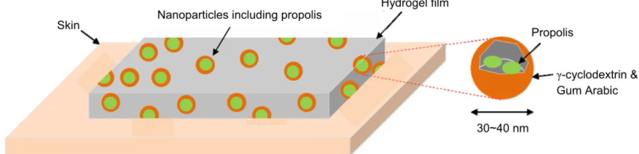
Fig. 1은 propolis 나노입자가 포함된 하이드로젤의 형태 를 나타내는 모식도이다. Propolis (Biopropolis Technology Co., Ltd., Gwangju, Korea)는 γ-cyclodextrin (Wacker Chemie AG, Iowa, USA)과 gum arabic (Sigma-aldrich Co., St. Louis MO)을 이용하여 나노입자 형태로 제형화 하였다. γ- cyclodextrin (0.8 g)과 gum arabic (0.2 g)을 50 mL 증류수에 첨가하고 5시간 동안 교반하여 용해하였다. Propolis (1.0 g)를 첨가한 후 Ultra Homogenizer (M-110S Microfluidizer, Microfluidics Co. Newton, USA)를 이용하여 10,000 rpm에 서 15분간 균질화하여 나노입자를 제조하였고, HITACHI 사의 S-4700 모델 주사전자현미경 (scanning electron microscope, HITACHI Co., Kyoto, Japan)을 이용하여 나노
입자의 형태와 입자크기를 관찰하였다. 제조한 나노입자 용액은 동결 건조를 통해 분말화하였고 냉동보관 하였다.
나노입자에 포함된 propolis의 양을 측정하기 위해 10 mg 나노입자를 증류수와 에탄올 (1:1 v/v) 혼합 용액에 분산시킨 후 ultrasonicator (VCX500, Sonics and Materials Inc., Newtown, CT, USA)로 분쇄하고 원심 분리하였다. 상 등액을 취해 플라보노이드 측정방법을 이용하여 UV/VIS spectrophotometer (UV-160A, Shimadzu Co., Kyoto, Japan)로 420 nm에서 흡광도를 측정하여 정량 분석하였다.
창상피복재의 형태로 제조하기 위해 propolis가 포함된 나노입자를 하이드로젤 (propolis-하이드로젤) 제조 시 첨 가하여 필름 형태로 제조하였다. 하이드로젤 제조에 사 용된 시약은 모두 Sigma-aldrich Chemical Co. (St. Louis, MO)에서 구입하여 사용하였다. 100 mL 증류수가 포함된 250 mL 삼각 플라스크에 0.75 g carboxymethyl cellulose (CMC), 0.5 g pullulan, 2.0 g gellan gum과 1.8 g agar를 넣은 후 70℃로 가열하였다. 하이드로젤 용액을 상온에서 냉각 시켜 50℃에 도달하였을 때 0.1 g propolis가 포함된 나노 입자를 첨가한 후 균질기 (Ultra Turrax T-50, Ika, Staufen, Germany)로 5~10분간 균일화하였다. 제조한 혼합 용액 을 petri-dish에 부은 후 실온에서 2일간 건조하였다. 하이 드로젤 필름 형성 시 젤화에 참여하지 않은 고분자를 제 거하기 위해 제조된 하이드로젤을 50℃의 증류수에 48 시간 이상 충분히 침지시키고 젤 표면의 물기를 제거하 였다. 하이드로젤 필름을 진공건조기에 넣어 60℃에서 2일간 건조하여 다음 실험에 사용하였다.
제조된 하이드로젤 필름의 압축강도는 만능물성시험기 (Compac-100, Sun Scientific Co., Ltd, Tokyo, Japan)를 이용 하여 상온에서 평가하였다. 하이드로젤 필름의 시편 두께 는 3±0.5 mm이고 지름은 10 mm로 준비하였고 압축강도 측정 시 크로스 헤드 (cross head) 속도는 120 mm/min이였 으며, 시편이 50% 이상 변형이 이루어 질 때의 값을 측
Nanoparticles including propolis
γ-cyclodextrin &
Gum Arabic Hydrogel film
Propolis Skin
30~40 nm
Fig. 1. Schematic diagram showing the design of the hydrogel film containing propolis nanoparticles.
정하였다.
하이드로젤 필름의 팽윤도 측정은 건조된 젤 필름을 상온에서 증류수에 침지시키며 시간 경과에 따른 무게변 화를 측정하였고, 팽윤도 (Qs,%)는 다음 식으로부터 계산 되었다.
Propolis-하이드로젤 필름으로부터 propolis의 방출 패 턴을 살펴보기 위해 하이드로젤 필름 (3 g)을 40 mL의 pH 7.4와 pH 5.5 용액에 각각 담근 후 shaking incubator (VS-8480SF, Vision Scientific Co., Daejon, Korea)에 80 rpm 으로 37℃에서 일정한 시간 간격으로 상등액 1 mL을 취 하여 UV/VIS spectrophotometer (shimadzu-160A, Shimadzu Co., Kyoto, Japan)를 이용하여 측정하였다.
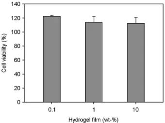
하이드로젤 필름의 세포 독성을 측정하기 위해 멸균 된 하이드로젤 필름을 세포배양액 (DMEM, Dulbecco's modified Eagle's medium, Gibco)에 0.1, 1, 10% (w/v)의 농도 로 넣어 37℃에서 24시간 이상 배양하여 용출액을 제조 하였다. 96 well tissue culture plate에 1 × 10⁵ cells/mL로 각 well 당 100 μL씩 분주하여, 24시간 동안 배양한 RAW 264.7에 하이드로젤 필름 용출액 (100 μL)를 처리하였 다. 24시간 후 약물이 포함되어 있는 배지에 MTT 용액 (Sigma-aldrich Chemical Co., St. Louis, MO)을 처리하여 37℃에서 4시간 동안 반응시킨 후 570 nm에서 ELISA reader (ELX808, Biotek Instruments, Vermont, USA)를 이용 하여 측정하였다.
모든 실험 결과는 평균값 (mean)과 표준편차 (standard deviation, SD)로 표시하였다. 대조군과 실험군 사이의 통 계학적 유의성 검정은 Student's t-test로 비교하였으며 P<
0.05일 때 유의한 차이가 있는 것으로 판단하였다.
γ-cyclodextrin과 gum arabic을 이용하여 제조한 propolis 나노입자는 주사전자현미경으로 관찰하였을 때 구 (sphere) 의 형태였으며 30~40 nm의 크기를 가지고 있음을 확인 할 수 있었다 (Fig. 2). 나노입자 내 propolis의 담지효율 을 평가한 결과, 처음 첨가량의 50 ± 2.3%가 담지되었 다. γ-cyclodextrin은 화학적으로 하이드록실기가 외부로 배향되어 있어 겉으로는 친수성을 띠고 있으나, C-H 기 와 에테르 결합은 내부로 배향되어 있어 내부는 소수성
을 띠는 캡슐 형태의 모양을 하고 있다 (Lee et al., 2002).
따라서, γ-cyclodextrin은 소수성 생리활성 물질을 내부에 담지할 수 있는 특징이 있어서 화장품, 식품, 의약품, 및 농약 등 많은 분야에서 이용되고 있다. 소수성을 가진 propolis는 γ-cyclodextrin의 내부에 쉽게 담지될 수 있다.
Gum arabic은 비이온성 계면활성제 역할로 propolis가 담 지된 γ-cyclodextrin을 구형으로 제형화하는데 도움을 줄 수 있다 (Charoen et al., 2011).
Propolis-하이드로젤 필름의 강도는 51.60 g · cm이었고, propolis 나노입자가 포함되지 않은 하이드로젤 필름의 강도는 68.97 g · cm였다. 하이드로젤 필름 사이에 propolis 나노입자는 필름의 압축강도를 감소시키는 역할을 하는 것으로 생각된다.
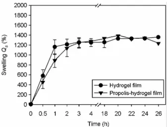
Fig. 3은 하이드로젤 필름의 팽윤 실험 결과를 보여주 Swelling (Qs, %)
(the weight of the swollen hydrogels - the weight of the dried hydrogels
= the weight of the dried hydrogels ×100
Fig. 2. Scanning electron microscopy (SEM) image showing the nanoparticles containing propolis.
Fig. 3. Swelling properties of the hydrogel film containing propolis nanoparticles.
고 있다. 하이드로젤 필름은 증류수에 담근 후 20시간 만에 건조 무게의 13.2배까지 팽윤하였고, 26시간까지 무 게의 감소는 관찰되지 않았다. Propolis-하이드로젤 필름 은 팽윤 실험 20시간에 건조 무게의 13.8배까지 증가하 였고 그 이후로 무게가 감소하여 26시간에 12.2배였다.
Propolis-하이드로젤 필름으로부터 propolis의 방출특성 을 Fig. 4에 나타내었다. 정상 조직과 염증 병소의 pH를 고려하여 방출 실험은 pH 7.4와 pH 5.5 조건에서 각각 수 행하였다. Propolis는 하이드로젤 필름으로부터 pH 7.4와 pH 5.5 버퍼에서 9시간 동안 각각 86.0 ± 2.0%와 64.6 ± 1.0%의 방출을 보였다 (P<0.001).
Propolis-하이드로젤 필름의 세포 독성을 살펴보기 위 해 RAW264.7 세포에 propolis-하이드로젤 필름 용출액을 처리한 후 세포 생존율을 평가하였다. Fig. 5에서 볼 수 있 듯이 propolis-하이드로젤 필름은 세포 독성을 유발하지 않았으며 세포 생존율은 propolis-하이드로젤 필름을 처리 하지 않은 세포와 크게 차이를 보이지 않았다 (P>0.05).
본 연구의 결과, propolis가 담지된 30~40 nm 크기와 구형의 나노입자를 성공적으로 제조할 수 있었다. 창상피 복재로의 응용은 propolis 나노입자를 하이드로젤 필름에 혼합 첨가함으로써 피부에 적용할 수 있는 형태로 제조 할 수 있었다. Propolis-하이드로젤 필름은 세포 독성을 보이지 않았으며, 하이드로젤 필름으로부터 propolis를 9시간 동안 서서히 방출하는 특징을 보였다. 이러한 결과 들을 종합해 볼 때, propolis 나노입자를 포함한 하이드로 젤 필름은 손상된 피부 부위와 외부 환경 사이의 물리적 인 막을 형성할 수 있을 뿐만 아니라 propolis의 항균, 항
염증과 같은 생리활성을 기대할 수 있어서 창상피복재로 의 응용이 기대된다.
감사의 글
본 연구는 교육과학기술부와 한국연구재단의 지역혁신 인력양성사업에 의해 지원되었으며 이에 감사 드립니다.
REFERENCES
Amoros M, Simoes CM, Girre L, Sauvager F, Cormier M.
Synergistic effect of flavonoids and flavonols against herpes simplex virus type 1 in cell culture. Comparison with the antiviral activity of propolis. J Nat Prod. 1992. 55: 1732-1740.
Ayvazyan A, Morimoto N, Kanda N, Takemoto S, Kawai K, Sakamoto Y, Taira T, Suzuki S. Collagen-gelatin scaffold impregnated with bFGF accelerates palatal wound healing of palatal mucosa in dogs. J Surg Res. 2011. 171: e247-257.
Barros MP, Lemos M, Maistro EL, Leite MF, Sousa JP, Bastos JK, Andrade SF. Evaluation of antiulcer activity of the main phenolic acids found in Brazilian Green Propolis. J Ethnopharmacol. 2008. 120: 372-377.
Burdock GA. Review of the biological properties and toxicity of bee propolis. Food Chem Toxicol. 1998. 36: 347-363.
Charoen R, Jangchud A, Jangchud K, Harnsilawat T, Naivikul O, McClements DJ. Influence of biopolymer emulsifier type on formation and stability of rice bran oil-in-water emulsions:
whey protein, gum arabic, and modified starch. J Food Sci.
2011. 76: E165-172.
Fig. 4. Propolis release properties from the hydrogel film
containing propolis nanoparticles in pH 5.5 or 7.4 buffers. Fig. 5. Cell viability of RAW 264.7 cells treated with the extracts of the hydrogel film containing proppolis nanoparticles.
Dimov V, Ivanovska N, Manolova N, Bankova V, Nikolov N, Popov S. Immunomodulatory action of propolis: influence on anti-infectious protection and macrophage function. Apidologie.
1991. 22: 155-161.
Durán N, Marcato PD, Buffo CM, De Azevedo MM, Esposito E.
Poly (epsilon-caprolactone)/propolis extract: microencapsu- lation and antibacterial activity evaluation. Pharmazie. 2007.
62: 287-290.
Grinstaff MW. Designing hydrogel adhesives for corneal wound repair. Biomaterials 2007. 28: 5205-5014.
Isla MI, Moreno MIN, Sampietro AR, Vattuone MA. Antioxidant activity of argentina propolis extracts. J. Ethnopharmacol.
2001. 76: 165-170.
Kalogeropoulos N, Konteles S, Mourtzinos I, Troullidou E, Chiou A, Karathanos VT. Encapsulation of complex extracts in beta-cyclodextrin: an application to propolis ethanolic extract.
J Microencapsul. 2009. 26: 603-613.
Lee CM, Lee KY, Choi CN, Kim DW, Lee IY. Encapsulation of β-cyclodextrin including DHA using curdlan. Korean J Biotechnol Bioeng. 2002. 17: 54-58.
Madhubala MM, Srinivasan N, Ahamed S. Comparative evaluation of propolis and triantibiotic mixture as an intracanal
medicament against Enterococcus faecalis. J Endod. 2011.
37: 1287-1289.
Markham KE, Mitchel KA, Wilkins AL, Daldy JA, Lu Y. HPLC and GC-MS identification of the major organic constituents in New Zealand propolis. Phytochemistry 1996. 42: 205-211.
Sartori G, Pesarico AP, Pinton S, Dobrachinski F, Roman SS, Pauletto F, Junior LC, Prigol M. Protective effect of brown Brazilian propolis against acute vaginal lesions caused by herpes simplex virus type 2 in mice: involvement of anti- oxidant and anti-inflammatory mechanisms. Cell Biochem Funct. 2011. Epub ahead of print.
Sun G, Zhang X, Shen YI, Sebastian R, Dickinson LE, Fox-Talbot K, Reinblatt M, Steenbergen C, Harmon JW, Gerecht S.
Dextran hydrogel scaffolds enhance angiogenic responses and promote complete skin regeneration during burn wound healing. Proc Natl Acad Sci USA 2011. 108: 20976-20981.
Velikova M, Bankova V, Tsvetkova I, Kujumgiev A, Marcucci MC. Antibacterial entkaurene from brazilian propolis of native stingless bees. Fitoterapia. 2000. 71: 693-690.
Watanabe MA, Amarante MK, Conti BJ, Sforcin JM. Cytotoxic constituents of propolis inducing anticancer effects: a review.
J Pharm Pharmacol. 2011. 63: 1378-1386.