Introduction
Tissue conditioners (TC) have been commonly used to enhance the recovery of denture-bearing tissues from trauma, residual ridge resorption or to manage inflammations usually caused by denture stomatitis. The demands on their antimicrobial actions are highly needed because TC are easily degradable or inevitably roughen with times which often compromise the hygiene, eventually; they are sus- ceptible to microbial colonization.1,2 TC could be kept clean by me- chanical or chemical methods, however, these can cause considerable their physical damages.2,3 Acrylic soft liners combined with antifun-
gal drugs were tried for the therapy of denture-induced stomatitis,4,5 there have been some following problems; short-term duration, costs of agents used, harmful reactions to elder patients with drug resis- tances. So far, an effective antimicrobial soft liner has not been de- veloped yet.5,6 Recently, the combination of silver nanoparticles with polysaccharide polymer such as chitosan has attracted a lot of atten- tions due to its potential applications.7,8 Chitosan (Ch) is a polysac- charide derived from chitin, whose unique chemical structure forms a linear polycation with high charge density and reactive chemical groups. Ch has been previously used to transport several drugs with sustained release and permanence in the action site due to its muco-
키토산-은나노 복합체가 함유된 의치 연성이장재 특성에 관한 연구
남기영1* 이철재2
1
계명대학교 의과대학 치과학교실 및 계명대학교 동산병원 치과,
2영남이공대학교 화장품화공계열
Characterization of tissue conditioner containing chitosan-doped silver nanoparticles
Ki Young Nam
1*, Chul Jae Lee
21Department of Dentistry, College of Medicine, Keimyung University, Daegu, Republic of Korea
2Division of Cosmetics Chemistry, Yeungnam College of Science & Technology, Daegu, Republic of Korea
Purpose: Development of a latent antimicrobial soft liner is strongly needed to overcome a possible inflammation related with its dimensional degrade or surface roughness.
Modified tissue conditioner (TC) containing chitosan-doped silver nanoparticles (ChSN) complexes were synthesized and assessed for their characterizations. Materials and methods: ChSN were preliminarily synthesized from silver nitrate (AgNO3), sodium borohydride (NaBH4) as a reducing agent and chitosan biopolymer as a capping agent.
Ultraviolet-visible and Fourier transform infrared spectroscopy were conducted to confirm the stable reduction of nanoparticles with chitosan. Modified TC blended with ChSN by 0 (control), 1.0, 3.0 and 5.0 % mass fraction were mechanically tested by ultimate tensile strength (UTS), silver ion elution and color stability (n=7). Results: At 24 hour and 7 day storage periods, UTS values were not significant (P>.05) as compared with pristine TC (control) and silver ion was detected with the dose-dependent values of ChSN incorporated. Color stability of TC were influenced by ChSN add, with the higher doses, the significantly greater color changes (P<.05). Conclusion: A stable syn- thesized ChSN was acquired and modified TC loading ChSN was characterized as silver ion releasing without detrimental physical property. For its clinical application, anti- microbial test, color control and multifactor investigations are still required. (J Korean Acad Prosthodont 2020;58:275-81)
Keywords: Characterization; Chitosan; Silver nanoparticles; Tissue conditioner
*Corresponding Author: Ki Young Nam
Department of Dentistry, Dongsan Hospital, College of Medicine, Keimyung University, 1035 Dalgubeol-daero, Dalseo-gu, Daegu 42601, Republic of Korea
+82 (0)53 258 4681: e-mail, [email protected]
Article history: Received February 3, 2020 / Last Revision August 20, 2020 / Accepted September 11, 2020
2020 The Korean Academy of Prosthodontics
This is an Open Access article distributed under the terms of the Creative Commons Attribution Non-Commercial License (http://creativecommons.org/
licenses/by-nc/4.0) which permits unrestricted non-commercial use, distribution, and reproduction in any medium, provided the original work is properly cited.
c cc
adhesive property.9 For these reasons, Ch has received great interests from medical fields, food, chemicals, pharmaceuticals and agricul- ture.10 Silver nanoparticles (SN) are already being used due to their effective antimicrobial with good silver ion release. SN could reduce microbial infections in skin and burn wounds or prevent bacterial colonization on various types of material surfaces such as catheters and prostheses.11,12 Despite potent antibacterial activity of silver, its cytotoxic effect against mammalian cells remains still controversial, thus, the balance between antibacterial effect and toxicity in the body has become a very important challenge. Chitosan complex with high infiltration of silver component (ionic or nanoparticles) are broadly represented as an enhanced and effective against bacteria than pure chitosan.13-15 Mei et al.16 suggested the synergistic effect; silver ions improve antibacterial effect and chitosan help stabilize SN or pre- vent its agglomeration. Furthermore, Ribeiro et al.17 demonstrated that chitosan-based nanoparticles showed no significant cytotoxicity against mammalian cells at the highest concentration of 100 ppm (µg/
mL). This study focuses on the synthesis of a novel agent, chitosan- doped silver nanoparticles (ChSN) and mechanical behaviors of the modified TC containing ChSN for challenge to clinical application as an antimicrobial denture material.
Materials and methods
1. Synthesis of chitosan-silver nanoparticles (ChSN) complex The synthesis of SN stabilized with chitosan was performed ac- cording to Peng et al.18 with minor modifications (Fig. 1). Chitosan
(low molecular type, 448869, Sigma-Aldrich Co., St. Louis, MO, USA) with degree of deacetylation and molecular weights are 75%
was used without further purifications. As for ChSN preparation, chitosan (8.0 g) was mixed with 1.0 g AgNO3 (Junsei Chemical Co., Tokyo, Japan) in 100 mL deionized water and stirred until homog- enous under 50℃ under 200 rpm for 1 hour using homomixer (TK homomixer II, Tokushu kika Co., Tokyo, Japan). To reduce the silver salt, NaBH4 stock solution (0.05 g) was added to the AgNO3 solution containing chitosan until the solution became cloudy, followed by centrifugation at 2,000 rpm for 30 minutes (Fig. 2A). The superna- tants were transferred to 200 mL of 100% methanol and they were dried by vacuum at 65℃ for 2 hours. The color of desiccated mixed powder was changed from colorless to light yellow and finally to yel- lowish brown indicating the formation of SN (Fig. 2B).
Fig. 2. Aqeous ChSN (A). Desiccated ChSN (B) powder with brownish pigment.
A B
Fig. 1. Schematic diagram for synthesis of ChSN and process of sample fabrication.
Chitosan-Ag sol AgNO3 1.0 g Chitosan 8.0 g Deionized H2O 100 mL
Homo-mixer 2000 rpm / 30 min
Aqeous ChSN
Reducing agent
NaBH4 0.05 g
Purification (100 % of ethanol) Vacuum desccation
Loading to pristine PEMA powder by weight %
2. Characterizations of ChSN
Ultra violet and visible spectroscopy (UV-vis) (Shimadzu- UV 2500, Kyoto, Japan) was used to characterize the synthesized nanoparticles at a resolution of 1 nanometer (nm) ranged 300 to 700 nm. To confirm the interaction of SN to chitosan, a finely dried ChSN powder was examined under Fourier transform infrared spectroscopy (FT-IR) (Agilent 660, Mulgrave, Australia) and their infrared spectra were recorded on the wavenumber of 600 - 1800 cm-1 region.
3. Modified TC samples containing ChSN
The acrylic TC selected for this study was Soft-Liner (GC, Tokyo, Japan) supplied as powder and liquid. Completely dried ChSN were passed through a sieve (60 mesh) and homogenized in a ball mill for 1 hour. ChSN was preliminarily combined to PEMA (polyethyl methacrylate) powder homogeneously according to three different mass ratios of 1.0, 3.0 and 5.0 wt% (weight %) corresponding to 110, 320, 500 ppm of silver amounts respectively. Unmodified, pristine TC was designated as the control (0 w% of ChSN). Secondarily, blended powders were mixed with EMA (ethyl methacrylate) liquid at designated powder/liquid ratio of 2.2/1.8 (g/mL) by manufactur- er’s instruction. When mixtures became a dough-staged, they were packed into custom-made moulds by two sample specifications; a disc shape (10 mm diameter × 3.0 mm depth) and a dumbbell-shape (ASTM D412)19 with a central cross-sectional area of 33.0 × 6.0 × 3.0 mm. Cured samples were trimmed and stored under 60% humidity for pending mechanical tests.
4. Mechanical properties of TC-ChSN
All of specimens fabricated were divided into 4 groups (n = 7) according to doses of ChSN added (mass ratios of 1.0, 3.0 and 5.0 wt%) with pristine TC designated as control (0 w%).
1) Ultimate tensile strength (UTS)
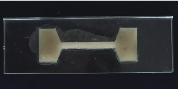
Dumbbell-shaped specimens (Fig. 3) were stored for either 24 hours or 7 days in 100 ml of distilled water at 37℃. They were sub- jected to tension in universal testing machine (MTS Model 4200, Instron Inc., Norwood, MA, USA) at a rate of 40 mm/min. A specific claw (20 mm × 20 mm) was made so the central cross-sectional area could stand exposed, while both ends would stay confined by the claw. The values of UTS were calculated in MPa unit.
2) Elution of silver ion
AAS (atomic absorption spectrophotometer, Analyst 100, Perkin- Elmer, Waltham, MA, USA) and shaking incubator (SI-600R, JEIO
TECH, Seoul, Korea) were used. Each disc specimen (n = 7) was put into 100 mL of sterile distilled water and stored at 37℃ under agita- tion. Eluted ions were determined at 24 hour and 7 days with daily replacing the storage water. The quantity of elution was scored as the amounts of silver ion in the solution per unit of surface area of the disc (cm2).
3) Color difference (ΔE*)
Color changes were measured with disc specimens (n = 7) using spectrophotometer (Minolta CR-10, Kyoto, Japan) at 24 hour and 7 day elapse from the onset of curing process. The mean of three read- ing was recorded and the values were calculated with CIELAB scale recommended by Commission Internationale de l’Eclairage. ΔE*
was calculated by the followed relationship and 2.69 was designated as the perceptible threshold for clinical acceptability.20
ΔE* = [(ΔL*)2 + (Δa*)2 + (Δb*)2]½,
where ΔL* = L1 - L0, Δa* = a1 - a0, Δb* = b1 - b0 (L1, a1, b1: ChSN impregnated, L0, a0, b0: control) 5. Statistics
The data were analyzed using the statistical software (SPSS v.24.0, SPSS Inc., Chicago, IL, USA). The experimental groups were com- pared through Two-way ANOVA and Tukey’s test for post-hoc at a significance level of 0.05.
Results
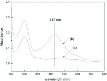
For ChSN synthesis, AgNO3 was successfully reduced and stabi- lized with chitosan. In UV-vis, a typical peak at 412 nm, the surface plasmon band of silver was detected in ChSN (Fig. 4) and molecular structure of chitosan was secured by similar bending shifts with ChSN in FT-IR spectra comparison (Fig. 5). All of modified TC samples did not exhibit the significant UTS differences in relation
Fig. 3. Dumbbell-shaped specimens (ASTM D412)19 for UTS.
to the control (P > .05) after two storage times (Table 1). Early and prolonged silver ion releases were detected from TC-ChSN with the dose-dependent manner and the amounts of ions were significantly decreased at 7 day storage (P < .05) (Table 2). ChSN additions influ- enced the color stability on pristine TC with the higher, the signifi- cantly greater ΔE* (P < .05) and no significant difference between two intervals were shown (P > .05) (Table 3).
Discussion
The SN doped to biopolymer was introduced to exert the antimi- crobial activity and little cytotoxicity with slow silver release from the matrix. Chitosan-silver complex in present work is reported to be less toxic to human fibroblasts, better biocompatibility and lower body absorption than uncoated SN.14 UV-vis spectrum of aqueous ChSN was presented in Figure 4 and the peak at 412 nm was identi- fied as the surface plasmon band of SN synthesized by the in situ re- duction. A plasmon band of spherical SN typically appears at around the 400 nm region, implying a stable conversion of AgNO3 to SN with chitosan. FT-IR spectra results clearly identified that some func- tional groups of chitosan acted as the capping sites for SN stabiliza- tion leading to the stable interaction between two agents. Similarity in bending shifts were observed at the range of 1320 cm-1 to 1376 cm-1 for methyl group, at 1645 cm-1 for carbonyl bonds of amide group, at 1575 cm-1 foramine group and the spectra ranged from 1160 cm-1 to 1000 cm-1 for carbon group (Fig. 5).21 These shifts may indicate the binding or coupling of SN to chitosan, and the ChSN polymerization did not change the molecular structure of chitosan during the present process.
Table 3. Mean color differences (ΔE*) of TC-ChSN to pristine PEMA
ChSN doses ΔE*
1.0% 3.0% 5.0%
24 hour 2.9a, (0.3) 5.2b (0.5) 7.3c (0.5) 7 day 3.1a (0.2) 5.6 b (0.3) 7.9c (0.6) Different letter indicates a statistical difference (P < .05).
ΔE* of 2.69 is designated as perceptible threshold for clinical application.
Table 1. Mean UTS values (MPa ± SD) of TC-ChSN
ChSN doses Control (0%) 1.0% 3.0% 5.0%
24 hour 4.24 ± 0.24a 4.20 ± 0.25 a 4.04 ± 0.15 a 3.98 ± 0.23 a 7 day 3.07 ± 0.17 b 3.06 ± 0.07 b 2.99 ± 0.18 b 2.99 ± 0.08 b Different letter indicates a statistical difference (P < .05).
SD: standard deviation.
Table 2. Mean (SD) concentrations of silver elution (ppm) from tested samples
ChSN doses Control 1.0% 3.0% 5.0%
24 hour ND 0.068a, (0.001) 0.161b (0.002) 0.294c (0.002) 7 day ND 0.032A, (0.001) 0.086B (0.001) 0.203C (0.001) Different letter indicates a statistical difference (P < .05).
ND: non detectable.
Fig. 4. UV-Vis absorption spectra of ChSN (b) aqueous solution and the peak at 412 nm is the surface plasmon band of SN. The spectrum of pure chitosan (a) is also shown for comparison.
Fig. 5. FT-IR spectra of pure chitosan (a) and ChSN (b) synthesized with 1.0%
AgNO3. Similarity in typical bending shifts ranged 600 to 1800 cm-1 wave- numbers.
ChSN addition to a PEMA might hinder the plasticizers from penetrating and forming a weak gel, consequently, it could affect the physical properties of pristine tissue conditioner. Generally, the ratio of nanofiller to matrix were regulated to be less than 5.0% for the mechanical security and ChSN doses below 5.0 w% in this study did not affect UTS value of control at two storage times (Table 1). UTS, one of fundamental properties of elastomer, can provide the informa- tion on the ultimate strength of a rubber in tension.22 UTS were sig- nificantly decreased at 7-day storage as compared to 24 hour for both control and ChSN group, the natural loss of plasticizer led to gradual hardening of TC samples and wet storing environment allowed ethanol and ester to be leached into water. Though a limitation of this study, only one brand of tissue conditioner was used, our results corroborate other studies involving the incorporation of functional agents into tissue conditioners. Ueshige et al.23 did not find changes in the dynamic viscoelastic properties of one tissue conditioner (Shofu, Shofu Inc., Kyoto, Japan) containing silver-zeolite. Schneid24 demonstrated significantly greater tensile strength of the tissue con- ditioner (Lynal, Dentsply, Mississauga, ON, Canada) associated with nystatin agent.
The antimicrobial mechanisms of nanocomposite have not been fully elucidated yet. One possible contribution to those effects is the release of silver ions from matrix diffused by water molecules to the aqueous medium.25 Considering the initial ChSN doses of 1.0 - 5.0 wt% (110 - 500 ppm of silver), small quantities of silver ions (0.032 - 0.294 ppm) were detected from cured samples (Table 2). Restricted ion elution could be explained by the facts that PEMA is a rather hydrophobic polymer which may have generated a barrier for water diffusion, and its water uptake may not be sufficient for ion release.
Leaching was significantly decreased at 7 days as comparing 24 hour storage, gradual hardening of TC by the loss of plasticizer might lead to strongly entrapped ChSN in PEMA bulk. Regarding the clinical recommendation of TC, low ion releasing would be preferable for the prevention of inflammations rather than high elution that might cause toxic or undesirable interactions to host tissues. Our previous results26 showed that the E. coli were killed by silver elution at the dose of 0.21 ppm within 3 days, while pristine soft liner (0 ppm) did not show any antimicrobial effect against that strain. Thus, though elution tests were not combined with antimicrobial assay against oral microbes to confirm the direct correlation, TC- ChSN samples can be expected to show both fast and prolonged antimicrobial actions.
Future studies are also needed for the direct contact between the TC surface and microbes as another possible contribution. Regardless of inorganic ions leaching, the bacterial inhibitory effects could be exhibited due to direct contact of SN immobilized on the surface of solid matrix.27
Color stability is an important clinical behavior for denture base
since it may provide critical information on the serviceability of this material. The whole TC- ChSN samples become brownish and darker with increased concentration of ChSN and exhibited the rapid discoloration within 24 hours from onset of curing. Color differences (ΔE*) were proven to clinically unacceptable according the threshold value (ΔE* > 2.69) when compared to pristine TC (Table 3). The oxidative reaction or plasmon by metal nanoparticles is known to influence the color instability28 and the unique color of chitosan pow- der might also contribute to these discolorations. Though TC is ap- plied or masked within hard denture base, a concern should be made to lessen the possibility on the cosmetic hazards for denture wearers.
Conclusion
In present study, the development of novel antimicrobial tissue conditioner containing chitosan doped silver nanoparticles was dis- cussed. Stably synthesized chitosan-silver nanoparticles complexes were achieved by the in situ reduction and their incorporation to tissue conditioner exhibited acceptable mechanical properties with evident silver ion elution. For clinical candidates, further studies including in vivo, practical microbial assays, multi-brands and color tests are still required.
ORCID
Ki Young Nam https://orcid.org/0000-0003-0481-0687 Chul Jae Lee https://orcid.org/0000-0002-4575-7539
References
1. Okita N, Orstavik D, Orstavik J, Ostby K. In vivo and in vitro studies on soft denture materials: microbial adhesion and tests for antibacterial activity. Dent Mater 1991;7:155-60.
2. Harrison A, Basker RM, Smith IS. The compatibility of tem- porary soft materials with immersion denture cleansers. Int J Prosthodont 1989;2:254-8.
3. Nikawa H, Iwanaga H, Hamada T, Yuhta S. Effects of denture cleansers on direct soft denture lining materials. J Prosthet Dent 1994;72:657-62.
4. Schneid TR. An in vitro analysis of a sustained release system for the treatment of denture stomatitis. Spec Care Dentist 1992;12:245-50.
5. Truhlar MR, Shay K, Sohnle P. Use of a new assay technique for quantification of antifungal activity of nystatin incorpo- rated in denture liners. J Prosthet Dent 1994;71:517-24.
6. Chow CK, Matear DW, Lawrence HP. Efficacy of antifungal agents in tissue conditioners in treating candidiasis. Gerodon- tology 1999;16:110-8.
7. Sanpui P, Murugadoss A, Prasad PV, Ghosh SS, Chattopad-
hyay A. The antibacterial properties of a novel chitosan- Ag-nanoparticle composite. Int J Food Microbiol 2008;31;
124:142-6.
8. Kalaivani R, Maruthupandy M, Muneeswaran T, Singh M, Sureshkumar S, Anand M, Ramakritinan CM, Quero F, Kumaraguru AK. Synthesis, characterization, fluorescence, photocatalytic and antibacterial activity of CdS nanoparticles using schiff base. J Fluoresc 2015;25:1481-92.
9. Nascimento EG, Sampaio TB, Medeiros AC, Azevedo EP.
Evaluation of chitosan gel with 1% silver sulfadiazine as an alternative for burn wound treatment in rats. Acta Cir Bras 2009;24:460-5.
10. Liu X, Hu Q, Fang Z, Zhang X, Zhang B. Magnetic chitosan nanocomposites: a useful recyclable tool for heavy metal ion removal. Langmuir 2009;25:3-8.
11. Samuel U, Guggenbichler JP. Prevention of catheter-related infections: the potential of a new nano-silver impregnated catheter. Int J Antimicrob Agents 2004;23:S75-8.
12. Wright JB, Lam K, Hansen D, Burrell RE. Efficacy of topi- cal silver against fungal burn wound pathogens. Am J Infect Control 1999;27:344-50.
13. Wei D, Sun W, Qian W, Ye Y, Ma X. The synthesis of chito- san-based silver nanoparticles and their antibacterial activity.
Carbohydr Res 2009;344:2375-82.
14. Rhim JW, Hong SI, Park HM, Ng PK. Preparation and char- acterization of chitosan-based nanocomposite films with anti- microbial activity. J Agric Food Chem 2006;54:5814-22.
15. Nguyen VQ, Ishihara M, Mori Y, Nakamura S, Kishimoto S, Fujita M, Hattori H, Kanatani Y, Ono T, Miyahira Y, Matsui T. Preparation of size-controlled silver nanoparticles and chitosan-based composites and their anti-microbial activities.
Biomed Mater Eng 2013;23:473-83.
16. Mei L, Xu Z, Shi Y, Lin C, Jiao S, Zhang L, Li P. Multivalent and synergistic chitosan oligosaccharide-Ag nanocomposites for therapy of bacterial infection. Sci Rep 2020;19;10:10011.
17. Ribeiro TG, Franca JR, Fuscaldi LL, Santos ML, Duarte MC, Lage PS, Martins VT, Costa LE, Fernandes SO, Cardoso VN, Castilho RO, Soto M, Tavares CA, Faraco AA, Coelho EA, Chávez-Fumagalli MA. An optimized nanoparticle delivery system based on chitosan and chondroitin sulfate molecules reduces the toxicity of amphotericin B and is effective in treat- ing tegumentary leishmaniasis. Int J Nanomedicine 2014;9:
5341-53.
18. Peng Y, Song C, Yang C, Guo Q, Yao M. Low molecular weight chitosan-coated silver nanoparticles are effective for the treatment of MRSA-infected wounds. Int J Nanomedicine 2017;12:295-304.
19. American Society for Testing and Materials. Standard test methods for vulcanized rubber and thermoplastic elastomers tension. West Conshohocken, ASTM, 2002. D412-98a.
20. Chang J, Da Silva JD, Sakai M, Kristiansen J, Ishikawa- Nagai S. The optical effect of composite luting cement on all ceramic crowns. J Dent 2009;37:937-43.
21. Duarte ML, Ferreira MC, Marvão MR, Rocha J. An opti- mised method to determine the degree of acetylation of chitin and chitosan by FTIR spectroscopy. Int J Biol Macromol 2002;31:1-8.
22. Waters MG, Jagger RG. Mechanical properties of an experi- mental denture soft lining material. J Dent 1999;27:197-202.
23. Ueshige M, Abe Y, Sato Y, Tsuga K, Akagawa Y, Ishii M.
Dynamic viscoelastic properties of antimicrobial tissue condi- tioners containing silver-zeolite. J Dent 1999;27:517-22.
24. Schneid TR. An in vitro analysis of a sustained release system for the treatment of denture stomatitis. Spec Care Dentist 1992;12:245-50.
25. Kumar R, Münstedt H. Silver ion release from antimicrobial polyamide/silver composites. Biomaterials 2005;26:2081-8.
26. Lee CJ, Nam KY, Kim DY, Kim HJ. New routes to the prepa- ration of silver-soft liner nanocomposites as an antibacterial agent. J Industrial Eng Chem 2014;20:1276–9.
27. Imazato S, Ebi N, Takahashi Y, Kaneko T, Ebisu S, Russell RR. Antibacterial activity of bactericide-immobilized filler for resin-based restoratives. Biomaterials 2003;24:3605-9.
28. Chladek G, Mertas A, Barszczewska-Rybarek I, Nalewajek T, Zmudzki J, Król W, Lukaszczyk J. Antifungal activity of denture soft lining material modified by silver nanoparticles-a pilot study. Int J Mol Sci 2011;12:4735-44.
키토산-은나노 복합체가 함유된 의치 연성이장재 특성에 관한 연구
남기영1* 이철재2
1
계명대학교 의과대학 치과학교실 및 계명대학교 동산병원 치과,
2영남이공대학교 화장품화공계열
목적: 의치 연성이장재 적용기간 경과에 따른 물성저하 및 표면거침성이 의치구내염 발생을 야기할 수 있으며 이 논문의 목적은 항균물질인 키토산-은 나노 복합체를 환원법으로 합성하고 이것을 연성이장재에 투여 후 그 특성을 평가하는 것이다.
재료 및 방법: 질산은과 키토산 분말로 혼합 정제된 키토산-은나노 복합체를 자외선 가시광선 및 적외선 분광법으로 분석하고 연성이장재 분말에 각 각 0(대조군), 1.0, 3.0 및 5.0의 질량 분율로 첨가 후 단량체 용액과 각각 중합하였다. 항균복합체가 첨가된 연성이장재 시편의 특성은 중합완료 24 시 간과 7 일 후 미세인장강도, 은 이온 용출 그리고 색조변화 등을 통하여 각각 평가하였다.
결과: 분광분석을 통하여 안정적인 키토산-은나노 복합체의 합성을 확인하였다. 대조군과 비교 시 복합체첨가에 따른 연성이장재의 유의한 인장강도
변화는 나타내지 않았고 (P > .05) 은 이온 용출은 복합체 투여량에 대하여 농도비례적으로 측정되었으며 색조변화량 또한 농도비례적으로 증가되었
다 (P < .05).
결론: 키토산-은나노 복합체가 투여된 연성이장재는 적절한 물성과 은 이온 용출 특성을 가진 보철생체재료의 가능성을 도출하였고 임상 적용을 위한 항균실험 및 색조 안정성 등의 연구들이 추후 필요할 것으로 사료된다. (대한치과보철학회지 2020;58:275-81)
주요단어: 특성; 키토산; 은나노; 연성이장재
*교신저자: 남기영
42601 대구 달서구 달구벌대로 1035 계명대학교 동산병원 치과 053 258 4681: e-mail, [email protected]
원고접수일: 2020년 2월 3일 / 원고최종수정일: 2020년 8월 20일 / 원고채택일: 2020년 9월 11일
2020 대한치과보철학회
이 글은 크리에이티브 커먼즈 코리아 저작자표시-비영리 4.0 대한민국 라이선스에 따라 이용하실 수 있습니다.
c cc