Epithelial Wound Healing after Cataract Surgery Comparing Two Different Topical Fluoroquinolones
Kyung Eun Han,
1Woo Suk Chung,
2Tae-im Kim,
3Sekyung Kim,
3Terry Kim,
4and Eung Kweon Kim
3,5,61Department of Ophthalmology, Hallym University College of Medicine, Chuncheon Sacred Heart Hospital, Chuncheon;
2Siloam Eye Hospital, Seoul; 3Corneal Dystrophy Research Institute, Department of Ophthalmology, Yonsei University College of Medicine, Seoul, Korea;
4Department of Ophthalmology, Duke University, Durham, NC, USA; 5Severance Biomedical Science Institute;
6Brain Korea 21 Project for Medical Science, Yonsei University College of Medicine, Seoul, Korea.
Received: March 15, 2013 Revised: May 13, 2013 Accepted: May 14, 2013
Corresponding author: Dr. Eung Kweon Kim, Department of Ophthalmology,
Yonsei University College of Medicine, 50 Yonsei-ro, Seodaemun-gu, Seoul 120-752, Korea.
Tel: 82-2-2228-3570, Fax: 82-2-312-0541 E-mail: [email protected]
∙ The authors have no financial conflicts of interest.
© Copyright:
Yonsei University College of Medicine 2014 This is an Open Access article distributed under the terms of the Creative Commons Attribution Non- Commercial License (http://creativecommons.org/
licenses/by-nc/3.0) which permits unrestricted non- commercial use, distribution, and reproduction in any medium, provided the original work is properly cited.
Purpose: To compare the epithelial wound healing response of two preservative- free fluoroquinolones, moxifloxacin and levofloxacin, in patients who underwent cataract surgery. Materials and Methods: In this prospective, evaluator-masked, randomized clinical trial, 59 eyes of 50 patients who underwent cataract surgery were enrolled. Patients were randomized to receive moxifloxacin 0.5% (n=32 eyes) or levofloxacin 0.5% (n=27 eyes). All patients instilled moxifloxacin or levoflox- ain four times daily for 1 week prior to surgery and 2 weeks after surgery. The epi- thelial wound healing status in the corneal incision site was scanned with a raster scan mode of fourier-domain optical coherence tomography (FD-OCT). The num- ber of eyes showing epithelial defect images and average number of corneal epi- thelial defect cuts per eye were compared between groups. All patients were evalu- ated on postoperative days 1, 2, 3, and 10. Results: On postoperative days 1, 2, and 3, the number of eyes showing epithelial defects in FD-OCT was not statisti- cally different (all p>0.05). The average number of corneal epithelial defect cuts was also not statistically different between the two groups (all p>0.05). No eyes showed epithelial defects on postoperative day 10 in either group. Conclusion:
There were no differences on epithelial wound healing comparing these two dif- ferent fluoroquinolones at the incision site of cataract surgery.
Key Words: Levofloxacin, moxifloxacin, cataract surgery, epithelial wound healing
INTRODUCTION
The selection of appropriate antibiotic eye drops is critical for preventing endo- phthalmitis, one of the most detrimental complications after cataract surgery. Fluo- roquinolones have long been used for the prevention of postoperative infections, but resistance to third-generations fluoroquinolones led to the introduction and de- velopment of fourth-generation fluoroquinolones (moxifloxacin or gatifloxacin).
Moxifloxacin and gatifloxacin have demonstrated 2- to 3-fold penetration into the anterior chamber, improved activity against atypical mycobacteria, and greater an- tibacterial effects against gram-positive bacteria compared to second-generation
surgeon (E.K.K.) performed all surgical procedures. A sin- gle investigator (W.S.C.), under the same conditions, evalu- ated the postsurgical wound.
The inclusion criteria for this study were patients >18 years age who were scheduled for removal of a cataract and implantation of a posterior chamber intraocular lens (IOL), intraocular pressure ≤20 mm Hg prior to surgery, and had otherwise normal and healthy eyes aside from cataracts as determined by ophthalmic examination. Patients with any of the following conditions were not eligible to participate in this clinical trial: fluorescein staining of the cornea be- fore surgery, history of any ocular diseases including ocular inflammatory diseases, ocular herpes infection, iritis, uve- itis, Sjögren’s syndrome, corneal dystrophy, uncontrolled diabetes and/or diabetic retinopathy, any history of systemic disease such as autoimmune diseases, history of treatment for an ocular infection within 30 days prior to study entry, use of topical or systemic steroids within 7 days prior to study entry, use of topical anti-inflammatory drugs within 7 days prior to study entry, known or suspected allergy or hy- persensitivity to levofloxacin or any related medicines, such as cinoxacin, ciprofloxacin, norfloxacin, ofloxacin, or nali- dixic acid, preservatives, dyes, or any component of the medications involved in the study, pregnancy, nursing/lacta- tion, or inadequate birth control methods. Oral contraceptive use was allowed. Patients with any cataract wounds requir- ing sutures or corneal burn secondary to the phacoemulsifi- cation handpiece were excluded in this study.
Surgical procedure
Patients were instructed to use the assigned antibiotics, mox- ifloxacin or levofloxacin, four times a day for 1 week prior to surgery. Standard phacoemulsification with a 2.2 mm clear corneal 3-plane incision was performed. Each corneal incision was made with a disposable phacoblade (Slu- 22AGF®, Kai medical, Gifu, Japan). The length of the cor- neal incision was set at 2 mm or greater for self-sealing. A 2.2 mm microcoaxial phaco-handpiece (No. 8065750853, Alcon, Fort Worth, TX, USA) was used for phacoemulsifi- cation. After irrigation and aspiration of the cortex, a poste- rior chamber intraocular lens (Acrysof SN60WF, Alcon, Fort Worth, TX, USA) was inserted into the capsular bag.
At the end of surgery, the corneal wound was sealed with stromal hydration. If any wound leak was observed, the corneal wound was sutured with 10-0 nylon sutures, and the patient was excluded from the study. An eye patch was then placed over the for 6 hours and then removed. Patients (ciprofloxacin and ofloxacin) or third-generation (levoflox-
acin) fluoroquinolones.1,2 Adverse epithelial cytotoxic ef- fects, however, should also be considered when choosing antibiotics because rapid re-epithelialization helps to pre- vent microbial invasion and other possible complications such as epithelial ingrowth.3,4
Moxifloxacin and levofloxacin are commonly used anti- biotic eye drops after cataract surgery or refractive proce- dures. Although there have been several reports comparing effects on epithelial wound healing of various fluoroquino- lones as well as their potential cytotoxicity,5-8 the results were not consistent. These studies also compared fluoroqui- nolones that contain the preservative, benzalkonium chlo- ride (BAK), with fluoroquinolones without BAK. BAK it- self has been reported beneficial in helping to prevent bacterial growth at a rate faster than preservative-free fluo- roquinolones, especially against gram-positive bacteria.9 However, BAK is known to cause corneal and/or conjunc- tival epithelial toxicity.10,11 Moreover, to our knowledge, there is no study comparing these two types of topical fluo- roquinolones in human eyes after cataract surgery.
To compare the intrinsic effects of third- and fourth-gener- ation fluoroquinolones without the influence of BAK on the re-epithelialization of corneal incision sites after cataract surgery, we compared the effects of two commercially avail- able preservative-free fluoroquinolones, moxifloxacin (Vi- gamox®, Alcon Laboratories, Fort Worth, TX, USA) and le- vofloxacin (Cravit®, Santen, Osaka, Japan).
MATERIALS AND METHODS
This study was a prospective, single-masked, randomized, comparative evaluation of patients undergoing phacoemul- sification. All participants signed an informed consent after a detailed explanation of the study. This study was prospec- tively approved by the Institutional Review Board of Sev- erance Hospital and followed the tenets of the Declaration of Helsinki.
The investigator assigned the operated eye of each pa- tient with an identification number that corresponded to a treatment regimen based on a randomization scheme creat- ed at baseline. The patients then received the appropriate treatment regimen for their identification number based on this randomization scheme. This allowed the investigator to be blinded as to which treatment regimens were allocated to patients. To minimize inter-surgeon variability, a single
without interruption were interpreted as a non-defect cut (Fig. 2B).
In eyes showing epithelial defects, the degree of wound healing was determined based on the number of defect cuts on the FD-OCT image (Fig. 1C). The proportion of eyes that showed epithelial defects and the average number of de- fect cuts (the total number of defect cuts found in each group/the number of patients with defect cuts in each group) were compared between the two groups.
Statistical analysis
All data was analyzed by a statistician not involved in the study design or collection of data. Statistical analyses were performed using PASW for Windows (version 18.0, SPSS Inc., Chicago, IL, USA). Average values of parameters were compared using either the chi-square test or Fisher’s exact test. A p value less than 0.05 was considered statisti- cally significant.
RESULTS
Participants
Eighty-four eyes of 72 patients were initially included. Nine patients receiving cataract surgery on both eyes had a peri- were asked to instill the assigned antibiotic four times per
day. Prednisolone acetate 1% (Pred-Forte®, Allergan, Inc., Irvine, CA, USA) was also instilled four times per day con- currently. After 2 weeks, eye drop use was discontinued. Pa- tients were evaluated on postoperative days 1, 2, 3, and 10.
Measurement of epithelial wound healing
Epithelial defect size can vary in accordance with elapsed time after staining and strength of illumination (Fig. 1A).
To obtain more objective information with regards to both the length and depth of epithelial defect, the corneal inci- sion site was scanned with the raster scan mode of FD- OCT, which provides 17 cross-sectional images in a size adjustable square that measured 3×3 mm2, resulting in 17 focal points each measured at 0.187 mm intervals in that square (Fig. 1B).
An epithelial defect cut was defined when an interruption or lack of epithelial growth (a black hollow space in the in- cision site, white arrows in Fig. 2A-1 and A-2) was found in the FD-OCT image above the extension line, an imagi- nary extension line between the cut edges of Bowman lay- er (red dotted line of Fig. 2A and B). Images showing a continuous epithelial growth (a dark gray space connected with the epithelium which showing similar dark gray col- or, black arrows in Fig. 2B-1 and B-2) above this line
A
B
C-1
C-2
C-3
C-4
Fig. 1. (A) A slit lamp photograph of the corneal incision after fluorescence dye staining. The margin of corneal defect is obscure (a or b) and can be interpreted differently according to varying strengths of illumination. (B) A capture window of raster scan mode in fourier-do- main optical coherence tomography (FD-OCT). By selecting a 3×3 mm2 sized capture window, 17 cuts of cross-sectional images (white and red arrows) could be acquired at the same gap. (C) On cross-sectional images of red arrows in Fig. 1B, the defect cuts (C-1 to 4) can be counted. The actual margin of corneal defect detected based on the FD-OCT images was determined to be “a” not “b” in Fig. 1A.
Defect cut
Defect cut Defect cut Defect cut
floxacin group; 64.6±8.7 years, p>0.05). There were no postoperative complications in these patients during the 10 days of follow-up after successful phacoemulsification and IOL implantation.
Incidence of epithelial defect
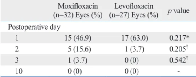
On the first day after the operation, 15 of 32 eyes (46.9%) of the moxifloxacin group and 17 of 27 eyes (63.0%) of the levofloxacin group showed at least one corneal epithelial defect cut, but the difference was not statistically significant (p=0.217, chi-square test) (Table 1). On postoperative day 2, five eyes (15.6%) in the moxifloxacin group and one eye (3.7%) in the levofloxacin group showed corneal epithelial defect cuts, but the difference was not statistically signifi- cant (p=0.205, Fisher’s exact test). On postoperative day 3, one eye (3.1%) in the moxifloxacin group showed corneal epithelial defect cuts and there was no eye showing an epi- thelial defect cut in the levofloxacin group. There was no statistically significant differences between the two groups (p=0.542, Fisher’s exact test). On postoperative day 10, no eye had an epithelial defect cut in either group.
od of at least two weeks between each cataract surgery, with each eye being treated as a new case. Among the initial 84 eyes, 25 eyes were excluded for the following reasons: can- cellation of surgery due to personal reasons (15 eyes), suture placement after surgery due to wound leakage (nine eyes), and occurrence of posterior capsule rupture (one eye). Final- ly, 59 eyes of 51 patients were enrolled upon completion of the study; 32 eyes received moxifloxacin and 27 eyes re- ceived levofloxacin. The mean age of the 51 patients was 65.1±8.0 years (moxifloxacin group; 65.4±7.4 years, levo-
A B
Fig. 2. (A) Representative images showing the defect and non-defect image cuts in images of the fourier-domain optical coherence to- mography. (A and B) An imaginary line (red dotted line) was drawn connecting Bowman layer on both ends of the sectional plane. (A-1 and A-2) Cases showing a lack of epithelium on the imaginary line and a discontinuation of this line were interpreted as a defect cut. The black hollow spaces without epithelial growth were noted (white arrows). The layer of powerfully bright light is considered to be a reflec- tion of the tear fluid (white hollow arrowheads). (B-1 and B-2) Cases showing epithelial growth both above this line and without interrup- tions were interpreted as a non-defect image cut. The spaces in the incision site were occupied with epithelial growth which showing similar dark gray color with the epithelial layer (black arrows).
A-1 B-1
A-2 B-2
Non-defect cut Defect cut
Table 1. Number of Eyes with Corneal Epithelial Defect Cuts after Cataract Surgery
Moxifloxacin
(n=32) Eyes (%) Levofloxacin
(n=27) Eyes (%) p value Postoperative day
1 15 (46.9) 17 (63.0) 0.217*
2 5 (15.6) 1 (3.7) 0.205†
3 1 (3.7) 0 (0) 0.542†
10 0 (0) 0 (0) -
*Chi-square test.
†Fisher’s exact test.
ofloxacin, levofloxacin, gatifloxacin, or ciprofloxacin on healthy rabbit corneas, and showed that moxifloxacin caused the least damage to the corneal epithelium. Never- theless, in the last two studies mentioned, moxifloxacin was the only solution that did not contain BAK. Because the ef- fect of BAK was not excluded in those studies, the authors presumed the cytotoxicity of fluoroquinolones was mainly a result of the preservatives and not of the fluoroquinolone itself.
To exclude these interfering effects of BAK, we have chosen commercially available and preservative-free mox- ifloxacin and levofloxacin, and our results showed there were no statistical differences in epithelial wound healing in clear corneal incision. This is consistent with an in vivo study that used the same preservative-free fluoroquino- lones as we studied, in which Watanabe, et al.14 treated healthy human volunteers with either moxifloxacin or le- vofloxacin three times per day for 1 week and reported that there were no differences in tear break-up time and mor- phological appearance of the corneal epithelium, stroma, and endothelium.
In this study, we used a clear corneal incision model to compare the effect of two different topical fluoroquinolones on wounded human corneas. As these patients were already scheduled for cataract surgery, the corneal incisions in this study posed no ethical problems and it was an appropriate model that could induce the standardized form of epithelial damage. Through this model, epithelial defects of the same length were made, and the rate of re-epithelialization could be compared.
Accurate epithelial defect measurements could not be as- certained when using fluorescein dye staining because sometimes distinguishing between true epithelial defect and wound irregularity after epithelial healing is difficult. In ad- dition, quantitative measurements of the degree of wound healing, as shown in Fig. 1A, can also be difficult. In this study, we used anterior segment FD-OCT to measure epi- thelial defects. Using FD-OCT, we could quantitatively measure the epithelial defect from focal points measured at 0.187 mm intervals. Thus, the length of epithelial defect could be measured more accurately.
The current study is the first to compare corneal epithelial wound healing in human eyes after cataract surgery with topical use of moxifloxacin and levofloxacin. Our study showed that there was no significant difference between these two antibiotics in terms of their effect on re-epitheliali- zation after cataract surgery.
The mean number of defect cuts per eye
Because individual variance of epithelial defect size exist- ed, we evaluated the mean number of epithelial defect cuts per eye (the total number of epithelial defect cuts/the num- ber of eyes with epithelial defect) and compared the values between the groups.
On postoperative day 1, the mean number of defect cuts per eye was 3.93±2.34 (range: 1-9 per eye) in the moxiflox- acin group and 2.82±1.67 (range: 1-7 per eye) in the levo- floxacin group. On postoperative day 2, the mean number of defect cuts per eye was 1.60±1.34 (range: 1-5 per eye) in the moxifloxacin group and 1 (1 cut/1 eye) in the levofloxa- cin group. On postoperative day 3, the mean number of de- fect cuts per eye was two (2 cuts/1 eye) in the moxifloxacin group and none (0 cut/0 eye) in the levofloxacin group. On postoperative day 10, there was no defect cut in either group.
There were no statistically significant differences through the follow-up examinations (all p>0.05).
DISCUSSION
In this study, there were no significant differences in the number of eyes showing epithelial defects between the moxifloxacin and levofloxacin groups on days 1, 2, 3, and 10 after cataract surgery. There was also no statistical dif- ference in the average number of defect cuts per eye be- tween the two groups on follow-up examinations.
Numerous experimental and clinical studies have com- pared the cytotoxicity of different fluoroquinolones, includ- ing levofloxacin and moxifloxacin, but the results have been inconsistent. In contrast to our results, Kim, et al.12 re- ported that levofloxacin (Cravit®) was less cytotoxic than moxifloxacin (Vigamox®) when these eyedrops were ex- posed to cultured human corneal epithelial cells over 2 hours. Tsai, et al.10 incubated human corneal epithelial cells with different fluoroquinolones (norfloxacin, ciprofloxacin, ofloxacin, levofloxacin, moxifloxacin and gatifloxacin) us- ing both commercial ophthalmic formulations and raw ma- terials (standard powders) of each antibiotic, and reported that levofloxacin and ofloxacin showed the least cytotoxici- ty. In contrast, Sosa, et al.11 reported that among moxifloxa- cin, gatifloxacin, ofloxacin, and levofloxacin, moxifloxacin showed the lowest degree of cytotoxicity in immortalized conjunctival and human corneal epithelial cells. In a clini- cal study, Kovoor, et al.13 compared in vivo confocal mi- croscopy images after instillation of either moxifloxacin,
Epithelial healing rates with topical ciprofloxacin, ofloxacin, and ofloxacin with artificial tears after photorefractive keratectomy. J Cataract Refract Surg 2000;26:690-4.
7. Moreira LB, Lee RF, de Oliveira C, LaBree L, McDonnell PJ. Ef- fect of topical fluoroquinolones on corneal re-epithelialization af- ter excimer laser keratectomy. J Cataract Refract Surg 1997;23:
845-8.
8. Moshirfar M, Chew J, Werner L, Meyer JJ, Hunter B, Stevens S, et al. Comparison of the effects of fourth-generation fluoroquino- lones on corneal re-epithelialization in rabbit eyes. Graefes Arch Clin Exp Ophthalmol 2008;246:1455-61.
9. Kowalski RP, Kowalski BR, Romanowski EG, Mah FS, Thomp- son PP, Gordon YJ. The in vitro impact of moxifloxacin and gati- floxacin concentration (0.5% vs 0.3%) and the addition of benzal- konium chloride on antibacterial efficacy. Am J Ophthalmol 2006;142:730-5.
10. Tsai TH, Chen WL, Hu FR. Comparison of fluoroquinolones: cy- totoxicity on human corneal epithelial cells. Eye (Lond) 2010;24:
909-17.
11. Sosa AB, Epstein SP, Asbell PA. Evaluation of toxicity of com- mercial ophthalmic fluoroquinolone antibiotics as assessed on im- mortalized corneal and conjunctival epithelial cells. Cornea 2008;
27:930-4.
12. Kim SY, Lim JA, Choi JS, Choi EC, Joo CK. Comparison of anti- biotic effect and corneal epithelial toxicity of levofloxacin and moxifloxacin in vitro. Cornea 2007;26:720-5.
13. Kovoor TA, Kim AS, McCulley JP, Cavanagh HD, Jester JV, Bugde AC, et al. Evaluation of the corneal effects of topical oph- thalmic fluoroquinolones using in vivo confocal microscopy. Eye Contact Lens 2004;30:90-4.
14. Watanabe R, Nakazawa T, Yokokura S, Kubota A, Kubota H, Nishida K. Fluoroquinolone antibacterial eye drops: effects on normal human corneal epithelium, stroma, and endothelium. Clin Ophthalmol 2010;4:1181-7.
ACKNOWLEDGEMENTS
This research was supported by the Converging Research Center Program funded by the Ministry of Education, Sci- ence and Technology (2012K001354) and by the educational grant from Alcon Laboratories, Inc. (Fort Worth, TX, USA).
All patients gave informed consent for participation in re- search. This study is registered at http://www.clinicaltrials.
gov (NCT00840580).
Terry Kim is a surgical consultant for Alcon, Inc.
REFERENCES
1. Hooper DC, Wolfson JS. Mode of action of the quinolone antimi- crobial agents. Rev Infect Dis 1988;10 Suppl 1:S14-21.
2. Mather R, Karenchak LM, Romanowski EG, Kowalski RP.
Fourth generation fluoroquinolones: new weapons in the arsenal of ophthalmic antibiotics. Am J Ophthalmol 2002;133:463-6.
3. Vargas LG, Vroman DT, Solomon KD, Holzer MP, Escobar-Go- mez M, Schmidbauer JM, et al. Epithelial downgrowth after clear cornea phacoemulsification: report of two cases and review of the literature. Ophthalmology 2002;109:2331-5.
4. Lee BL, Gaton DD, Weinreb RN. Epithelial downgrowth follow- ing phacoemulsification through a clear cornea. Arch Ophthalmol 1999;117:283.
5. Pollock GA, McKelvie PA, McCarty DJ, White JF, Mallari PL, Taylor HR. In vivo effects of fluoroquinolones on rabbit corneas.
Clin Experiment Ophthalmol 2003;31:517-21.
6. Patel GM, Chuang AZ, Kiang E, Ramesh N, Mitra S, Yee RW.