저작자표시-비영리-변경금지 2.0 대한민국 이용자는 아래의 조건을 따르는 경우에 한하여 자유롭게 l 이 저작물을 복제, 배포, 전송, 전시, 공연 및 방송할 수 있습니다. 다음과 같은 조건을 따라야 합니다: l 귀하는, 이 저작물의 재이용이나 배포의 경우, 이 저작물에 적용된 이용허락조건 을 명확하게 나타내어야 합니다. l 저작권자로부터 별도의 허가를 받으면 이러한 조건들은 적용되지 않습니다. 저작권법에 따른 이용자의 권리는 위의 내용에 의하여 영향을 받지 않습니다. 이것은 이용허락규약(Legal Code)을 이해하기 쉽게 요약한 것입니다. Disclaimer 저작자표시. 귀하는 원저작자를 표시하여야 합니다. 비영리. 귀하는 이 저작물을 영리 목적으로 이용할 수 없습니다. 변경금지. 귀하는 이 저작물을 개작, 변형 또는 가공할 수 없습니다.
Master's Thesis in Medicine
Optimal Fluence and Duration of Low-Level
Laser Therapy for Efficient Wound Healing in
Mice
Graduate School of Ajou University
Department of Medical Sciences
Optimal Fluence and Duration of Low-Level
Laser Therapy for Efficient Wound Healing in
Mice
You Chan Kim, M.D., Ph.D., Advisor
I submit this thesis as the
Master's thesis in medicine.
February, 2020
Graduate School of Ajou University
Department of Medical Sciences
The Master's thesis of Jisun Yoon in medicine is hereby approved.
Thesis Defense Committee Chair
You Chan Kim
Member
Seonghyang Sohn
Member
You-sun Kim
Graduate School of Ajou University
i
-ABSTRACT-
Optimal Fluence and Duration of Low-Level Laser Therapy for Efficient Wound
Healing in Mice
Background/Purpose: Low-level laser (light) therapy is a promising technology that stimulates healing,
relieves pain and inflammation, and restores function in injured body parts. However, few studies have compared the effects of light-emitting diodes of different fluence levels or different treatment durations. Here, we investigated the effects of various fluence levels and treatment durations on wound closure in mice.
Methods: Full-thickness wounds were created on the dorsal skin using an 8-mm diameter punch, and
the wounds were irradiated at 1, 4, or 40 J/cm2 for 5 consecutive days starting on day 1. To determine the optimal irradiation duration, wounds were irradiated at the most potent fluence of previous study for 5, 10 or 15 days. Photographic documentation, skin biopsies, and wound measurements were performed to compare the effects of different treatment parameters.
Results: The most effective fluence level was 40 J/cm2, as determined by monitoring wound closure.
There were no statistically significant differences in wound healing with different durations.
Conclusion: We have shown that repeated exposure to low levels of light significantly stimulates
wound healing in mice, and demonstrated more efficient wound closure with certain fluences of 830 nm irradiation.
Keywords: wound healing, low-level light therapy
ii
TABLE OF CONTENTS
ABSTRACT ... i
TABLE OF CONTENTS ... ii
LIST OF FIGURES ... iii
LIST OF TABLES ... iv
INTRODUCTION ...1
MATERIAL AND METHODS ...2
A. Animal selection and care ...2
B. Wound creation and LED irradiation ...2
C. Photo documentation and wound closure analysis ...2
D. Statistical analysis ...3
RESULTS ...4
A. Effect of LED irradiation fluence on wound closure ...4
B. Effect of the duration of LED irradiation on wound closure ...6
DISCUSSION ...9
CONCLUSION ...11
REFERENCES ...12
iii
LIST OF FIGURES
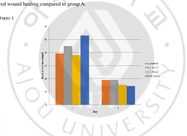
Figure 1. Wound area after treatment with different fluences of irradiation of Day 1 and Day 5 ...5
Figure 2. Percent wound closure at different fluences of irradiation of Day 1 and Day 5 ...10
Figure 3. Wound area after treatment with different durations of irradiation of Day 1 and Day 5 ...13
iv
LIST OF TABLES
Table. 1. Wound closure (%) at different fluences of irradiation ...3
1
INTRODUCTION
Low-level laser (light) therapy (LLLT) is a promising technology used in various fields to stimulate healing, relieve pain and inflammation, and restore function to injured body parts. Since the initial experiments in 1983 studying the effects of low-level HeNe laser irradiation on wounds in rats, many studies have investigated wound healing by LLLT [1].
Low-level lasers can affect lymphocytes, increasing their proliferation and activation; macrophages, increasing their phagocytosis; and fibroblasts, increasing their growth factor secretion and enhancing the uptake of both fibrin and collagen [2]. In addition, LLLT increases the motility of epithelial cells and the amount of granulation tissue produced during healing, and may reduce the synthesis of inflammatory mediators [3, 4], resulting in reductions in skin wound area in both humans and animals. However, the optimal physical variables for LLLT still lack consensus [5].
A few studies have directly compared the effects of different fluences of LLLT. Silva et al. investigated the effects of a 670 nm-wavelength laser on rats, by irradiating skin lesions with 0, 2, or 4 J/cm2 for 10 consecutive days [6]. At 4 J/cm2, the re-epithelialization process was significantly faster than that in the other groups. A study using a 632.8 nm-wavelength laser reported that 3-6 J/cm2 photostimulation facilitates the tissue repair process in diabetic wound healing by accelerating the rates of contraction and collagen production [7]. With irradiation at 830 nm, a preliminary investigation demonstrated that 5 J/cm2 LLLT improved wound healing, as measured by increased wound tensile strength [8]. A study in mice comparing the influences of 632.8, 785, and 830 nm lasers on burn wound healing found that treatment with 830 nm light at a fluence of 3 J/cm2 had profound effects on healing compared to untreated controls and mice treated with lasers of other wavelengths [9]. However, no studies have compared the effects of different fluences and irradiation durations on wound size reduction.
In this study, we describe the effects of LLLT on wound size reduction in a standardized model of full-thickness excisional wound healing in mice, using an 830-nm diode laser with various fluence levels and durations of irradiation.
2
MATERIALS AND METHODS
A. Animal selection and care
Eight-week-old female albino hairless mice (Skh:hr-1) weighing 25–30 g were maintained in individual ventilated cage systems. Constant temperature, humidity, and a 12-h light/dark cycle were maintained, and the mice were fed a standard diet. All experimental protocols were approved by the Committee for Animal Care and Use of Ajou University.
B. Wound creation and LED irradiation
After anesthesia by intraperitoneal injection of tiletamine/zolazepam (Virbac, Seoul, Korea) and xylazine (Bayer, Seoul, Korea), four full-thickness wounds were created on the dorsal skin of each mouse using a 8-mm-diameter punch. The wounds were left exposed without sutures or dressings. A total of 40 mice were used to compare the effects of different irradiation fluences on wound healing. The mice were divided into untreated control (group A) and treated groups (n=10 per group). The wounds of the treated groups were irradiated at fluences of 1 (group B), 4 (group C), or 40 J/cm2 (group D) for five consecutive days starting on day 1 when the wounds were made. To determine the optimal duration of treatment, 30 mice (n=10 per group) were treated with 40 J/cm2 infrared light for 5 (IR5), 10 (IR10), or 15 (IR15) consecutive days . A low-intensity LED irradiation device named SHINeY (WON TECH Co., Seoul, Korea) was used as the light source. The distance between the light source and the dorsal skin was approximately 3 cm. Nonirradiated (control) mice were maintained under similar conditions.
C. Photo documentation and wound closure analysis
On days 1, 5, 10, and 15, images of the wounds were acquired, and the wound areas were measured using Image-Pro Plus 6.0 software (Media Cybernetics, Silver Spring, MD, USA). The wound size immediately after wound creation was designated the original wound area. The percentage of wound closure at each time point was calculated using the following formula:
(original wound area−area on day x) × 100 original wound area
3 D. Statistical analysis
A linear mixed effect model was used to compare wound size reduction between treatment groups. Wound size reduction data were analyzed using R software, version 3.5.2. P values < 0.05 were considered statistically significant. Summary data are expressed as the mean ± standard deviation.
4
RESULTS
A. Effect of LED irradiation fluence on wound closure
Our first goal was to identify the optimal fluence of LED irradiation to reduce the time required for wound closure. There were some variations in the sizes of the initial wounds, due to the difficulty in creating wounds in the flexible skin of the mice. Therefore, the percentage of wound closure was analyzed along with the absolute values. Compared to the baseline values for each group, the wound areas in all groups steadily decreased over time (p < 0.001; Figure 1, 2). When comparing the wound closure percentages in groups A (control) and B (1 J/cm2), more efficient wound closure was observed in group B (Table 1). However, there was no significant difference in wound healing between groups A and C (4 J/cm2). The most efficient fluence was 40 J/cm2 (group D), which demonstrated significantly improved wound healing compared to group A.
5
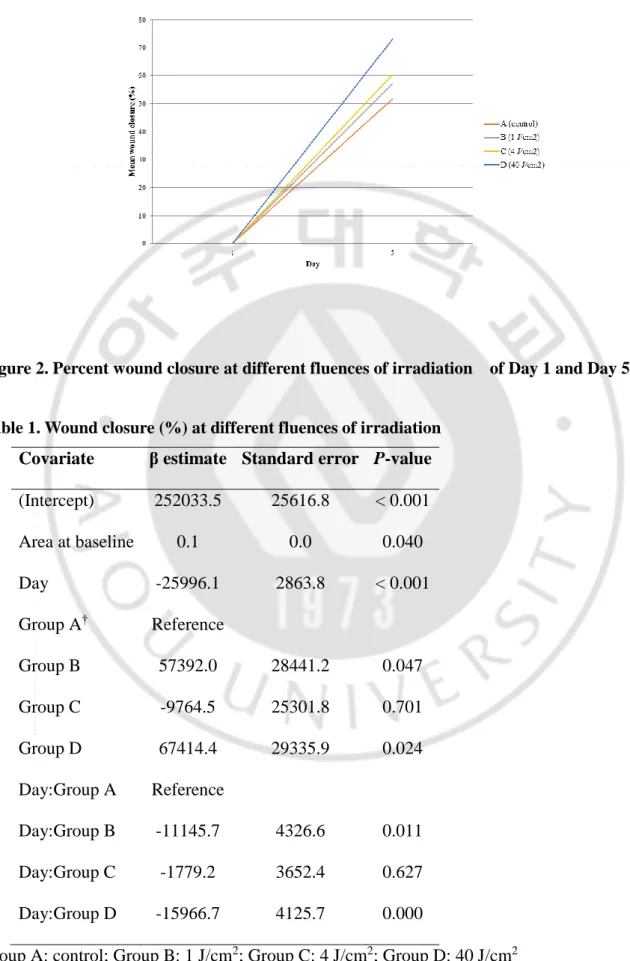
Figure 2. Percent wound closure at different fluences of irradiation of Day 1 and Day 5
Table 1. Wound closure (%) at different fluences of irradiation Covariate β estimate Standard error P-value
(Intercept) 252033.5 25616.8 < 0.001 Area at baseline 0.1 0.0 0.040 Day -25996.1 2863.8 < 0.001 Group A† Reference Group B 57392.0 28441.2 0.047 Group C -9764.5 25301.8 0.701 Group D 67414.4 29335.9 0.024 Day:Group A Reference Day:Group B -11145.7 4326.6 0.011 Day:Group C -1779.2 3652.4 0.627 Day:Group D -15966.7 4125.7 0.000
6
B. Effect of the duration of LED irradiation on wound closure
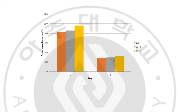
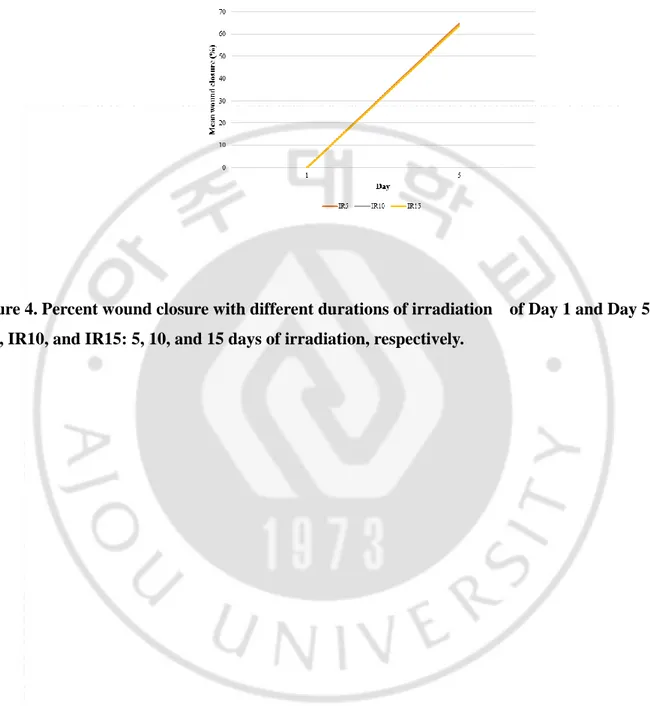
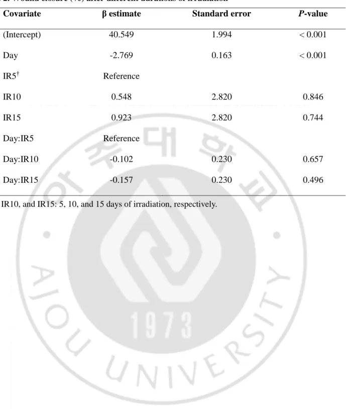
The other goal of our study was to identify the best duration of LED irradiation for wound healing. The effects of 5, 10, and 15 days of radiation (groups IR5, IR10, and IR15, respectively) were compared. No mice died during this experiment. Compared to their baseline values, wound areas steadily decreased in all groups (p < 0.001; Figure 3, 4). There was no significant difference in wound area reduction between the IR5 and IR10 groups, nor between the IR5 and IR15 groups (Table 2).
Figure 3. Wound area after treatment with different durations of irradiation of Day 1 and Day 5 IR5, IR10, and IR15: 5, 10, and 15 days of irradiation, respectively.
7
Figure 4. Percent wound closure with different durations of irradiation of Day 1 and Day 5 IR5, IR10, and IR15: 5, 10, and 15 days of irradiation, respectively.
8
Table 2. Wound closure (%) after different durations of irradiation
Covariate β estimate Standard error P-value
(Intercept) 40.549 1.994 < 0.001 Day -2.769 0.163 < 0.001 IR5† Reference IR10 0.548 2.820 0.846 IR15 0.923 2.820 0.744 Day:IR5 Reference Day:IR10 -0.102 0.230 0.657 Day:IR15 -0.157 0.230 0.496
9
DISCUSSION
Wound closure involves the migration of the boundaries of an injury towards its center, and can be assessed through related parameters, such as the percentage of wound contraction [10]. In this study, all treated mice except those in the 4 J/cm2 group displayed more effective wound healing than the
untreated mice, and the most potent fluence was 40 J/cm2. Therefore, our wound model demonstrated a biphasic dose response to 830-nm light: 1 and 40 J/cm2 improved healing, while 4 J/cm2 had no effect. Tatiana et al. evaluated the effects of laser therapy on excisional wounds and found that the dose effects are not linear for various fluences of 635-nm light, with a maximum positive effect at 2 J/cm2 [11]. They reported that intensities of 1 and 10 J/cm2 improved healing to a lesser extent, while 50 J/cm2 had a negative effect on wound healing. Using 670-nm laser therapy, treatment at 4 J/cm2 displayed superior wound healing than treatment at 8 J/cm2 [12]. Inadequate doses can result in weak and insignificant effects; while excessive doses can cause negative or minimal effects [13]. With even higher doses, a biosuppressive or inhibitory effect may be observed [14]. In contrast to these studies, we used 830-nm light and observed an optimal fluence of 40 J/cm2. As light at this wavelength can penetrate the skin more deeply, we hypothesize that a higher fluence of irradiation might be required for wound healing at 830 nm.
We also investigated the effects of treatment duration, and observed no statistically significant differences between the groups. Wound closure begins with an inflammatory phase and re-epithelialization, followed by the remodeling phase, which generally begins 5-7 days after injury. Therefore, 5 days of irradiation could be adequate to reduce the wound area. In a previous study, while healing curves generated for control mice demonstrated an initial decrease in wound size during days 1-4 after injury, the wounds of LLLT-treated mice started to contract immediately after illumination [11]. The basic biological mechanism behind the effects of LLLT is thought to involve the absorption of red and near-infrared light by mitochondrial chromophores, in particular cytochrome c oxidase (CCO), a component of the mitochondrial respiratory chain [15-17]. CCO activation results in increased production of ATP, which provides both the energy and phosphate required to regulate a variety of cellular functions. Consistent with this notion, the addition of exogenous ATP stimulated wound healing in an animal model [18]. Although wound contraction was not increased in mice treated with external ATP, in vitro observations suggest that ATP increases wound contraction by serving as an energy source for motility and contractile force generation, and as a phosphate donor for kinases regulating contraction [19, 20].
10
Hypertrophic scars and keloids are benign skin tumors that usually form following surgery, trauma, or acne, and are difficult to eradicate. Fibroblastic proliferation and excess collagen deposits are their two main characteristics, and imbalances in the rates of collagen biosynthesis and degradation, along with individual genetic predisposition, have been implicated in their pathogenesis [21]. It was recently proposed that poor regulation of interleukin (IL)6 signaling and TGFβ1 expression may play a significant role in this process [22-25]. LLLT can decrease IL6 mRNA levels [26], and has been proposed as an alternative therapy for hypertrophic scars. In three case studies, Barolet and Boucher reported significant improvements to scars after LLLT following scar revision by surgery or CO2 laser ablation [27].
The effects of laser irradiation on collagen metabolism are controversial. Studies by Abergel et al. and Yu et al. reported increased procollagen, collagen, and basic fibroblast growth factor production and fibroblast proliferation after exposure to low-energy laser irradiation in in vitro and in vivo animal models [28, 29]. Conversely, van Breugel and Bär reported decreased collagen synthesis and cell proliferation after irradiation [13]. Ma et al. demonstrated that in vitro, dual-wavelength light (635/830 nm) and infrared light (830 nm) have stimulating effects on proliferation and collagen synthesis in human fibroblasts whereas visible red light (635 nm) does not [30]. With its ability to increase collagen synthesis, LLLT is often used clinically for skin rejuvenation [31-34].
Wound contraction is thought to be facilitated by SMA-expressing myofibroblasts in the dermis surrounding the injured area [35]. There is evidence that LLLT induces fibroblast-myofibroblast transformation both in vitro and in vivo [12, 36, 37]. A previous study revealed a significant number of SMA-positive cells in tissues surrounding LLLT-treated wounds, but not in nonilluminated control wounds [11]. The presence of contractile myofibroblasts at the edge of illuminated wounds explains the lack of wound expansion one day after injury.
11
CONCLUSION
We have shown that repeated exposure to low levels of light significantly stimulates wound healing in mice, and demonstrated more efficient wound closure with certain fluences of 830 nm irradiation. Cnversely, the duration of irradiation did not significantly affect wound healing. Further studies
regarding human wound healing will be required to examine the applicability of these results to clinical LLLT.
12
REFERENCES
1. Surinchak, J.S., et al., Effects of low-level energy lasers on the healing of full-thickness skin defects. Lasers Surg Med, 1983. 2(3): p. 267-74.
2. Andrade Fdo, S., R.M. Clark, and M.L. Ferreira, Effects of low-level laser therapy on wound healing. Rev Col Bras Cir, 2014. 41(2): p. 129-33.
3. Bashardoust Tajali, S., et al., Effects of low power laser irradiation on bone healing in animals: a meta-analysis. J Orthop Surg Res, 2010. 5: p. 1.
4. Channual, J., et al., Vascular effects of photodynamic and pulsed dye laser therapy protocols. Lasers Surg Med, 2008. 40(9): p. 644-50.
5. Al-Watban, F.A.H. and B.L. Andres, LASER PHOTONS AND PHARMACOLOGICAL TREATMENTS IN WOUND HEALING. LASER THERAPY, 2000. 12(1): p. 3-11.
6. da Silva, T.S., et al., Estudo microscópio da lesão tecidual em pele de ratos Wistar, tratados com laser de baixa potência. Revista Brasileira de Biociências, 2010. 8(3).
7. Maiya, A.G., P. Kumar, and S. Nayak, Photo-stimulatory effect of low energy helium-neon laser irradiation on excisional diabetic wound healing dynamics in Wistar rats. Indian J Dermatol, 2009. 54(4): p. 323-9.
8. Stadler, I., et al., 830-nm irradiation increases the wound tensile strength in a diabetic murine model. Lasers Surg Med, 2001. 28(3): p. 220-6.
9. Rathnakar, B., et al., Photo-biomodulatory response of low-power laser irradiation on burn tissue repair in mice. Lasers Med Sci, 2016. 31(9): p. 1741-1750.
10. Hegde, V.N., et al., Effect of laser dose and treatment schedule on excision wound healing in diabetic mice. Photochem Photobiol, 2011. 87(6): p. 1433-41.
11. Demidova-Rice, T.N., et al., Low-level light stimulates excisional wound healing in mice. Lasers Surg Med, 2007. 39(9): p. 706-15.
12. Medrado, A.R., et al., Influence of low level laser therapy on wound healing and its biological action upon myofibroblasts. Lasers Surg Med, 2003. 32(3): p. 239-44.
13. van Breugel, H.H. and P.R. Bar, Power density and exposure time of He-Ne laser irradiation are more important than total energy dose in photo-biomodulation of human fibroblasts in vitro. Lasers Surg Med, 1992. 12(5): p. 528-37.
14. Coombe, A.R., et al., The effects of low level laser irradiation on osteoblastic cells. Clin Orthod Res, 2001. 4(1): p. 3-14.
15. Karu, T.I. and S.F. Kolyakov, Exact action spectra for cellular responses relevant to phototherapy. Photomed Laser Surg, 2005. 23(4): p. 355-61.
16. Greco, M., et al., Increase in RNA and protein synthesis by mitochondria irradiated with helium-neon laser. Biochem Biophys Res Commun, 1989. 163(3): p. 1428-34.
17. Karu, T.I., L.V. Pyatibrat, and G.S. Kalendo, Photobiological modulation of cell attachment via cytochrome c oxidase. Photochem Photobiol Sci, 2004. 3(2): p. 211-6.
18. Chiang, B., et al., Enhancing skin wound healing by direct delivery of intracellular adenosine triphosphate. Am J Surg, 2007. 193(2): p. 213-8.
19. Kreis, T.E. and W. Birchmeier, Stress fiber sarcomeres of fibroblasts are contractile. Cell, 1980. 22(2 Pt 2): p. 555-61.
20. Ehrlich, H.P., et al., Vanadate and the absence of myofibroblasts in wound contraction. Arch Surg, 1999. 134(5): p. 494-501.
21. Uitto, J. and D. Kouba, Cytokine modulation of extracellular matrix gene expression: relevance to fibrotic skin diseases. J Dermatol Sci, 2000. 24 Suppl 1: p. S60-9.
22. Wolfram, D., et al., Hypertrophic scars and keloids--a review of their pathophysiology, risk factors, and therapeutic management. Dermatol Surg, 2009. 35(2): p. 171-81.
23. Uitto, J., IL-6 signaling pathway in keloids: a target for pharmacologic intervention? J Invest Dermatol, 2007. 127(1): p. 6-8.
13
24. Ghazizadeh, M., et al., Functional implications of the IL-6 signaling pathway in keloid pathogenesis. J Invest Dermatol, 2007. 127(1): p. 98-105.
25. Liu, W., D.R. Wang, and Y.L. Cao, TGF-beta: a fibrotic factor in wound scarring and a potential target for anti-scarring gene therapy. Curr Gene Ther, 2004. 4(1): p. 123-36.
26. Lee, S.Y., et al., A prospective, randomized, placebo-controlled, double-blinded, and split-face clinical study on LED phototherapy for skin rejuvenation: clinical, profilometric, histologic, ultrastructural, and biochemical evaluations and comparison of three different treatment settings. J Photochem Photobiol B, 2007. 88(1): p. 51-67.
27. Barolet, D. and A. Boucher, Prophylactic low-level light therapy for the treatment of hypertrophic scars and keloids: a case series. Lasers Surg Med, 2010. 42(6): p. 597-601.
28. Abergel, R.P., et al., Biostimulation of wound healing by lasers: experimental approaches in animal models and in fibroblast cultures. J Dermatol Surg Oncol, 1987. 13(2): p. 127-33.
29. Yu, W., J.O. Naim, and R.J. Lanzafame, The effect of laser irradiation on the release of bFGF from 3T3 fibroblasts. Photochem Photobiol, 1994. 59(2): p. 167-70.
30. Ma, H., et al., Effect of Low-Level Laser Therapy on Proliferation and Collagen Synthesis of Human Fibroblasts in Vitro. J Wound Manag Res, 2018. 14(1): p. 1-6.
31. Weiss, R.A., et al., Clinical experience with light-emitting diode (LED) photomodulation. Dermatol Surg, 2005. 31(9 Pt 2): p. 1199-205.
32. Weiss, R.A., et al., Clinical trial of a novel non-thermal LED array for reversal of photoaging: clinical, histologic, and surface profilometric results. Lasers Surg Med, 2005. 36(2): p. 85-91.
33. Barolet, D., et al., Regulation of skin collagen metabolism in vitro using a pulsed 660 nm LED light source: clinical correlation with a single-blinded study. J Invest Dermatol, 2009. 129(12): p. 2751-9.
34. Geronemus, R., et al., Non-ablative LED photomodulation-light activated fibroblast stimulation clinical trial. Vol. 25. 2003.
35. Tomasek, J.J., et al., Myofibroblasts and mechano-regulation of connective tissue remodelling. Nat Rev Mol Cell Biol, 2002. 3(5): p. 349-63.
36. Pourreau-Schneider, N., et al., Helium-neon laser treatment transforms fibroblasts into myofibroblasts. Am J Pathol, 1990. 137(1): p. 171-8.
37. Powell, D.W., et al., Myofibroblasts. I. Paracrine cells important in health and disease. Am J Physiol, 1999. 277(1): p. C1-9.
14 - 국문요약 -