I. Introduction
Some therapeutic agents and medicines such as phenytoin(PHT), nifedipine and cyclosporine may lead to adverse effects causing pathologic changes in tissue, especially on the gingivae1-8). The com- mon histological finding is a marked gingival hyper- plasia, with proliferation of gingival fibroblasts and an increase of collagen fibers in connective tissue.
PHT-induced human gingival overgrowth may be genetically programmed for increased synthesis of collagen9). PHT-induced gingival overgrowth was preceded by the inflammation of gingivae10-11). Previous studies reported that PHT, nifedipine and cyclosporine increased the cell activity of human gingival fibroblasts12-17).
Some differentiation factors such as glycyrrhetinic acid(GA), retinoic acid, ursolic acid and oleanolic acid inhibited18-25). Previous studies reported that GA inhibited intercellular communication between human fibroblasts26-27).
Glycyrrhizin(GL), a major component of licorice (Glycyrrhiza glabra L.), is ingested orally as a sweet- ener or as an ingredient in oriental medicine. GL has a steroid-like action and anti-viral activity (Pompei et al., 1979). Indeed, use of GL to treat chronic viral hepatitis has shown that it can reduce hepatocellular injury by immunopathologic mecha- nisms and hepatotoxins, possibly through its ability to inhibit phospholipase A2 activity preventing liver lysosome labilization29-30). GL is metabolized to GA, and then 3-epi-18-GA via 3-keto-18-GA by human intestinal flora31). GA shows pronounced anti- inflammatory activity and anti-estrogenic action via alteration of cortisol metabolism32-35). GA inhibits the growth and stimulates melanogenesis in cultured melanoma cells36). There are many reports about the use of differentiation factors for the resolution of uncontrolled growth such as in tumors, but there are few report about its use in the dental field including gingival hyperplasia.
The purpose of this study was to investigate the 대한치주과학회지 : Vol. 28, No. 3, 1998
Effect of glycyrrhetinic acid on the viability of human gingival fibroblasts
Soo-Kyoung Yoo1, Ki-Young Kim2, Yong-Ouk You3, Seon-Il Jang1, Kang-Ju Kim1, Jong-Keun Park4, Chong-Pyoung Chung5 and Hidemi Kurihara6
1Dept. of Oral Microbiology and Immunology, 2Dept. of Medical Pathology,
3Dept. of Oral Biochemistry, 4Dept. of Molecular Biology, Wonkwang University,
5Dept. of Periodontology, Seoul National University,
6Dept. of Endodontology and Periodontology, Hiroshima University School of Dentistry
This paper was supported (in part) by research fund, Wonkwang University, 1997.
effects of GA on human gingival fibroblasts by mon- itoring cell growth and proliferation, total collagen synthesis, and nuclear acridine orange binding in human gingival fibroblasts.
II. Materials and Methods
1. Culture of human gingival fibroblastsHuman gingival fibroblasts were isolated with explant culture technique from patients undergoing orthodontic treatment by previously described meth- ods and characterized by alkaline phosphatase activ- ity37-38). Briefly, these gingival tissues were cut into 1mm3explants and placed on a 100mm culture dishes (Falcon, Division of Becton Dickinson and Co., Lincoln Park, NJ, U.S.A.) containing 10,000 U/ml of penicillin G sodium, 10,000 ㎍/ml of strep- tomycin sulphate, 25 ㎍/ml of amphotericin B, and 10% heat-inactivated fetal bovine serum(FBS) at 37
℃ in humidified atmosphere of 5% CO2and 95%
air. After 2 or 3 days, fibroblasts started to outgrow from the explants. When the primary cell culture reached confluence, cells were detached with 0.025% trypsin and 0.05% EDTA, diluted with cul- ture medium, and then subcultured in a ratio of 1:4.
Cell cultures between the 5th and 6th passage were used in this study. The viability of cells was verified utilizing the trypan blue exclusion method39). Over 95% of viable cells were used in this study.
2. Cell morphology
Cells were plated in 35 mm dishes(Falcon) at 2×
104cells per dish and allowed to attach for 48 h. GA (Sigma Chemical Co., St. Louis, MO, U.S.A) was first dissolved in small volume of ethanol and diluted with culture medium to 0, 50 ㎍/ml and 100 ㎍/ml.
This diluted GA was added to each culture plate and
the cells were incubated for 24 or 48 h. The cell morphology was observed under an inverted-micro- scope(Olympus Optical Co., Tokyo, Japan) at the end of each experiment.
3. MTT assay
The cell growth and proliferation assay is depen- dent on the cellular reduction of 3-(4,5-dimethylthia- zol-2-yl)-2,5-diphenyltetrazolium bromide(MTT) by the mitochondrial dehydrogenase of viable cells to a blue formazan product which can be measured spectrophotometrically40). The fibroblast cells were plated in 96-well dishes(Falcon) at 1×10⁴cells per well. Monolayer cultures in microtitration plates were incubated at 0, 50 and 100 ㎍/ml concentra- tions of GA. After 24 or 48 h incubation of fibroblast cells, 100㎍ of MTT was added to each well and then the plates were incubated at 37℃ in the dark for 4 h prior to use. The supernatant of each well was vacuum-aspirated, and 1 ml of Dimethyl sulfox- ide(DMSO) was added to each well. The plates were agitated to enhance the dissolution of the MTT-formazan. The mitochondrial dehydrogenase activity at each GA concentration was calculated as the percentage of the control activity from the absorbance values. The absorbance was measured at 570 nm in a microplate reader(Molecular Devices Co., Menlo Park, CA, USA). 0.1 M of phosphoric acid was used as a positive control.
4. Quantitative measurement of 4-hydroxypro- line
To measure the collagen synthesis, 4-hydroxypro- line in the cell and medium was measured. The human gingival fibroblasts were homogenated in 6 N-HCl for the hydrolysis of cells and heated at 110℃
for 12-18 h41). After hydrolysis, the sample was then
filtered through a No. 4 filterpaper(Whatman Co., Maidstone, U.K.) on a funnel, and 50 ㎕ of filtrated fluid was transferred to a 5 ml glass vial. One ml of 60% isopropylalcohol and 84 mg chloramine T (Sigma) in 0.2 N acetate citric buffer were added to the vial and then incubated for 10 min at room tem- perature. After addition of p-Dimethylaminoben- zaldehyde as chromogen, the vial was further incu- bated at 50℃ for 90 min, and the absorbance of supernatant measured at 558 nm. 0.1 M of phos- phoric acid was used as a positive control.
5. Acridine orange binding assay
After treatment with GA for 24 h in vitro, the cells were centrifuged, resuspended in α- minimal essen- tial medium(α- MEM), and maintained at 4℃. Just before the flow cytometric analysis, cells were stained for 2-3 min with 0.1 mM acridine orange (Sigma) in α- MEM. This procedure permits cellular staining of DNA without affecting cell viability42-43). Acridine orange binding to DNA(>50,000 nucleated cells) was analyzed using a flow cytometer (Becton- Dickinson, Mountain View, CA, U.S.A.).
6. Flow cytometry
Acridine orange binding of DNA was measured using the flow cytometer instrument with an excita- tion wavelength of 488 nm. Green fluorescence emitted was measured at 515-560 nm. DNA con- tents per nucleus were measured by using the fluo- rescence of individual cells. A flow cytometric para- meter was adjusted such that the mean green fluo- rescence of diploid cells was arbitrarily assigned a value between channels 90-100. The quantity of viable cells was measured using the fluorescence of individual cells. The cytometric parameter was adjusted such that the mean green fluorescence of
viable cells was arbitrarily assigned a value between channels 180-200.
7. Statistics
Using the ANOVA and student's t-test, the statisti- cal significance of the diffenences in the proportion of cell growth, cell proliferation and collagen pro- duction.
III. Results and Discussion
Most of the control human gingival fibroblasts in culture exhibited normal stretched cytoplasmic processes and only a few cells were round . Treatment with 100 ㎍/ml of GA for 24 h made human gingival fibroblasts round . Some cells detached from monolayer and floated into the growth medium. GA treatment might be related to the disappearance of cytoplasmic processes and the change into round shape .
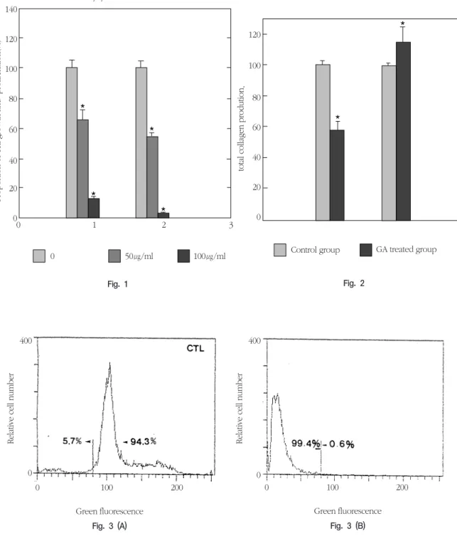
The inhibitory effects of GA on human gingival fibroblasts were significantly higher than those of the control, and cell growth and proliferation was significantly inhibited at 50 ㎍/ml and 100 ㎍/ml of GA(Fig. 1).
The inhibition of cell growth and proliferation might result in the morphological change in gingival fibroblasts. This result supports the hypothesis that GA changes the morphotype of human gingival fibroblasts and inhibits the cell growth and prolifera- tion in gingival fibroblasts by blocking selectin(a family of adhesion receptors) modulating signal transduction, and inhibiting arachidonic acid metab- olism44-46).
GA inhibits the cell activity of human gingival fibroblasts in a concentration-dependent manner.
The total collagen in the extracellular medium was increased and that in the cell was decreased in the
presence of GA(Fig. 2). Hence, GA enhanced the release of collagen from cells into the extracellular medium. The released extracellular collagen might bind the membrane receptors for collagen47). After binding, the extracellular collagen may be related to the change of cell morphology, and regulate cell activity such as cell growth and proliferation, and total collagen synthesis in human gingival fibrob- lasts.
To investigate the molecular mechanism for change of cell morphology and the inhibition of cell activity, we carried out acridine orange binding assay by flow cytometry. This type of analysis is based on both morphometric and biochemical crite- ria42) and permits cellular staining of DNA without affecting cell viability. Furthermore, this technique permits the analysis of treatment effects on a sub- population of cells as opposed to biochemical pro- cedures that quantitate an average effect for an entire cell population(see in materials and meth- ods).
In the study of acridine orange binding by flow cytometry in human gingival fibroblasts at 24 h after GA treatment, diploid cells with a mean fluores- cence over channel 80 decreased and cells with reduced acridine orange binding with a fluores- cence below channel 80 increased(Fig. 3A and Fig.
3B). The decrease in cell viability and the acridine orange binding suggest that DNA degradation might occur in human gingival fibroblasts treated with GA
42). These results indicate that GA might be related to cytotoxic effect of fibroblast via DNA degradation
48-49). Further study is needed to monitor the expres- sion of HLA class I antigens in human gingival fibroblasts associated with cytotoxic effector func- tion. Our previous study also showed that GA decreased the β- blocker induced calcium ion influx in human gingival cells50). These effects of GA might be useful to modulate the drug-induced gingival
change.
All these results suggest that GA may inhibit the cell activity of human gingival fibroblasts including collagen synthesis, and might be useful for the reso- lution of uncontrolled growth in dental field such as gingival hyperplasia at the concentration of about 50
㎍/ml.
IV. Conclusion
We examined the effect of glycyrrhetinic acid (GA), a cell differentiation factor for the resolution of uncontrolled growth, on the viability of cultured human gingival fibroblasts. Human gingival fibrob- lasts were harvested and isolated from patients undergoing orthodontic treatment. Cell growth and proliferation, collagen synthesis, and acridine orange binding of the nucleus of cultured fibroblasts in the presence of GA were examined by 3-(4,5- dimethylthiazol-2-yl)-2,5-diphenyltetrazolium bro- mide (MTT) assay, the amount of 4-hydroxyproline, and acridine orange binding by flow cytometry, respectively.
Morphologically, the fibroblasts treated with 100㎍
/ml of GA became round in shape. GA inhibited the growth and proliferation of human gingival fibrob- lasts at concentrations over 50 ㎍/ml. The total colla- gen inside the cell decreased in the presence of GA, and that in the extracellular medium increased in the presence of GA. When human gingival fibroblasts were cultured with 100 ㎍/ml of GA for 24 h, diploid cells with a mean fluorescence over channel 80 decreased and cells with reduced acridine orange binding (fluorescence below channel 80) increased.
These results suggest that GA can inhibit cell growth, proliferation and collagen synthesis, DNA degradation may be induced by GA in human gingi- val fibroblasts.
V. Acknowledgement
We gratefully acknowledge Dr H. R. Kim in Dental School, Wonkwang Univ. and Dr. K. Shibata in the Dept. of Endodontology and Periodontology, Dental School, and Dr. T. Clifford in the School of Medicinal Chemistry, Hiroshima Univ. for their valu- able advice and Dr H. T. Chung and Dr C. D. Chun in Medical School for their help with flow cytometry to analyse the acridine orange binding of the DNA in human gingival fibroblasts.
VI. Referenxes
1. Sklans S., Taylor R. G. and Shklar G.: Effect of diphenylhydantoin sodium on healing of experimentally produced features in rabbit mandible. J. Oral Surg. 25: 310-319, 1967.
2. Hassell T. M., Page R. C. and Lindhe J.:
Histologic evidence for impaired growth con- trol in diphenylhydantoin gingival growth in man. Archs. Oral Biol. 23: 381-384, 1978.
3. Hassell T. M.: Evidence for production of an inactive collagenase by fibroblasts from phenytoin-enlarged human gingivae. J. Oral Pathol. 11: 310-317, 1982.
4. David L., Harry L., Stephen R., Paul D. F. and Flushing N. Y.: Gingival hyperplasia associat- ed with nifedipine therapy. Oral Surg. Oral Med. Oral Pathol. 57: 620-622, 1984.
5. Lucas R. M., Howell L. P. and Wall B. A.:
Nifedipine-induced gingival hyperplasia- A histochemical and ultrastructural study. J.
Periodontol. 56: 211-215, 1985.
6. Sooriyamoorthy M., Grower D. B. and Eley B. M.: Androgen metabolism in gingival hyperplasia induced by nifedipine and cyclosporin. J. Periodont. Res. 25: 25-30,
1990.
7. Salo T., Oikarinen K. S. and Oikarinen A. I.:
Effect of phenytoin and nifedipine on colla- gen gene expression in normal human gingi- val fibroblasts. J. Oral Pathol. Med. 19: 404- 407, 1990.
8. Xu Y. Q., Arthur S. P. and Craig W. C.:
Evidence for developmental changes in sodi- um channel inactivation gating and sodium channel blockage by phenytoin in rat cardiac myocytes. Circulation Res. 69: 644-656, 1991.
9. Hassell T. M., Page R. C. Narayanan A. S. and Cooper C. G.: Diphenylhydantoin (Dilantin) gingival hyperplasia: drug-induced abnormali- ty of connective tissue. Proc. Natl. Acad. Sci.
U.S.A. 73: 2909-2912, 1976.
10. Do Nascimento A., De Castro Barreto R., Bozz L. and Paes de Almeida O. (1985) Interaction of phenytoin and inflammation induces gingi- val overgrowth in rats. J. Periodont. Res. 20:
386-391.
11. Morisaki I., Mihara J., Kato K., Kitamura K., Adachi C., Sobue S. and Hamada S.:
Phenytoin-induced gingival overgrowth with Streptococcus sorbrinus6715. Archs Oral Biol.
35: 753-758, 1990.
12. Modeer T., Dahllof G., Karsten J. and Otteskog P.: Potentiation of fibroblast DNA synthesis by a phenytoin-induced mononu- clear cell derived factor in vitro. Scan. J. Dent.
Res. 97: 186-187, 1989.
13. Willershausen Zonnchen B., Lemmen C. and Hamm G.: The effect of cyclosporin A on the growth and metabolic activity of gingival fibroblasts. Schweiz-Monatsschr-Zahnmed 101: 18-23, 1991.
14. Willershausen Zonnchen B., Lemmen C., Zonnchen B., Hamm G. and Schumacher U.:
Influence of nifedipine on the metabolism of
gingival fibroblasts. Biol. Chem. Hoppe Seyler 375: 299-303, 1994.
15. Williamson M.S., Miller E.K., Plemons J., Rees T. and Iacapino A.M.: Cyclosporine A upregu- lates interleukin-6 gene expression in human gingiva: possible mechanism for gingival over- growth. J. Periodontol. 65: 895-903, 1994.
16. Nares S., Ng M.C., Dill R.E., Park B., Cutler C.W. and Iacopino AM.: Cyclosporine A upregulates platelet derived growth factor B Chain in hyperplastic human gingiva. J.
Periodontol. 67(3):271-278, 1996.
17. Iacopino A.M., Doxey D., Cutler C.W., Nares S., Stoever K., Fojt J., Gonzales A. and Dill R.E.: Phenytoin and cyclosporine A specifical- ly regulate macrophage phenotype and expression of platelet-derived growth factor and interleukin-1 in vitro and in vivo: possible molecular mechanism of drug-induced gingi- val hyperplasia. J. Periodontol. 68:73-83, 1997.
18. Tamura Y.: Study of effects of glycyrrhetinic acid and its derivatives on 4-5αand 5β- reduc- tase by rat liver preparations. Folia endocrinol. Jpn 51: 589-600, 1975.
19. Odashima S., Nakayabu Y., Honjo N., Abe H.
and Arichi S.: Induction of phenotypic reverse transformation by ginsenosides in cultured Morris hepatoma cells. Eur. J. Cancer 15: 885- 892, 1979.
20. Abe N., Ebina T. and Ishida N.: Interferon induction by glycyrrizin and glycyrrhetinic acid in mice. Microbiol. Immunol. 26: 535- 539, 1982.
21. Sporn M. B. and Roberts A. B.: Role of retinoids in differentiation and carcinogenesis.
Cancer Res. 43: 3034-3040, 1983.
22. Odashima S., Ohta Y. and Kohno H.: Control of phenotypic expression of cultured B16
melanoma clones isolated from cultured cell lines, subcutaneous tumors and individual lung metastasis. Cancer Res. 42: 2770-2778, 1985.
23. Tokuda H., Ohigashi H., Koshimizu K. and Ito Y.: Inhibitory effects of ursolic aid and oleanolic acid on skin tumor promotion by 12-o-tetradecanoyl-phobol-13-acetate. Cancer Lett. 33: 279-285, 1986.
24. Grippo J. F. and Gudas L. J.: The effect of dibutyl cyclic AMP and butyrate on F9 terato- carcinoma cellular retinoic acid-binding pro- tein activity. J. Biol. Chem. 262: 4492-4509, 1987.
25. Stoner C. M. and Gudas L. J.: Mouse cellular retinoic acid binding protein: cloning, comple- mentary DNA sequences, and messenger RNA expression during the retinoic acid-induced differentiation of F8 wild type and RA-3-10 mutant tetracarcinoma. Cancer Res. 49: 1497- 1504, 1989.
26. Davidson J. S., Baumgarten I. M. and Harley E. H.: Reversible inhibition of intercellular junctional communication by glycyrrhetinic acid. Biochem. Biophys. Res. Commun. 134:
29-36, 1986.
27. Davidson J. S. and Baumgarten I. M.:
Glycyrrhetinic acid derivatives: a novel class of inhibitors of gap-junctional intercellular com- munication. Structure-activity relationships. J.
Pharmacol. Exp. Ther. 246: 1104-1107, 1988.
28. Pompei R., Flore O., Marccialis M. A., Pani A.
and Loddo B.: Glycyrrizinic acid inhibits virus growth and inactivates virus particles. Nature 281: 689-690, 1979.
29. Crance J.M., Leveque F., Biziagos E., van Cuyck-Grandre H., Jouan A. and Deloince R.:
Studies on mechanism of action of gly- cyrrhizin against hepatitis A virus replication in
vitro. Antiviral Res. 23: 63-76, 1994.
30. Shiki Y., Ishikawa Y., Shirai K. and Yoshida S.: Effect of glycyrrhizin on lysosomes labiliza- tion by phospholipase A2. American J.
Chinese Medicine 14: 131-137, 1986.
31. Akao T., Hattori M., Namba T. and Kobashi K.: Inhibitory effects of glycyrrhetinic acid and its related compounds on 3 alpha-hydroxys- teroid dehydrogenase of rat liver cytosol.
Chem. Pharm. Bull. Tokyo 40: 1208-1210, 1992.
32. Kumagai A., Nishino K., Shimomura A., Kin T. and Yamamura Y.: Effect of glycyrrhizin on estrogen action. Endocrinol. Jpn 14: 34-38, 1967.
33. Ishikawa A., Kanamaru R., Wakui A., Kanno S. and Ohtsuki K.: Characterization of gly- cyrrhizin-binding protein kinase from the crude membrane fraction of rat liver.
Biochem. Biophy. Res. Commun. 167: 876- 882, 1990.
34. Benediktsson R., Calder A.A., Edwards C.R.
and Seckl J.R.: Placental 11 beta-hydroxys- teroid dehydrogenase: a key regulator of fetal glucocorticoid exposure. Clin. Endocrinol. 46:
161-6, 1997.
35. Sakamoto K. and Wakabayashi K.: Inhibitory effect of glycyrrhetinic acid on testosterone production in rat gland. Endocrinol. Jpn 35:
333-342, 1988.
36. Abe H., Ohya N. and Yamamoto K. F., Shibuya T., Arichi S. and Odashima S.: Effects of glycyrrhizin and glycyrrhetinic acid on growth and melanogenesis in cultured B16 melanoma cells. Eur. J. Cancer Clin. Oncol.
23: 1549-1555, 1987.
37. Mckevitt K. M. and Irwin C. R.: Phenotypic differences in grow, matrix synthesis and response to nifedipine between fibroblasts
derived from clinically healthy and overgrown gingival tissue. J. Oral Pathol. Med. 24: 66-71, 1995.
38. Bowers G.N. Jr. and McComb R.B.: A contin- uous spectrophotometric method for measur- ing the activity of serum alkaline phosphatase.
Clin. Chem. 12: 70-88, 1966.
39. Patterson M.K. Jr.: Measurement of growth and viability of cells in culture. Methods Enzymol. 58: 141-152, 1979.
40. Mosmann T.: Rapid colorimetric assay for cel- lular and survival: Application to proliferation and cytotoxic assays. J. lmmunol. Methods 65:
55-63, 1983.
41. Jamall I. S., Finelli V. N. and Hee S. S.: A sim- ple method to determine nanogram levels of 4-hydroxyproline in biological tissues. Anal.
Biochem. 112: 70-75, 1981.
42. Compton M. M., Haskill J. S. and Cidlowski J.
A.: Analysis of glucocorticoid actions on rat thymocyte deoxyribonucleic acid by fluores- cence-activated flow cytometry. Endocrinology 122: 2158-2164, 1988.
43. Myc A., Traganos F., Lara J., Melamed M.R.
and Darzynkiewicz Z.: DNA stainability in aneuploid breast tumors: comparison of four DNA fluorochromes differing in binding prop- erties. Cytometry 13: 389-394, 1992.
44. Kelloff G. J., Boone C. W., Steele V. E., Fay J.
R., Lubet R. A., Crowell J. and Sigman C. C.:
Mechanistic considerations in chemopreven- tive drug development. J. Cell Biochem. 20:
1-24, 1994.
45. Rao B.N., Anderson M. B., Musser J. H. and Gilbert J. H., Schaefer M. E., Foxall C. and Brandley B.K.: Sialyl Lewis X mimics derived from a pharmacophore search are selectin inhibitors with anti-inflammatory activity. J.
Biol Chem. 269: 19663-19666, 1994.
46. Wayner E. A. and Carter W. G., Piotrowicz R.
S. and Kunicki T. J.: The function of multiple extracellular matrix receptors in mediating cell adhesion to extracellular matrix: preparation of monoclonal antibodies to the fibronectin receptor that specifically inhibit cell adhesion to fibronectin receptor that specifically inhibit cell adhesion to fibronectin and react with platelet glycoproteins Ⅰc-Ⅱa. J. Cell Biol.
107: 1881-1891, 1988.
47. Pitaru S., Madgar D., Metzger Z. and Hekmati H.: Mechanism of endotoxin inhibition of human gingival fibroblast attachment to type I collagen. J. Dent. Res. 69: 1602-1606, 1990.
48. Yoo S.K., Kim K.J., Shin H.S., Park J.K. and Jeon B.H.: Development of in vitro gingival hyperplasia model and drug to treat gingival hyperplasia. J. Oral Biol. 18: 121-126, 1994.
49. Su Z. Z., Lin J., Prewett M., Goldstein N. I.
and Fisher P. B.: Apoptosis mediates the selective toxicity of caffeic acid phenethyl ester (CAFE) toward oncogene-transformed rat embryo fibroblast cells. Anticancer Res. 15:
1841-1848, 1995.
50. Yoo S. K., Han M. K., Kim K. J. and Byun H.
Y.: Effects of diltiazem and glycyrrhetinic acid on intracellular calcium in human gingival cells. J. Wonkwang Biomaterial·lmplant 4:
91-96, 1995.
사진부도
*
*
*
*
*
*
140 120 100 80 60
40 20 0
120 100 80 60 40 20
0 1 2 3 0
0 50㎍/ml 100㎍/ml Control group GA treated group
Proportion of cell growth and proliferation(%) total collagen prodution.
Glycyrrhetinic acid
Fig. 1 Fig. 2
Relative cell number
400
0
0 100 200 0 100 200
Relative cell number
400
0
Green fluorescence
Fig. 3 (A) Fig. 3 (B)
Green fluorescence
사진부도 설명
Fig. 1. Proportional change of cell growth and proliferation by GA in human gingival fibroblasts (%). GA- induced decrease of cell growth and proliferation was shown in human gingival cells. Cells(1×10⁴ cells) were treated with 0, 50 and 100 ㎍/ml concentrations of GA for 24 or 48 h in α-MEM and then analysed by ELISA reader using 100 ㎍ of MTT. The proportion of cell growth and proliferation was cal- culated as a percentage of the control activity from the absorbance values at 570 nm [Cell activity (%)=(Optical density of experimental group/Optical density of control group)×100]. Each column and vertical bar represent the mean + 1 SD of 12 samples. Significant differences from control, *P<0.01.
Fig. 2. Proportional change of total collagen production by GA in human gingival fibroblasts (%). GA-induced release of total collagen from human gingival fibroblasts into medium was shown. Cells(1×10⁴cells) were treated with 100 ㎍/ml concentrations of GA for 24 h in α-MEM and then analysed by ELISA read- er using 0.4% of SRB. The proportion of total collagen synthesis was calculated as a percentage of the control collagen synthesis from the absorbance values at 558 nm [Relative proportion of total collagen synthesis (%)=(Optical density of experimental group/Optical density of control group)×100]. Each col- umn and vertical bar represent the mean + 1 SD of 3 samples. Significant difference between medium and cell, *P<0.01.
Fig. 3. DNA histogram showing GA-induced loss of acridine orange binding in human gingival fibroblasts.
Cells(2×10⁶/ml) were treated with 100 ㎍/ml of GA for 24 h in α-MEM and then analysed by flow cytometry using acridine orange staining method. Relative cell number was indicated on the vertical axis, and green fluorescence was indicated on the horizontal axis. Diploid cells had a mean fluores- cence at channel 100 and cells with reduced acridine orange binding had a fluorescence below channel 80.