Print ISSN 1738-6586 / On-line ISSN 2005-5013 10.3988/jcn.2010.6.3.152 CASE REPORT
J Clin Neurol 2010;6:152-155
Takotsubo Cardiomyopathy Following Cerebral Infarction Involving the Insular Cortex
Hyun-Ji Cho, MDa; Hahn Young Kim, MDb; Seol Heui Han, MD, PhDb; Hyun Joong Kim, MD, PhDc; Yeon Sil Moon, MDb; Jeeyoung Oh, MD, PhDb
aDepartment of Neurology, Ewha Womans University School of Medicine, Seoul, Korea
bDepartments of Neurology and cCardiology, Konkuk University School of Medicine, Seoul, Korea
Received May 20, 2009 Revised June 16, 2009 Accepted June 16, 2009 Correspondence Jeeyoung Oh, MD, PhD Department of Neurology, Konkuk University School of Medicine,
1 Hwayang-dong, Gwangjin-gu, Seoul 143-701, Korea Tel +82-2-2030-7564 Fax +82-2-2030-7469 E-mail [email protected]
BackgroundzzTakotsubo cardiomyopathy is characterized by clinical features similar to those of acute myocardial ischemia, but without angiographic evidence of obstructive coronary artery disease. We present a patient with takotsubo cardiomyopathy following acute infarction involv- ing the left insular cortex.
Case ReportzzA 52-year-old man was admitted with acute infarction of the left middle cerebral artery territory and acute chest pain. Acute myocardial infarction was suspected because of ele- vated serum troponin levels and hypokinesia of the left ventricle on echocardiography. However, a subsequent coronary angiography revealed no stenosis within the coronary arteries or ballooning of the apical left ventricle.
ConclusionszzWe postulated that catecholamine imbalance due to the insular lesion could be responsible for these interesting features. J Clin Neurol 2010;6:152-155 Key Wordszz takotsubo cardiomyopathy, insula, infarction.
Introduction
Transient left ventricular apical ballooning syndrome, also known as takotsubo cardiomyopathy, is an emerging disease entity that mimics acute myocardial infarction. This syn- drome is characterized by the clinical features of acute chest pain and dyskinesia of the left ventricular apical or midven- tricular segment on echocardiography, but there is no angio- graphic evidence of obstructive coronary artery disease.1 We present a patient with takotsubo cardiomyopathy following acute infarction of the left insular cortex.
Case Report
A 52-year-old man was transferred to the emergency room presenting with decreased mentality, chest pain, speech distur- bance, and right hemiparesis. His medical history was unre- markable except for a 30-pack-year history of current smok- ing. His initial blood pressure and heart rate were 132/100 mmHg and 78 bpm, respectively. Phonemic paraphasia, left gaze preponderance, and right hemiparesis were noted on
neurological examination. Diffusion weighted magnetic res- onance (MR) imaging revealed a hyperintense lesion over the left frontoparietal lobe including the insular cortex, and MR angiography revealed no significant stenosis in the rele- vant arteries (Fig. 1). On admission, the patient complained of anterior chest pain and an irregular heart rate (110-140 bpm).
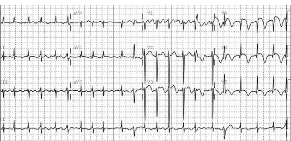
Levels of the myocardial enzyme levels creatine phosphoki- nase and troponin I were increased to 424 U/l (normal range 58-348 U/l) and 12.09 ng/mL (normal range 0-2 ng/mL), re- spectively. Electrocardiography (ECG) revealed atrial fibril- lation and ST-segment elevation at leads I, aVL, and V2-5, sug- gesting anterolateral wall ischemia (Fig. 2). Emergent transth- oracic echocardiography demonstrated hypokinesia of the mid- dle and apical walls of the left ventricle (LV) and an ejection frac- tion of 36% (Fig. 3A). We concluded that the patient had ex- perienced an acute myocardial infarction with new onset atrial fibrillation and cardioembolic stroke. Digitalization and hep- arinization were commenced. However, subsequent coronary angiography did not disclose any stenosis within the coronary arteries (Fig. 3B). Left ventriculography revealed severe hypo- kinesia of the anterolateral and apical walls of the LV with vig- 152 Copyright © 2010 Korean Neurological Association
Cho HJ et al.
www.thejcn.com 153 orous contraction of the basal segment, mimicking a takotsubo,
a round-bottomed, narrow-necked Japanese fishing pot used for trapping octopus (Fig. 3C).
The patient’s condition had improved with the cardiac en- zyme levels having declined to within the normal range by 4 days after the initial event. A follow-up echocardiography
A B
Fig. 1. Brain magnetic resonance image and angiography of the patient. A: Acute infarction of the middle cerebral artery territory involving the left insular cortex. B and C: No significant stenosis was noted in relevant arteries on magnetic resonance angiography.
Fig. 2. Electrocardiography showing atrial fibrillation with ST-segment elevation at leads I, aVL, and V2-5, suggesting acute anterolateral wall ischemia.
Fig. 3. Echocardiography showing hypokinesia of the middle and apical walls of the left ventricle (A); no stenotic lesion was found on coro- nary angiography (B). Note the characteristic appearance of “apical ballooning” on the left ventriculogram (C), which is similar to that of a takotsubo, a Japanese octopus-trapping pot.
A B C
C
Takotsubo Cardiomyopathy Following Cerebral Infarction
154 J Clin Neurol 2010;6:152-155
performed 6 days after the attack demonstrated improved wall motion of the apical LV. ECG performed on the same day showed T wave inversion and QTc prolongation at the anterolateral leads. The patient was discharged 2 weeks after the initial event after achieving an International Normalized Ratio within the therapeutic range.
Discussion
Takotsubo cardiomyopathy is a rare syndrome that mimics acute myocardial infarction with chest pain, dyspnea, elec- trocardiographic ST-segment changes, elevated troponin, and left ventricular dysfunction. Cardiac catheterization should reveal a typical left ventricular wall motion pattern without any sign of coronary heart disease. A hypokinetic apical seg- ment and hyperkinetic basal segment, which looks like a takot- subo, is observed on end-systolic ventriculography.1
Takotsubo cardiomyopathy is observed in 0.7-2.5% of pa- tients with suspected acute coronary syndrome, affecting post- menopausal women in most cases.1,2 Although the exact mech- anism underlying this pathology is not clear, reduced estrogen levels seem to cause endothelial dysfunction or changes in catecholamine metabolism.2
The prognosis seems to be favorable and recurrence oc- curs in <10% of patients.2 The common complication is left- heart failure, left ventricular intracavitary obstruction, and ar- rhythmia.1 In extreme and neglected cases it can lead to sudden and unexpected death.
The etiology of this syndrome is still unclear, but catechol- amine-mediated neurogenic myocardial stunning has been proposed,3 given that physical or emotional stress usually provokes this syndrome.4,5 Some medicolegal problems as- sociated with any procedure-related cardiac complication is suspected with this emotional or physical stress-induced car- diomyopathy.
Serum catecholamine levels are elevated, even beyond those of patients with acute myocardial infarction6 and ab- normal sympathetic nerve function, as shown by single-pho- ton-emission computed tomography using 123I-metaiodoben- zylguanidine, a radioiodinated analog of norepinephrine.7 No consensus or guideline for treatment exists; however, beta- blocker administration can be helpful, given that emotional- stress-induced apical LV ballooning was normalized by pre- treatment with adrenoreceptor blockade.8
Our patient had chest pain on admission, which indicates takotsubo cardiomyopathy preceding ischemic infarction as a possible scenario. However, this patient had no other under- lying cardiac or systemic disease or stressful emotional event that could have caused left ventricular dysfunction. In addi- tion, atrial fibrillation had resolved by the follow-up ECG
and cardiac wall motion had improved, suggesting a condi- tion of a transient rather than a permanent nature. Therefore, we postulate that autonomic imbalance secondary to infarc- tion involving the insular cortex might have caused the tran- sient left ventricular dysfunction observed in this patient.
Whilst cardiac abnormalities including takotsubo cardio- myopathy have been reported in cases of all types of massive ischemic stroke, the insular cortex has attracted the attention of physicians. The reason for this is that the viscerotopic sen- sory area of the insular cortex is a convergent center that re- ceives input from the limbic and autonomic systems in the brain.9 Lesion or stimulation of the insular cortex causes var- ious cardiovascular responses, including changes in blood pressure, QT dispersion, T wave inversion, and elevated tro- ponin levels.10-13
Sympathetically mediated takotsubo-like syndrome has been reported in a few cases of subarachnoid hemorrhage.14,15 However, takotsubo cardiomyopathy following cerebral in- farction has been reported only rarely. The mechanism un- derlying the pathology of each case might be different.15 In ad- dition, neurologists tend to assign a chest pain in recent stroke patients to concurrent acute myocardial ischemia.
Cardiac autonomic dysfunction in various neurological and medical conditions has not attracted the neurologists’ at- tention. We believe that research into and treatment of these dysfunctions by neurologists is necessary, because the hu- man cardiac organ is simple and regulated only by the ner- vous system. More precise studies of the neural regulation of cardiac function would elucidate the underlying pathophysi- ologic mechanism of this enigmatic syndrome, enabling the development of a therapeutic regimen.
Conflicts of Interest
The authors have no financial conflicts of interest.
Acknowledgements
The work described in this paper was supported by Konkuk University in 2006.
REFERENCES
1. Pilgrim TM, Wyss TR. Takotsubo cardiomyopathy or transient left ventricular apical ballooning syndrome: A systematic review. Int J Cardiol 2008;124:283-292.
2. Prasad A, Lerman A, Rihal CS. Apical ballooning syndrome (Tako- Tsubo or stress cardiomyopathy): a mimic of acute myocardial in- farction. Am Heart J 2008;155:408-417.
3. Kume T, Kawamoto T, Okura H, Toyota E, Neishi Y, Watanabe N, et al. Local release of catecholamines from the hearts of patients with tako-tsubo-like left ventricular dysfunction. Circ J 2008;72:106-108.
4. Bybee KA, Prasad A, Barsness GW, Lerman A, Jaffe AS, Murphy JG, et al. Clinical characteristics and thrombolysis in myocardial in- farction frame counts in women with transient left ventricular apical ballooning syndrome. Am J Cardiol 2004;94:343-346.
5. Parodi G, Del Pace S, Carrabba N, Salvadori C, Memisha G, Sim-
Cho HJ et al.
www.thejcn.com 155
onetti I, et al. Incidence, clinical findings, and outcome of women with left ventricular apical ballooning syndrome. Am J Cardiol 2007;99:182-185.
6. Wittstein IS, Thiemann DR, Lima JA, Baughman KL, Schulman SP, Gerstenblith G, et al. Neurohumoral features of myocardial stunning due to sudden emotional stress. N Engl J Med 2005;352:539-548.
7. Akashi YJ, Nakazawa K, Sakakibara M, Miyake F, Musha H, Sasaka K. 123I-MIBG myocardial scintigraphy in patients with “takotsubo”
cardiomyopathy. J Nucl Med 2004;45:1121-1127.
8. Ueyama T, Kasamatsu K, Hano T, Yamamoto K, Tsuruo Y, Nishio I.
Emotional stress induces transient left ventricular hypocontraction in the rat via activation of cardiac adrenoceptors: a possible animal model of ‘tako-tsubo’ cardiomyopathy. Circ J 2002;66:712-713.
9. Eickhoff SB, Lotze M, Wietek B, Amunts K, Enck P, Zilles K. Seg- regation of visceral and somatosensory afferents: an fMRI and cyto- architectonic mapping study. Neuroimage 2006;31:1004-1014.
10. Colivicchi F, Bassi A, Santini M, Caltagirone C. Cardiac autonomic derangement and arrhythmias in right-sided stroke with insular in- volvement. Stroke 2004;35:2094-2098.
11. Ay H, Koroshetz WJ, Benner T, Vangel MG, Melinosky C, Arsava EM, et al. Neuroanatomic correlates of stroke-related myocardial in- jury. Neurology 2006;66:1325-1329.
12. Bagaev V, Aleksandrov V. Visceral-related area in the rat insular cor- tex. Auton Neurosci 2006;125:16-21.
13. Cheshire WP Jr, Saper CB. The insular cortex and cardiac response to stroke. Neurology 2006;66:1296-1297.
14. Banki NM, Kopelnik A, Dae MW, Miss J, Tung P, Lawton MT, et al.
Acute neurocardiogenic injury after subarachnoid hemorrhage. Cir- culation 2005;112:3314-3319.
15. Mayer SA, Lin J, Homma S, Solomon RA, Lennihan L, Sherman D, et al. Myocardial injury and left ventricular performance after sub- arachnoid hemorrhage. Stroke 1999;30:780-786.