BackgroundzzWe describe herein the application of whole exome sequencing (WES) for the molecular genetic diagnosis of a large Korean family with dominantly inherited myopathy.
Case ReportzzThe affected individuals presented with slowly progressive proximal weak- ness and ankle contracture. They were initially diagnosed with limb-girdle muscular dystro- phy (LGMD) based on clinical and pathologic features. However, WES and subsequent capil- lary sequencing identified a pathogenic splicing-site mutation (c.1056+1G>A) in COL6A1, which was previously reported to be an underlying cause of Bethlem myopathy. After identi- fication of the genetic cause of the disease, careful neurologic examination revealed subtle contracture of the interphalangeal joint in the affected members, which is a characteristic sign of Bethlem myopathy. Therefore, we revised the original diagnosis from LGMD to Beth- lem myopathy.
ConclusionszzThis is the first report of identification of COL6A1-mediated Bethlem myopathy in Korea, and indicates the utility of WES for the diagnosis of muscular dystrophy.
Key Wordszz Bethlem myopathy, collagen type VI alpha 1 (COL6A1), whole exome sequencing.
Molecular Genetic Diagnosis of a Bethlem Myopathy Family with an Autosomal-Dominant COL6A1 Mutation,
as Evidenced by Exome Sequencing
INTRODUCTION
Muscular dystrophy is a clinically and genetically heterogeneous inherited disorder char- acterized by progressive muscle weakness and wasting. A step-by-step approach with as- sessment of medical history, clinical examination, laboratory evaluation, muscle patholo- gy, muscle immunoanalysis, and mutational analysis is typically used for the diagnosis of muscular dystrophy.1,2 However, this serial approach often fails to identify causative mu- tations due to high phenotypic and pathologic variability, small pedigrees, and the limited power of traditional linkage analyses.3 Recent advances in next-generation sequencing has made it possible to selectively sequence only the protein-coding exons of the genome, a process termed ‘whole exome sequencing’ (WES). The application of WES not only saves time but is also cost-effective for the identification of causative genes in Mendelian diseases.4 Therefore, WES is being increasingly adopted for the identification of causative genes in muscular dystrophy research.5-8
Herein we report a mutation in the gene encoding collagen type VI α1 (COL6A1) in a large Korean family with autosomal-dominant Bethlem myopathy that was detected us- ing WES.
Hyung Jun Parka* Young-Chul Choib* Seung Min Kimb Se Hoon Kimc Young Bin Hongd Bo Ram Yoone Ki Wha Chunge Byung-Ok Choid
a Department of Neurology, Mokdong Hospital, Ewha Womans University School of Medicine, Seoul, Korea
bDepartments of Neurology and
c Pathology, Yonsei University College of Medicine, Seoul, Korea
d Department of Neurology, Samsung Medical Center, Sungkyunkwan University School of Medicine, Seoul, Korea
e Department of Biological Science, Kongju National University, Gongju, Korea
pISSN 1738-6586 / eISSN 2005-5013 / J Clin Neurol 2015;11(2):183-187 / http://dx.doi.org/10.3988/jcn.2015.11.2.183
Received May 16, 2013 Revised September 3, 2013 Accepted September 6, 2013 Correspondence Byung-Ok Choi, MD Department of Neurology, Samsung Medical Center, Sungkyunkwan University School of Medicine, 81 Irwon-ro, Gangnam-gu, Seoul 135-710, Korea Tel +82-2-3410-1296 Fax +82-2-3410-0052 E-mail bochoi@skku.edu Ki Wha Chung, PhD
Department of Biological Science, Kongju National University, 56 Gongjudaehak-ro, Gongju 314-701, Korea Tel +82-41-850-8506 Fax +82-41-850-0957 E-mail kwchung@kongju.ac.kr
*Hyung Jun Park and Young-Chul Choi contributed equally to this work.
cc This is an Open Access article distributed under the terms of the Creative Commons Attribution Non-Com- mercial License (http://creativecommons.org/licenses/by-nc/3.0) which permits unrestricted non-commercial use, distribution, and reproduction in any medium, provided the original work is properly cited.
JCN
Open Access CASE REPORTBethlem Myopathy with COL6A1 Mutation
JCN
CASE REPORT
Eighteen members of a large Korean family with domi- nantly inherited myopathy (7 affected and 11 unaffected) were enrolled (Fig. 1A). Written informed consent to par- ticipate was obtained from all participants and from the parents of participants younger than 18 years, according to a protocol approved by the Institutional Review Board for Ewha Womans University Mokdong Hospital, Seoul, Korea.
Patients
The proband, a 38-year-old woman (III-4), presented with progressive proximal weakness and ankle contractures (Sup- plementary Table 1 in the online-only Data Supplementary).
Her initial development after birth was reportedly normal, but she did not begin walking until she was 16 months old.
She recalled that she had always been weaker than her peers.
Her motor function remained relatively stable until her mid- 20s. However, she experienced slowly progressive muscle weakness after the delivery of her first child at an age of 27 years. Her neck flexors [Medical Research Council (MRC)
grade 3] appeared to be more damaged than her neck ex- tensor (MRC grade 4+). The proximal muscles of her upper and lower limbs were more severely involved than the distal muscles, and her ankle joints were affected by contracture.
In addition, she appeared to exhibit mild facial weakness and absent tendon reflexes in the upper and lower limbs.
However, sensory examination revealed no abnormalities.
Laboratory studies revealed a serum creatine kinase level of 66 IU/L (normal, <135 IU/L), and her vital capacity was 3,120 mL. Electrocardiography and echocardiography find- ings were normal. Nerve conduction studies and needle electromyography revealed active generalized myopathy. A muscle biopsy sample obtained from the left biceps brachii revealed nonspecific muscular dystrophic changes. Hema- toxylin and eosin staining revealed variation in muscle fiber size (Fig. 2A), and modified Gomori trichrome staining re- vealed a few ragged red fibers (Fig. 2B). Architectural changes of disorganized intermyofibrillar networks, such as lobulated fibers, were accentuated in staining with nicotinamide ade- nine dinucleotide tetrazolium reductase (Fig. 2C). In addi- tion, adenosine triphosphatase (pH 9.4) staining demonstrat-
1 I
II
III
IV
1
1
1
3
3
3
2
2
2
2
4
4
4
5
5
5
6
6
6
7
7
7
8
8
9
9
10
10
11
11 12 13 14
G/G G/G
G/G G/G
Wild allele Mutated allele (c.1056+1G>A)
Exon 14 Intron Exon 14 Intron
G/G G/G G/G
G/G
G/G G/G G/A G/A G/A G/G
G/A G/A
G/A G/A
*
* * *
A
B
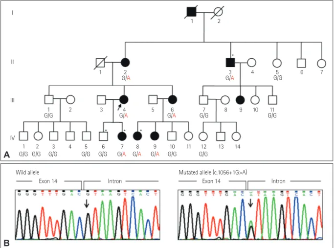
Fig. 1. A: Pedigree of an autosomal-dominant Bethlem myopathy family. Asterisks (*) indicate individuals whose DNA was used for exome se- quencing. Genotypes of COL6A1 (c.1056+1G>A) are indicated under each subject (arrow, proband; square, male; circle, female; filled, affected; not filled, unaffected; diagonal bar across symbol, deceased). B: Sequencing chromatograms of the c.1056+1G>A splicing-site mutation in COL6A1. Ar- row indicates the polymorphic site. The COL6A1 mutations detected by exome sequencing were confirmed by capillary sequencing. The heterozy- gous c.1056+1G>A mutation was completely cosegregated with the affected individuals within this family, and was not found in a sample of 200 healthy controls.
Park HJ et al.
JCN
ed a 69% predominance of type I fibers (Fig. 2D). Electron microscopy revealed many mitochondria, and immunohis- tochemical analyses of the muscle specimens revealed nor- mal staining patterns for the C-terminal of dystrophin, rod domain of dystrophin, N-terminal of dystrophin, dysferlin, α-sarcoglycan, β-sarcoglycan, γ-sarcoglycan, δ-sarcoglycan, α-dystroglycan, and caveolin.
The other affected members of the family had similar clin- ical presentations (Supplementary Table 1 in the online-on- ly Data Supplementary). They experienced very slow pro- gressive muscle weakness and lived without significant disability until old age. Subject II-2 required aid for ambu- lation after the age of 50 years, while subject II-3 (61 years old) was able to walk independently. The ankle joints were affected by contracture in all seven affected patients, while the elbow joints were involved only in subject II-2. Based on the clinical and pathologic features, the family was initially diagnosed with autosomal-dominant limb-girdle muscular dystrophy.
After identification of the causative gene, all affected mem- bers underwent a second neurologic examination. This iden- tified contracture of the interphalangeal joint–which is a
characteristic sign of Bethlem myopathy–in five family mem- bers (II-2, II-3, III-4, III-6, and IV-7). These contractures were very subtle and were not found on routine neurologic examination; they were only apparent when the wrist and fin- gers were extended passively.
Genetic analysis
Whole exome sequencing was performed for five members of the family, including four affected members (II-3, III-4, IV-7, and IV-8) and one unaffected member (IV-6) to iden- tify the genetic causes of the disease, following the method described by Choi et al.9 The exome sequencing data are summarized in Supplementary Table 2 (in the online-only Data Supplementary). The mean total sequencing yield was 9.3 Gbp/sample, and the coverage rate of the targeted exon regions (≥10×) was 93.56%. The average read depth of the target regions was 69.3 reads, and the average number of observed variants per sample was 92,174 SNPs and 9,321 in- dels. By comparing the exome data between 4 affected and 1 unaffected family members, we found that 15 functionally significant cosegregated variants (Supplementary Table 3 in the online-only Data Supplementary). Subsequent capillary Fig. 2. Histopathologic observations of biceps brachii muscle samples taken from the proband (III-4). A: Hematoxylin and eosin staining revealed variations in muscle fiber size and some fibers with internal nuclei (×200). B: Modified Gomori trichrome staining revealed a few ragged red fibers (×200). C: Staining with nicotinamide adenine dinucleotide tetrazolium reductase revealed architectural changes of disorganized intermyofibrillar networks, such as lobulated fibers (×400). D: Adenosine triphosphatase (pH 9.4) staining demonstrated a 69% predominance of type I fibers (×100).
A
C
B
D
Bethlem Myopathy with COL6A1 Mutation
JCN
sequencing analysis of control samples and other family members who were not included in the exome sequencing excluded most variants as the underlying cause of myopathy.
However, a c.1056+1G>A splicing-site mutation in COL6A1 completely cosegregated with affected status within the fam- ily (Fig. 1B), and was not found in 200 healthy controls.
This mutation has been reported to be the underlying cause of Bethlem myopathy.10-13 Thus, we determined that the c.1056+1G>A mutation in COL6A1 was the underlying cause of the disease in this family.
DISCUSSION
Whole exome sequencing of five members from a single fam- ily identified a splice donor site mutation at c.1056+1G>A of COL6A1. This mutation causes the formation of abnormal collagen VI protein by skipping of exon 14 and consequent in-frame deletion of amino acids from the triple helical do- main of the α1 chain.12
Bethlem myopathy is a dominantly inherited myopathy caused by mutations in one of three genes encoding colla- gen type VI alpha (COL6A1, COL6A2, and COL6A3).14 The phenotype is characterized by slowly progressive proximal weakness and multiple contractures. Prominent contracture in the early stages of the disease is one of the most important clinical features in Bethlem myopathy, Emery-Dreifuss mus- cular dystrophy, and Ullrich congenital muscular dystrophy.
Among these conditions, Bethlem myopathy demonstrates the most benign clinical course and mildest contractures.
Bethlem myopathy is often difficult to diagnose and its frequency may be underestimated for several reasons. First, mild contractures often lead to confusion in the diagnosis.15,16 In the present family, even though ankle contracture was initially detected, this is a common nonspecific finding in many other neuromuscular diseases. Contracture of the in- terphalangeal joint is a hallmark of Bethlem myopathy, but is often so subtle that it goes unrecognized. Second, muscle biopsy is not typically used for confirmatory diagnosis of Bethlem myopathy due to nonspecific myopathic changes and lack of detected abnormalities of collagen VI, even in immunohistochemical analyses. Both Ullrich congenital muscular dystrophy and Bethlem myopathy are collagen-IV- related myopathies. Immunohistochemistry in Ullrich con- genital muscular dystrophy exhibits complete absence or unequivocal reduction of collagen VI compared to normal; it can thus be used for diagnostic purposes. However, in Beth- lem myopathy, immunostaining of muscle biopsy with vari- ous collagen VI antibodies is usually normal.14 Third, identi- fication of the causative genes by general sequencing is costly and time-consuming because it is necessary to screen all 107
exons in all 3 genes for molecular genetic diagnosis. For these reasons, careful clinical assessment and cost-effective, time-saving strategies for genetic analysis are important for the diagnosis of Bethlem myopathy.
Whole exome sequencing is a well-justified strategy for discovering the causative genes of muscular dystrophy. WES is based on next-generation sequencing, which reduces the cost and time relative to Sanger sequencing.17 In addition, WES focuses only on protein-coding regions, but it is still an effective diagnostic tool because more than 90% of the pathogenic mutations for Mendelian disorders are found in exons.4
In conclusion, we identified a COL6A1 mutation in a Ko- rean family with Bethlem myopathy; this is the first such re- port in Korea. Even though the causative mutation identi- fied in the present study has been reported previously, this work underscores the usefulness of WES for the diagnosis of muscular dystrophy.
Supplementary Materials
The online-only Data Supplement is available with this arti- cle at http://dx.doi.org/10.3988/jcn.2015.11.2.183.
Conflicts of Interest
The authors have no financial conflicts of interest.
Acknowledgements
This study was supported by the Korean Health Technology R&D Project (A120182), Ministry of Health & Welfare, Republic of Korea.
REFERENCES
1. Norwood F, de Visser M, Eymard B, Lochmüller H, Bushby K; EFNS Guideline Task Force. EFNS guideline on diagnosis and management of limb girdle muscular dystrophies. Eur J Neurol 2007;14:1305-1312.
2. Bushby K. Diagnosis and management of the limb girdle muscular dystrophies. Pract Neurol 2009;9:314-323.
3. Ku CS, Naidoo N, Pawitan Y. Revisiting Mendelian disorders through exome sequencing. Hum Genet 2011;129:351-370.
4. Bamshad MJ, Ng SB, Bigham AW, Tabor HK, Emond MJ, Nickerson DA, et al. Exome sequencing as a tool for Mendelian disease gene discovery. Nat Rev Genet 2011;12:745-755.
5. Davidson AE, Siddiqui FM, Lopez MA, Lunt P, Carlson HA, Moore BE, et al. Novel deletion of lysine 7 expands the clinical, histopatho- logical and genetic spectrum of TPM2-related myopathies. Brain 2013;136(Pt 2):508-521.
6. Klar J, Sobol M, Melberg A, Mäbert K, Ameur A, Johansson AC, et al. Welander distal myopathy caused by an ancient founder mutation in TIA1 associated with perturbed splicing. Hum Mutat 2013;34:572- 577.
7. Ronchi D, Di Fonzo A, Lin W, Bordoni A, Liu C, Fassone E, et al. Mu- tations in DNA2 link progressive myopathy to mitochondrial DNA in- stability. Am J Hum Genet 2013;92:293-300.
8. Weterman MA, Barth PG, van Spaendonck-Zwarts KY, Aronica E, Poll-The BT, Brouwer OF, et al. Recessive MYL2 mutations cause infan- tile type I muscle fibre disease and cardiomyopathy. Brain 2013;
136(Pt 1):282-293.
9. Choi BO, Koo SK, Park MH, Rhee H, Yang SJ, Choi KG, et al. Exome
Park HJ et al.
JCN
sequencing is an efficient tool for genetic screening of Charcot-Marie- Tooth disease. Hum Mutat 2012;33:1610-1615.
10. Pan TC, Zhang RZ, Sudano DG, Marie SK, Bönnemann CG, Chu ML.
New molecular mechanism for Ullrich congenital muscular dystrophy:
a heterozygous in-frame deletion in the COL6A1 gene causes a severe phenotype. Am J Hum Genet 2003;73:355-369.
11. Lucioli S, Giusti B, Mercuri E, Vanegas OC, Lucarini L, Pietroni V, et al.
Detection of common and private mutations in the COL6A1 gene of patients with Bethlem myopathy. Neurology 2005;64:1931-1937.
12. Baker NL, Mörgelin M, Pace RA, Peat RA, Adams NE, Gardner RJ, et al. Molecular consequences of dominant Bethlem myopathy collagen VI mutations. Ann Neurol 2007;62:390-405.
13. Lamandé SR, Shields KA, Kornberg AJ, Shield LK, Bateman JF. Beth-
lem myopathy and engineered collagen VI triple helical deletions pre- vent intracellular multimer assembly and protein secretion. J Biol Chem 1999;274:21817-21822.
14. Lampe AK, Bushby KM. Collagen VI related muscle disorders. J Med Genet 2005;42:673-685.
15. Scacheri PC, Gillanders EM, Subramony SH, Vedanarayanan V, Crowe CA, Thakore N, et al. Novel mutations in collagen VI genes: expansion of the Bethlem myopathy phenotype. Neurology 2002;58:593-602.
16. Merlini L, Morandi L, Granata C, Ballestrazzi A. Bethlem myopathy:
early-onset benign autosomal dominant myopathy with contractures.
Description of two new families. Neuromuscul Disord 1994;4:503-511.
17. Metzker ML. Sequencing technologies - the next generation. Nat Rev Genet 2010;11:31-46.