서 론
건에 발생하는 황색종은 인지질의 국소적인 퇴적 작용으로 지질 대사 장애 시 발생한다. 슬개건 또는 드물게 삼두건이나 발가락 의 신전건을 침범하기도 하는데1,2) 그 중 아킬레스건은 흔한 침범 부위 중 하나이다. 아킬레스건의 황색종의 크기가 클 경우 통증 과 보행 시 불편감을 유발할 수 있는데 이러한 경우에는 수술적 치료를 요한다. 수술적 치료 중의 하나로 병변의 전 절제술 후 아
Copyright © 2021 by The Korean Orthopaedic Association
“This is an Open Access article distributed under the terms of the Creative Commons Attribution Non-Commercial License (http://creativecommons.org/licenses/by-nc/4.0/) which permits unrestricted non-commercial use, distribution, and reproduction in any medium, provided the original work is properly cited.”
The Journal of the Korean Orthopaedic Association Volume 56 Number 2 2021 Received May 28, 2020 Revised June 15, 2020 Accepted June 22, 2020
Correspondence to: Sung Taek Jung, M.D., Ph.D.
Department of Orthopaedic Surgery, Chonnam National University Medical School, 42 Jebong-ro, Dong-gu, Gwangju 61469, Korea
TEL: +82-62-227-1640 FAX: +82-62-225-7794 E-mail: [email protected] ORCID: https://orcid.org/0000-0003-1255-9568
양측 아킬레스건에 발생한 거대 황색종의 쐐기형 절제술을 이용한 수술적 치료
김성민 • 안영섭 • 정동민 • 정성택
전남대학교 의과대학 정형외과학교실
Wedge-Shaped Resection for Massive Xanthomatosis of Achilles Tendon
Sungmin Kim, M.D., Yeong Seub Ahn, M.D., Dong-Min Jung, M.D., and Sung Taek Jung, M.D., Ph.D.
Department of Orthopaedic Surgery, Chonnam National University Medical School, Gwangju, Korea
Purpose: Xanthomatosis of the Achilles tendons is rare. In some patients, however, the lesions in the Achilles tendon need to be removed,
which may be painful and disfiguring. While studies of successful surgical outcomes for the total resection and reconstruction of the Achilles tendon have been reported, reconstruction surgery has a technical challenge, and extended surgical exposures are required. This study analyzed five cases of bilateral xanthoma of the Achilles tendon, which was treated surgically using a wedge-shaped tendon-sparing approach to eliminate the need for tendon reconstruction.Materials and Methods: From July 2010 to May 2018, five patients with xanthomatosis in both Achilles tendons underwent wedge-
shaped tendon preserving surgery. The average age was 49 years (range, 40–55 years), and the follow-up period was 21.4 months (range, 12–31 months). The patients consisted of three males and two females. Complications related to surgery were recorded. The outcome measures included the range of motion of the ankle joint, American Orthopaedic Foot and ankle Society (AOFAS) ankle/hindfoot score, and visual analogue scale (VAS) for overall satisfaction at the last follow-up. The availability of a single-limb heel raise and returning time to work were also measured.Results:
Wound dehiscence that did not require secondary surgery was noted in one patient. At the last follow-up, the range of motion of the ankle joint was normal in all patients. The mean AOFAS ankle/hindfoot score was 91 (range, 85–96) and the VAS for the overall satisfaction ranged from 8 to 10. The average time between surgery and return to work was 27.6 days (range, 17–58 days) and all patients could perform a single-limb heel raise test.Conclusion: The tendon-sparing technique, which can preserve the anatomical functioning of the Achilles tendon, could be an excellent
surgical approach because it has very promising functional and cosmetic surgical outcomes in patients with Achilles tendon xanthomatosis.Key words: xanthomatosis, Achilles tendon, wedge-shaped resection
킬레스건 재건술을 시행할 수 있으며 성공적인 결과들이 보고된 바 있다.3-5) 하지만 재건술은 높은 수술 숙련도를 요하며 큰 상처 로 인한 상처 회복과 늦은 수술 후 일상생활 복귀에 대한 우려가 있다. 또한 이식 종류와 재건 방식에 따라 감염, 공여부위 통증 등 의 합병증의 위험을 안고 있다. 이러한 이유로 저자들은 양측 아 킬레스건에 발생한 거대 황색종에 대해 자가 아킬레스건을 보존 하는 쐐기형 절제술로 치료하고자 하였으며 이에 대한 추시 결과 를 분석하고자 하였다.
대상 및 방법
1. 대상
저자들은 2010년 7월부터 2018년 5월까지 양측 아킬레스건에 발생한 황색종 환자 5명에 대해 수술적 치료를 시행하였다(Ta- ble 1). 평균 나이는 49세(범위, 40–55세)였고 추시 기간은 평균 21.4개월(범위, 12–31개월), 남자는 3명, 여자는 2명이었다. 모 든 환자는 양측 아킬레스건을 침범하였으며 그 외에 다른 부위에 서도 병변이 관찰되었다. 모든 환자는 점진적으로 커지는 종물을 Table 1. Clinical Findings of the Patients
Patient no. Age (yr) /sex Mass location Follow-up period (mo)
Dyslipidemia
Family history Total cholesterol (normal
range, 150–200 mg/dl)
LDL (normal range,0–100 mg/dl)
1 40/M Both Achilles tendon,left elbow triceps, foot extensor
18 262 192 Yes; two sisters
2 54/F Both Achilles tendon, TA, TP, PL tendon both wrist ECU
21 240 160 Yes; sister
3 46/M Both Achilles tendon, TA, TP, PL tendon
31 219 125 Yes; brother
4 55/F Both Achilles tendon, TA, TP, PL tendon, both wrist ecu, both patella tendon
25 168 98.4 No
5 50/M Both Achilles tendon, TA, TP, PL tendon
12 172 83 No
LDL, low-density lipoprotein; M, male; F, female; TA, tibialis anterior; TP, tibialis posterior; PL, peroneus longus; ECU, extensor carpi ulnaris.
A
B C
Figure 1. Clinical photographs of both ankles demonstrate fusiform swelling caused by the xanthoma. (A) Lateral aspect of the left ankle. (B) Lateral aspect of the right ankle. (C) Posterior aspect of the foot dorsum area showing multiple nodular masses.
호소하였으며 3명의 환자는 신발을 신을 때 불편감을 호소하였 고 2명은 불편감과 함께 통증을 호소하였다.
신체검사상 양측 아킬레스 건 주행방향을 따라 평균 13×6.0 cm (범위, 11×5.5 cm–15×6.5 cm)의 종물이 촉진되었으며 피 부 궤양 등은 관찰되지 않았다(Fig. 1). 무릎 신전 상태에서 족배 굴곡 운동 범위는 평균 10.5도로 제한은 없었으며 근력 약화도 없었다. 본 연구는 임상시험심사위원회 심사를 거쳐 승인을 받았 다(CNUH-EXP-2020-098).
2. 영상 검사
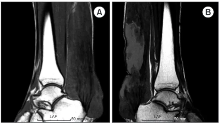
단순 방사선 사진상 아킬레스건 주위의 팽창된 연부조직의 음영 이 관찰되었으나 골조직의 변화는 없었다(Fig. 2). 자기공명영상 (magnetic resonance imaging) 촬영 사진상 아킬레스건 내로
광범위하게 지방조직이 침범하여 혼재된 신호와 함께 비대해진 아킬레스건이 관찰되었다(Fig. 3, 4). 3명의 환자(환자 1, 3, 4)에 서 아킬레스건 전장을 침범한 소견이 관찰되었다.
3. 혈액학적 검사
수술 전 시행한 생화학적 검사상 총콜레스테롤과 저밀도 지단백 (low-density lipoprotein)은 3명(환자 1, 2, 3)에서 증가되어 있 으며(Table 1) 트리글리세리드(triglyceride)는 1명(환자 4; 235 mg/dl, 정상 범위 0–150 mg/dl)을 제외하고 정상 범위 내였다.
4. 수술 방법
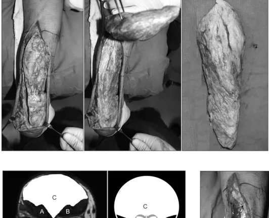
수술은 전신마취하 복와위 자세에서 허벅지에 지혈대(300 mmHg)를 사용하여 시행하였다. 종아리의 병변 부위에 종절개 를 가하고 아킬레스건을 노출시킨 후 아킬레스건 내부로 황색종 을 관찰하였다. 정상 조직 간의 경계가 불명확하여 황색종만을 완전히 제거할 수 없었다. 따라서 아킬레스건에 정중앙에 종절개 를 가하고 내부에 존재하는 황색종을 쐐기 형태로 충분히 제거하 여 종괴를 줄여주었다(Fig. 5). 그 후 아킬레스건의 바깥층을 봉합 하였다(with running locked stitches; Fig. 6, 7).
5. 수술 후 재활
수술 직후 단하지부목으로 고정하여 수술 2주까지 체중부하는 제한하였다. 수술 후 2주째부터 단하지부목 고정 대신 발목 부츠 (ankle boots)를 착용하며 목발을 이용한 부분 체중부하를 허용 하였다. 수술 후 4주째 부츠를 제거하면서 체중부하를 허용하였 다.
6. 임상적 평가
창상 문제, 아킬레스건 파열, 비복 신경(sural nerve) 손상, 감
A B
LAF 50 mm LAF 50 mm
Figure 3. Magnetic resonance imaging shows both ankles. Sagittal T1- weighted magnetic resonance image of both ankles displays a grossly enlarged mass along the Achilles tendon, which extends proximally to the gastrocnemius and soleus muscular portion, thereby helping it to distinguish between the normal Achilles tendon and the mass. (A) Right ankle. (B) Left ankle.
A B
Figure 2. Radiography of the ankles reveals a normal osseous architecture with increased soft tissue density posterior to the ankle joint along the expected course of the Achilles tendon. (A) Right ankle. (B) Left ankle.
A B
Figure 4. Axial proton density magnetic resonance imaging of both ankles demonstrates diffuse infiltration of the Achilles tendon with lipid and inflammatory tissue. (A) Right ankle. (B) Left ankle.
염 등의 수술 관련 합병증 등을 기록하였다. 마지막 추시에 족관 절 운동 범위, American Orthopaedic Foot & Ankle Society (AOFAS) ankle/hindfoot score, 치료 만족도 시각적 척도(visu- al analogue scale for overall satisfaction), single-limb heel raise 가능 여부와 직장으로의 복귀 시간을 측정하여 수술 후 임 상적인 평가를 시행하였다.
모든 분석에는 IBM SPSS Statistics ver. 23.0 (IBM Corp., Armonk, NY, USA)을 이용하였다
결 과
1. 합병증
환자 1에서 수술 후 4주째 열개창(wound dehiscence)이 발생 하였으나 압박드레싱과 항생제 투약 후 상처는 회복되었다. 모든 환자에게서 신경학적 문제는 없었으며 종물의 재발, 아킬레스건 의 파열은 관찰되지 않았다.
2. 임상적 결과
마지막 추시에서 환자들의 족관절의 운동 범위는 족배굴곡 평균 10도(범위, 5–15도)로 정상 범주였다(Table 2). AOFAS ankle/
A B
C
C
A B
A B
Figure 6. Schematic diagram of a wedge-shaped tendon-sparing technique shows the removal of the xanthoma and surgical suturing of
the remaining tissue. A, B: Achilles tendon. C: Xanthoma. Figure 7. Intraoperative photographs demonstrate the sutured remnant- mixed tendon where a continuous repair using running locked sutures was performed after a debulking resection.
Figure 5. Intraoperative photographs, taken during mass excision, show the maintenance of a wedge-shaped formation during the surgical process.
hindfoot score는 평균 91점(범위, 85–96)이었으며 치료 만족도 시각적 척도(1은 가장 낮은, 10은 가장 높은 만족도를 나타냄)는 2명이 10점을, 나머지는 8–9점을 보였다. 직장으로의 복귀는 평 균 27.6일(범위, 17–58일)이었으며 마지막 추시에서 모든 환자 들은 single-limb heel raise가 가능하였다. 수술 후 환자 1, 2, 3 은 atorvastatin calcium trihydrate 20 mg의 투여를 시작하였다.
고 찰
아킬레스건에 발생한 황색종은 드문 질환이며 가족성 고콜레스 테롤혈증과 밀접한 관련이 있다.6) 황색종은 발병학적으로 지방 대사장애에 의해 발생하며, 전기영동법에 의한 고지방혈증의 분 류중 type2 hyperbetalipoproteinemia는 가족력이 있으며 대 부분의 황색종이 이에 속한다.7) 혈액 검사상 콜레스테롤 수치가 상승되어 있고 트리글리세리드는 정상 범위 내에 있는 것이 특징 이다. 본 연구에서는 3명의 환자에서 황색종에 대한 가족력이 관 찰되었으나 전기영동법 등 정확한 진단은 시행되지 않았으며 3 명의 환자에서 총콜레스테롤 수치의 상승이 관찰되었고 트리글 리세리드는 1명(환자 4)에서 상승되어 있었다. 이러한 환자들에 게서는 심혈관계 동맥경화증 등의 발병률이 높기 때문에7,8) 이에 대한 교육과 정확한 진단이 필요하다.
황색종의 발생 위치는 하지보다 상지에서 2–3배 호발하며 상 지에서 수부와 완관절 부위의 건에 잘 발생하며, 하지에서 대부 분의 병변은 족부의 신전부에 발생한다.9) 본 연구에서 양측 아킬 레스건 이외에 족부, 완관절, 주관절 부위에서 관찰되었으며 상 지보다는 하지에서의 분포가 많았다.
내과적 치료는 식이조절과 함께 콜레스테롤 수치를 낮추는 약 물을 복용할 수 있다. 다불포화지방(polyunsaturated fat)과 저 콜레스테롤 함유 음식 등의 식이요법을 통해 혈중 콜레스테롤을 낮출 수 있으며, cholestyramine, Atromid-S, D-thyroxine 등 의 약물은 혈청지질을 낮추는 데 효과가 있다고 알려져 있다.7,10) 본 연구에서 환자들은 atorvastatin calcium trihydrate 20 mg 으로 치료를 시작하였다.
하지만 약물치료는 황색종의 크기를 줄이는 데 도움이 되지 못
한다.11-14) 통증이 발생하거나 신발을 신을 때 등의 일상생활에 불
편감이 지속되는 경우 수술적 제거가 필요하다. Fahey 등7)은 아 킬레스건에 발생한 황색종에 대한 문헌 고찰을 하였으며 173예 중에서 15명만이 수술적 치료를 요하였다고 기술한 바 있다.
수술적 치료로는 부분 절제술, 전 절제술, 재건술 등이 있다.10) 재발을 우려하여 병변 부위 전 절제술을 시행할 수 있으며 전 절 제술과 아킬레스건 근막 이전술 후 좋은 결과에 대한 보고가 있 다.3,5) 또한 Panman과 Hamming15)은 Dacron graft를 이용 한 재건술 후 만족할 만한 결과를 보고한 바 있으며 Moroney와 Besse16)는 전 절제술 후 장무지굴건(flexor hallucis longus)을 이용한 재건술 후 좋은 결과를 보고한 바 있다. 하지만 전 절제술 은 재건을 요하기 때문에 부담이 될 수 있으며 재건방식 또는 이 식의 종류에 따라 감염위험, 공여부위 합병증 등의 우려가 있다.
또한 재건술 후 활동 시기는 6주 이상으로 보고되고 있다. 본 연 구에서 시행한 황색종의 쐐기형 절제술은 아킬레스건의 재건에 대한 부담을 줄여주었으며 경한 합병증 이외의 아킬레스건의 파 열 등의 주요 합병증은 발생하지 않았다. 환자들은 수술 후 4주 째 체중부하를 함으로써 일상으로의 복귀가 재건술에 비해 빨랐
다.7,16,17) 또한 마지막 추시에서 모든 환자들은 single-limb heel
raise가 가능하였다. 부분 절제술 후 재발의 위험은 12%–15%로 보고되고 있으나,9) 대부분의 환자는 수술 후 불편감, 통증 등을 호소하지 않는 것으로 보고된다.7) 본 연구에서 황색종이 재발한 환자는 없었으며 모든 환자들은 수술 후 불편감, 통증 등의 주관 적인 증상을 호소하지 않았다.
본 연구는 적은 환자의 수에 대한 한계를 가지며 또한 부분 절 제술 후 아킬레스건의 추시 상태에 대한 영상의학적 증거가 없어 아킬레스건의 수술 후 상태에 대한 객관적 평가가 어려우며 약 물 복용 후 혈청 지질의 변화를 제시하지 못하였다. 또한 수술 방 법에 대한 대조군이 없어 수술 결과에 대한 한계점을 가지고 있 다. 하지만 본 연구에서는 AOFAS ankle/hindfoot score를 통하 여 기능적인 측면에 대한 분석을 시행하였으며 치료 만족도 시각 적 척도를 통해 환자 개개인의 만족도 평가를 하여 대조군 부재 에 대한 한계를 극복하고자 하였다. 추후 영상의학적 방법을 통 한 황색종의 재발이나 아킬레스건의 파열 등을 객관적으로 확인 Table 2. Postoperative Clinical Findings of the Patients
Patient no. Complication Ankle dorsiflexion (°) AOFAS score VAS score for overall
satisfaction Time to return to work (d)
1 Wound dehiscence 15 85 8 58
2 No 10 91 10 21
3 No 10 92 10 21
4 No 5 91 9 21
5 No 10 96 8 17
AOFAS, American Orthopaedic Foot & Ankle Society; VAS, visual analogue scale.
할 수 있는 연구가 필요하다고 판단된다.
결 론
본 연구에서는 양측 아킬레스건에 발생한 거대 황색종에 대해 쐐 기 모양 부분 절제술을 시행하였으며 좋은 결과를 보였다. 아킬 레스건에 황색종이 발생하였을 경우, 자가 아킬레스건을 보존하 며 시행하는 쐐기 모양 부분 절제술은 좋은 수술적 치료가 될 수 있을 것으로 판단된다.
CONFLICTS OF INTEREST
The authors have nothing to disclose.
ORCID
Sungmin Kim, https://orcid.org/0000-0002-7116-9503 Yeong Seub Ahn, https://orcid.org/0000-0002-1097-7814 Dong-Min Jung, https://orcid.org/0000-0002-7422-5160 Sung Taek Jung, https://orcid.org/0000-0003-1255-9568
REFERENCES
1. Kruth HS. Lipid deposition in human tendon xanthoma. Am J Pathol. 1985;121:311-5.
2. Harlan WR Jr, Graham JB, Estes EH. Familial hypercholester- olemia: a genetic and metabolic study. Medicine. 1966;45:77- 110.
3. Lee CK, Weiss AB. Xanthomas of the Achilles tendons. A case report of bilateral total resection and tendon reconstruc- tion using the fascia lata. J Bone Joint Surg Am. 1980;62:666- 9.
4. Saraf SK, Sharma SV. Reconstruction for xanthoma of the Achilles tendon. Int Orthop. 1992;16:37-8.
5. Tomita T, Ochi T, Fushimi H, Matsuzawa Y, Ono K. Recon- struction of the Achilles tendon for xanthoma: findings at operative re-exploration. A case report. J Bone Joint Surg
Am. 1994;76:444-7.
6. Brunzell JD. Disorders of lipoprotein metabolism. In: Wyn- gaarden JB, Smith LH Jr, Bennett JC, ed. Cecil textbook of medicine. 19th ed. Philadelphia: Saunders; 1992. 1084-5.
7. Fahey JJ, Stark HH, Donovan WF, Drennan DB. Xanthoma of the achilles tendon. Seven cases with familial hyperbetali- poproteinemia. J Bone Joint Surg Am. 1973;55:1197-211.
8. Fredrickson DS, Levy RI, Lees RS. Fat transport in lipopro- teins--an integrated approach to mechanisms and disorders.
N Engl J Med. 1967;276:148-56 contd.
9. Friedman MS. Xanthoma of the Achilles tendon. J Bone Joint Surg Am. 1947;29:760-6.
10. Park CS, Lee KH, Kim MK, Lee SN, Ryuh JW. Xanthoma of the Achilles tendon. J Korean Orthop Assoc. 1991;26:1-5.
11. Sperry WM, Schick B. Essential xanthomatosis: treatment with cholesterol-free diet in two cases. Am J Dis Child.
1936;51:1372-84.
12. Thannhauser SJ. Lipidoses: diseases of the cellular lipid me- tabolism. 2nd ed. New York: Oxford University Press; 1950.
13. Urbach F, Hildreth EA, Wackerman MT. The therapeutic uses of low fat, low cholesterol diets. I. Treatment of essential familial xanthomatosis. J Clin Nutr. 1952;1:52-65.
14. Young F, Harris CT. Complete excision and reconstruction of both Achilles tendons for giant cell xanthoma. Surg Gynecol Obstet. 1935;61:662-9.
15. Panman WF, Hamming JJ. Xanthoma of the Achilles tendon.
Neth J Surg. 1986;38:155-7.
16. Moroney PJ, Besse JL. Resection of bilateral massive Achil- les tendon xanthomata with reconstruction using a flexor hallucis longus tendon transfer and Bosworth turndown flap: a case report and literature review. Foot Ankle Surg.
2012;18:e25-8.
17. Dinçel YM, Arıkan Y, Özer D, Basılgan S. Reconstruction of the Achilles tendon using quadriceps tendon graft in bilateral xanthomas secondary to familial hypercholesterolemia: a case report. Eklem Hastalik Cerrahisi. 2018;29:117-22.
양측 아킬레스건에 발생한 거대 황색종의 쐐기형 절제술을 이용한 수술적 치료
김성민 • 안영섭 • 정동민 • 정성택
전남대학교 의과대학 정형외과학교실
목적:
아킬레스건의 황색종은 드물게 발생하며 증상이 심할 경우 수술적 치료가 필요한 경우가 있다. 전 절제술 후 재건술은 높은 수술의 숙련도를 요하며 다양한 합병증에 대한 우려가 있다. 본 연구에서는 양측 아킬레스건에 발생한 거대 황색종에 대해 자가 아킬레스건을 보존하는 쐐기형 절제술 후 추시 결과를 분석하고자 하였다.대상 및 방법:
2010년 7월부터 2018년 5월까지 양측 아킬레스건에 발생한 황색종 환자 5명에 대해 자가 아킬레스건을 보존하 는 쐐기형 절제술을 시행하였다. 평균 나이는 49세(범위, 40–55세)였고 추시 기간은 평균 21.4개월(범위, 12–31개월), 남자는 3 명, 여자는 2명이었다. 수술 후 발생한 합병증을 기록하였으며 족관절 운동 범위, American Orthopaedic Foot & Ankle Society (AOFAS) ankle/hindfoot score, 치료 만족도 시각적 척도(visual analogue scale for overall satisfaction), single-limb heel raise 가능 여부, 그리고 직장으로의 복귀 시간을 측정하여 수술 후 임상적인 평가를 시행하였다.결과:
1명에서 열개창(wound dehiscence)이 발생하였으며 추가적인 수술적 치료 없이 호전되었다. 마지막 추시에서 모든 환자들 의 족관절의 운동 범위는 정상이었으며 AOFAS ankle/hindfoot score는 평균 91점(범위, 85–96점)이었고 치료 만족도 시각적 척 도는 8–10점의 분포를 보였다. 직장으로의 복귀는 평균 27.6일(범위, 17–58일)이었으며 모든 환자는 single-limb heel raise가 가 능하였다.결론:
아킬레스건에 황색종이 발생하였을 경우 자가 아킬레스건을 보존하며 시행하는 쐐기형 절제술은 좋은 수술적 치료가 될 수 있을 것으로 판단된다.색인단어: 황색종, 아킬레스건, 쐐기형 절제술
접수일 2020년 5월 28일 수정일 2020년 6월 15일 게재확정일 2020년 6월 22일 책임저자 정성택
61469, 광주시 동구 제봉로 42, 전남대학교 의과대학 정형외과학교실
TEL 062-227-1640, FAX 062-225-7794, E-mail [email protected], ORCID https://orcid.org/0000-0003-1255-9568
Copyright © 2021 by The Korean Orthopaedic Association
“This is an Open Access article distributed under the terms of the Creative Commons Attribution Non-Commercial License (http://creativecommons.org/licenses/by-nc/4.0/) which permits unrestricted non-commercial use, distribution, and reproduction in any medium, provided the original work is properly cited.”