Kor. J. Pharmacogn.
45(3) : 214∼ 219 (2014)
214
청호의 Phytosterol 성분 분리 및 뇌세포 보호 활성
이지우1·원진배1·마충제1,2*
1강원대학교 의생명과학대학 생물의소재공학과, 2강원대학교 생명공학연구소
Neuroprotective Activity of Phytosterols Isolated from Artemisia apiacea
Jiwoo Lee1, Jin Bae Weon1, and Choong Je Ma1,2*
1Department of Medical Biomaterials Engineering, College of Biomedical Science, Kangwon National University, Chuncheon 200-701, Korea
2Research Institute of Biotechnology, Kangwon National University, Chuncheon 200-701, Korea
Abstract − Artemisia apiacea is a traditional herbal medicine using treatment of eczema and jaundice in Eastern Asia including China, Korea, and Japan. In this study, the three phytosterol constituents were isolated and identified from the hexane fraction of 80% aqueous methanol extract of A. apiacea. Compounds were isolated using open column chromatography (silica gel).
Their chemical structures were also established using 1H-NMR and 13C-NMR. Moreover, neuroprotective activity of each com- pound against glutamate-induced neurotoxicity in hippocampal HT-22 cell line was evaluated by 3-(4,5-dimethylthiazol-2-yl)- 2,5-diphenyltetrazolium bromide (MTT) assay. Furthermore, Inhibition of reactive oxygen species (ROS) and calcium ion (Ca2+) accumulation were measured for elucidation of neuroprotective mechanism of isolated compounds. They showed that stigmasterol had neuroprotective activity against the glutamate-induced toxicity by inhibition of ROS and Ca2+ production. In conclusion, isolated compound of A. apiacea might be useful for therapeutic agent against neurodegenerative diseases.
Key words − Artemisia apiacea, Neuroprotective activity, Reactive oxygen species (ROS)
최근 현대사회로 접어들면서 생활 수준 향상 및 의학의 발달로 인해 개인의 평균 수명이 증가하는 추세이다. 그러 나 이에따른 급격한 노령화로 인해 치매와 같은 퇴행성 뇌 신경계 질환 발생 역시 증가하여 사회적 문제로 대두되고 있다. 대부분의 퇴행성 뇌신경계 질환의 주요 발병 원인이 자유 라디칼의 생성 및 활성 산소종(reactive oxygen species, ROS) 축적에 기인한다는 사실이 보고된 바 있다.1-3) ROS의 축적은 세포의 nucleus, mitochondrial DNA, cytoplasmic protein 및 다른 주요 성분에 손상을 주어 사멸에 이르게 한 다고 알려져 있다.4-5) Glutamate는 중추신경계의 주요 흥분 성 신경전달물질로서 과도한 glutamate는 N-acetyl cystein 의 흡수를 억제하여 세포 내 산화적 스트레스를 유발한다.6) 마우스의 해마에서 기원한 세포주인 HT22 cell은 in vitro 모델에서 glutamate 유도성 뇌신경 질환의 기전 연구에 사 용되고 있다.7)
천연물은 예로부터 퇴행성 뇌 질환 등의 다양한 질병에
대한 치료 및 예방을 목적으로 사용되었다. 이러한 천연물 은 적은 부작용 및 높은 순응도로 인해 이용이 증가하는 추
세이다.8-10) 개사철쑥(Artemisia apiacea Hance)은 국화과
(Compositae)에 속하는 1년생 또는 2년생 초본으로서 강기 슭 및 냇가 모래땅 등지에 자생하며 한국, 중국, 일본 등에 분포한다. 잎이 거의 없고 꽃대가 있는 길이 60-90 cm의 줄 기를 가지며 줄기의 바깥 면은 황갈색이다. 잎은 녹갈색이 며 잔주름이 많고 꽃대는 황록색이다. 예로부터 개사철쑥의 지상부를 건조한 것을 청호(靑蒿)라고 하여 피부 진균증, 황 달(黃疸), 습진(濕疹), 욕창( 瘡) 및 탈모(脫毛) 치료에 사용 되어 왔다.11,12)최근 연구들에 의하면, 개사철쑥은 항 말라 리아, 항산화, 항염증 및 탈모 치료에 효능이 있는 것으로 밝혀졌다.13-16) 개사철쑥으로부터 campesterol, stigmasterol, β-sitosterol, daucosterol 및 artemisterol과 같은 식물 steroid 성분들 및 7-methoxycoumarin, 7,8-dimethoxycoumarin, 7,8-methylenedioxycoumarin, daphnetin 등의 coumarin 류, 그리고 α-pinene, artemisia ketone 등의 정유 성분들이 분리 된 바 있다.12,17-23)
본 연구에서는 청호의 hexane층으로부터 phytosterol인
*교신저자(E-mail):[email protected] (Tel): +82-33-250-6565
서 구입하여 사용하였으며, 대전대학교 서영배 교수님을 통 해 동정하였고 청호 표본은(NO. CJ064M)는 강원대학교에 보관하였다.
시약 및 기기 − 추출 및 분획에 사용된 methanol, n- hexane, chloroform, ethyl acetate, n-butanol은 DAE JUNG(Korea)에서 구입하였다. TLC plates(Silica gel 60 F254)와 silica gel(70-230 mesh)는 Merck(Darmstadt, Germany)에서 구입하였다. NMR 분석을 위해 Cambridge Isotope Laboratories, Inc.(U.S.A.)의 CDCl3와 DMSO-d6를 사용하였다. Dulbecco's modified Eagle's medium(DMEM) 과 fetal bovine serum(FBS)은 Gibco BRL. Co.(Eggenstein, Germany)로부터 구입하였다. Glutamate와 6-hydroxy- 2,5,7,8-tetramethylchroman-2-carbboxylic acid(trolox), 3- (4,5-dimethylthiazol-2-yl)-2,5-diphenyl tetrazolium bromide (MTT)는 Sigma(St. Louis, U.S.A.)로부터 구입하여 사용하 였다.
NMR spectrum은 Bruker Avance 400(400MHz, Germany) 을 사용하여 측정하였다.
추출 및 분획 − 구입한 청호 10.2 kg에 80% MeOH를 가 하고 90분씩 3회 초음파 추출 하였으며 감압 농축하여 445.22 g의 추출물을 얻었다. 얻어진 추출물을 증류수에 현 탁한 후 동량의 hexane을 가하고 진탕 방치하여 hexane 분 획(32.66 g)을 얻었다. 같은 방법으로 수층에 동량의 CHCl3 를 가하고 진탕 방치하여 CHCl3 분획(86.99 g)을 얻었으며, 다시 수층에 동량의 EtOAc를 가하여 EtOAc 분획(35.11 g) 을, 수층에 동량의 BuOH를 가하여 BuOH 분획(81.63 g)을 얻었다.
Hexane 분획(29.54 g)을 silica gel open column(40×5 cm, 70~230 mesh)에 걸어 CHCl3:MeOH(100:1→0:1, v/v)의 조 성으로 chromatography를 실시하여 6개의 소분획(A~F)으로 나누었다. 소분획 B(19 g)를 CHCl3:MeOH(100:1→0:1, v/v) 의 조성으로 silica gel column chromatography(74×3 cm, 70~230 mesh)를 실시하여 TLC 분석을 통해 9개의 소분획 (Ba~Bi)을 얻었다. 소분획 Bc를 hexane:EtOAc(70:1→1:2, v/v)의 조성으로 silica gel column chromatography(55×2.8 cm, 70~230 mesh)를 실시하여 20개의 소분획(Bc1~Bc20)을 얻었으며, 소분획 Bc9에서 화합물 1(122.3 mg)과 2(27.6 mg) 를 얻었다. 소분획 E를 MeOH에서 재결정하여 화합물 3 (47.2 mg)을 얻었다.
6), 71.83 (C-3), 21.23 (C-21), 21.09 (C-26), 19.84 (C-19), 19.42 (C-27), 12.27 (C-29), 11.88 (C-18).
Campesterol (2) − white powder. 1H NMR (400 MHz, CDCl3) δ 5.36 (1H, d, J = 5.2 Hz, H-6), 3.53 (1H, m, H- 3), 1.03 (3H, d, J = 6.4 Hz, H-21), 1.01 (3H, s, H-19), 0.85 (3H, d, J = 6.3 Hz, H-26), 0.81 (3H, t, J = 7.0 Hz, H-28), 0.70 (3H, d, J = 7.3 Hz, H-27), 0.65 (3H, s, H- 18); 13C NMR (400 MHz, CDCl3) δ 140.75 (C-5), 121.72 (C-6), 71.82 (C-3), 21.22 (C-21), 21.08 (C-26), 19.82 (C- 19), 19.40 (C-27), 12.25 (C-28), 11.86 (C-18).
Daucosterol (3) − Pale yellow crystals. 1H NMR (400 MHz, DMSO-d6) δ 5.33 (1H, d, J = 4.8 Hz, H-6), 4.88 (1H, d, J = 9.2 Hz, H-1'), 3.48 (1H, m, H-3), 1.02 (3H, d, J = 7.3 Hz, H-21), 0.96 (3H, s, H-19), 0.91 (3H, d, J
= 6.5 Hz, H-26), 0.85 (3H, d, J = 7.9 Hz, H-27), 0.80 (3H, t, J = 7.6 Hz, H-29), 0.65 (3H, s, H-18); 13C NMR (400 MHz, DMSO-d6) δ 140.80 (C-5), 121.59 (C-6), 101.13 (C-1'), 77.26 (C-3), 77.12 (C-3'), 73.83 (C-2'), 70.46 (C-4'), 61.45 (C-6'), 20.09 (C-26), 19.48 (C-27), 19.30 (C-19), 18.99 (C-21), 12.16 (C-29), 12.05 (C-18).
HT22 세포 배양 및 뇌신경세포 보호 활성 측정 − 마우 스 해마 유래 세포주인 HT22 세포는 서울대학교로부터 분 양받아 사용하였다. HT22 세포는 10% FBS가 첨가된 DMEM 배지를 이용하여 37oC의 5% CO2 배양기에서 배양 하였다. Glutamate에 의해 유도된 독성에 대한 뇌신경세포 보호 활성을 측정하고자 MTT assay를 시행하였다. HT22 세포를 48-well plate에 4.02×10³ cells/well의 농도로 분주하 여 23시간 배양한 후 시료(10, 50, 100 µM), trolox(positive control)를 30 µl씩 처리하였으며, 1시간 배양 후 1.8 mM glutamate 30µl를 처리하였다. 24시간 배양 후에 배지를 제 거하고 MTT assay를 시행하여 microplate reader를 사용하 여 570 nm에서 OD값을 측정하였다. 뇌세포 보호 활성은 relative protection(%)으로 나타내었으며 통계처리는 ANOVA test를 적용하였다.
세포 내 Reactive Oxygen Species(ROS) 생성 측정 − HT22 세포에서 glutamate에 의해 유발된 ROS를 2',7'- dichlorofluorescin diacetate(DCF-DA)를 사용하여 측정하였 다. HT22 세포를 48-well plate에 4.02×10³ cells/well의 농
도로 분주하여 37oC에서 24시간 배양한 후 시료, trolox (positive control), glutamate를 처리하여 8시간 배양하였다.
이후 10 µM DCF-DA를 처리하고 1시간 배양하고 1%
Triton X-100을 처리하여 형광 분석기를 통해 488 nm의 들 뜸 파장과 530 nm의 방출 파장에서 측정하였다.
세포 내 칼슘 이온(Ca2+) 생성의 측정 − HT22 세포 내 칼슘 이온(Ca2+)를 측정하기 위해, Fura-2AM을 사용하였다.
시료, trolox(positive control), glutamate를 처리한 후, 세포 를 수확하기 전에 2 µM Fura-2AM을 처리한다. 2시간 후에 세포를 PBS로 세척한 후, 1% Triton X-100를 처리하여 세 포를 추출한다. 칼슘 이온(Ca2+)형광 분석기를 통해 400 nm 의 들뜸 파장과 535 nm의 방출 파장에서 형광 측정하였다.
HPLC를 이용한 화합물 함량 분석 − HPLC를 이용하여 분리된 세 화합물의 함량을 분석하였다. 이동상은 HPLC 분 석용 water(A)와 acetonitrile(B)를 사용하였으며, 용매 조건 은 처음 5분간 10% B 조건으로, 40분간 10-90% B 조건으 로, 20분간 100% B 조건으로 분리하였다. flow rate는 1.0 ml/min으로 설정하였고, column은 Shiseido C18 column (4.6 mm I.D. x 250 mm, 5 µm pore size)을 통해 분석하였 다. 시료의 injection volume은 10 µl였으며, UV-wavelength 는 205 nm와 254 nm에서 분석하였다.
결과 및 고찰
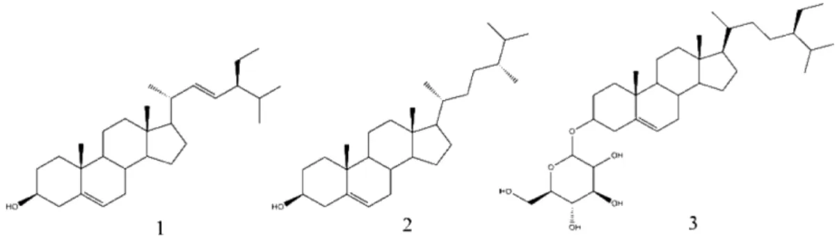
Phytosterol의 분리정제 − 많은 유효 성분의 보존과 추출 공정의 효율을 높이기 위해 80% MeOH에서 초음파 추출 법을 이용하여 청호 추출물을 얻었다. 얻어진 추출물을 감 압 농축한 후, 증류수에 현탁하여 동량의 hexane, CHCl3, EtOAc, BuOH를 순차적으로 가해 분획하였으며 hexane 분 획을 column chromatography로 분리하여 화합물 1-3을 얻 었다(Fig. 1).
Stigmasterol(1)은 백색의 powder 형태로 분리되었으며, silica gel TLC에 전개하고 10% 황산 발색 시 자색을 나타 내었다. Campesterol(2)은 stigmasterol이 분리된 소분획 Bc9 에서 백색의 powder 형태로 분리되었으며 silica gel TLC 분석을 통해서 stigmasterol과는 다른 화합물임을 확인하였
다. Daucosterol(3)은 연한 노란색 결정으로 분리되었다. 세 화합물은 1H 및 13C-NMR 데이터와 문헌 상의 데이터를 비 교하여 구조를 동정하였다.24-28)
HT22 세포 배양 및 뇌신경세포 보호 활성 측정 − 청호 추출물과 hexane, chloroform, ethyl acetate, n-butanol 층과 Fig. 1. Chemical structure of compounds 1-3 isolated from the A. apiacea.
Fig. 2. The neuroprotective effects of Artemisia apiacea frac- tions (A), compound 1, 2 and 3 (B) on glutamate-induced cytotoxicity in HT22 cells. HT22 cells were pretreated with Artemisia apiacea fractions and each compound. Then incu- bated for 24 h with glutamate (1.8 mM) after 1 hr. Each bar represents the Mean±SD of three independent experiments.
*p<0.05, **p<0.01, ***p<0.001 vs. glutamate-treated cells (ANOVA). <Control: control group, Vehicle: only glutamate treated group, Trolox: glutamate and trolox treated group, Compound 1: stigmasterol, Compound 2: campesterol and Compound 3: daucosterol>
청호에서 분리한 stigmasterol, campesterol 및 daucosterol의 glutamate로 유발된 독성에 의한 HT22 세포에 대한 보호 활 성을 측정하였다. 청호의 분획층 중 hexane 층이 뇌 신경세 포 보호 활성을 나타내었으며, 분리된 화합물 중 stigmasterol 이 50, 100 µM에서 각각 53.48%와 61.57%의 세포보호 활 성을 나타내어 농도 의존적으로 높은 활성을 나타내었다 (Fig. 2). Campesterol과 daucosterol은 glutamate 유도성 신 경세포 독성에 대한 보호 활성을 나타내지 않았다.
세포 내 ROS량 측정 − 청호에서 분리한 화합물 중 뇌신 경세포 보호 활성을 나타낸 stigmasterol에 대한 ROS 생성 억제 활성 측정하였다. 많은 연구들에서 고농도의 glutamate 는 ROS를 생성하며, 높은 농도의 ROS는 산화적 손상을 유 발한다는 사실이 보고된 바 있다. 측정 결과, glutamate를 처리한 세포의 ROS량은 대조군에 비해 113.12% 가 증가하 였으나, 50 µM와 100 µM 농도의 stigmasterol를 처리한 세 포에서는 각각 99.11%와 97.45%로 glutamate에 의해 증가 된 ROS량을 감소시켰다(Fig. 3).
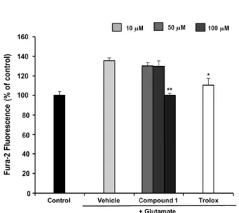
세포 내 칼슘이온(Ca2+) 측정 − ROS 활성을 유도하는 세 포 내 Ca2+이온 량을 측정하였다. glutamate를 처리한 세포 내의 칼슘 이온량 측정 결과, 135.25%로 증가하였으나, 100µM 농도의 stigmasterol을 처리시 100.27%의 칼슘 이 온량이 측정되어 glutamate에 의해 처리된 세포의 칼슘 이 온량과 비교하여 유의적으로 감소시켰다(Fig. 4).
HPLC를 이용한 화합물 함량 분석 − 청호에서 세 화합물, stigmasterol, campesterol 그리고 daucosterol의 함량을 분석 하기 위해 HPLC를 이용하였다(Fig. 5). 205 nm에서는 daucosterol 그리고 254 nm에서는 campesterol과 daucosterol
을 분석하였다. 분석 결과, stigmasterol은 0.42%, campesterol 은 1.95% 그리고 daucosterol은 0.23%를 포함하는 것으로 확인되었다.
결 론
본 연구에서 청호로부터 3종류의 phytosterol(stigmasterol, campesterol, daucosterol)을 분리하였으며 1H NMR, 13C NMR 등의 분광학적 방법 및 문헌 조사를 통해 그 구조를 규명하였다.
HPLC를 이용하여 세 화합물의 함량을 평가한 결과, campesterol이 가장 많이 포함되어 있었으며, daucosterol이 가장 적게 포함되어 있었다.
청호 추출물, 각 분획층 그리고 분리된 화합물을 대상으 로 HT22 세포에서 glutamate에 의해 유도된 뇌신경 세포 사멸에 대한 보호 활성을 평가하였다. 측정 결과, 청호 추 출물과 hexane 분획층은 glutamate에 의해 유도된 세포 사 멸을 억제하였으며, 세 화합물 중 stigmasterol이 glutamate 유도성 독성에 대해 100 µM에서 61.57%의 높은 보호 활 성을 나타내었다. 이전 연구에 의하면, stigmasterol은 HT22 세포에서 glutamate와 amyloid-β에 의해 유발된 독성에 대 해 보호활성이 보고된 바 있으며,29) stigmasterol의 뇌신경세 포 보호 활성은 3-OH group과 연관된 것으로 판단된다.30)
고농도의 glutamate는 세포 내 ROS의 량을 증가시키고 그로 인해 산화적 스트레스를 일으켜 뇌신경 세포의 손상 을 일으킨다. 신경 전달을 담당하는 요소 중인 하나인 칼슘 Fig. 3. The effect of compound 1 on glutamate-induced ROS
production in HT22 cells. Control: control group, Vehicle: only glutamate treated group, Trolox: glutamate and trolox treated group and Compound 1: stigmasterol. Data represent means ± S.E.M. (*p<0.05, **p<0.01, and ***p<0.001 versus the glutamate treated group).
Fig. 4. The effect of compound 1 on glutamate-induced Ca2+
influx in HT22 cells. Control: control group, Vehicle: only glutamate treated group, Trolox: glutamate and trolox treated group and Compound 1: stigmasterol. Data represent means±
S.E.M. (*p<0.05, **p<0.01, and ***p<0.001 versus the glutamate treated group).
이온의 경우, 증가된 glutamate에 의해 세포 내로의 유입되 어 축적되는 현상을 일으켜 뇌 세포 손상을 유도한다.6,31) Stigmasterol의 뇌신경세포 보호 기전을 밝히기 위해 뇌신경 세포 내의 ROS량과 Ca2+량을 측정하였으며, stigmasterol은 ROS량과 세포 내 Ca2+량을 유의적으로 억제시켰다. 위 결 과를 바탕으로, stigmasterol의 뇌신경세포 보호 활성은 ROS 축적 및 세포 내 Ca2+량의 억제를 통해 활성을 나타내는 것 으로 사료된다. 위의 결과를 통해, 청호의 뇌신경 세포 보 호 활성은 stigmasterol의 뇌신경 세포 보호 활성과 관련 있 는 것으로 사료된다. 추가적으로 stigmasterol의 glutamate 대한 신경 세포 보호의 추가적인 기전 연구가 필요하다고 사료된다.
사 사
2013년도 강원대학교 학술연구조성비로 연구하였음(관리 번호-120131861).
인용문헌
1. Christen, Y. (2000) Oxidative stress and Alzheimer disease.
Am. J. Clin. Nutr. 71: 621S-629S.
2. Halliwell, B. (2001) Role of free radicals in the neurode- generative diseases: therapeutic implications for antioxidant treatment. Drugs & Aging 18: 685-716.
3. Emerit, J., Edeas, M. and Bricaire, F. (2004) Neurodegen- erative diseases and oxidative stress. Biomed. Pharmacother.
58: 39-46.
4. Choi, D. W. (1992) Excitotoxic cell death. J. Neurochem. 23:
1261-1276.
5. Jin, D. Q., Lim, C. S., Hwang, J. K., Ha, I. and Han, J. S.
(2005) Anti-oxidant and anti-inflammatory activities of macelignan in murine hippocampal cell line and primary cul- ture of rat microglial cells. Biochem. Biophys. Res. Commun.
331: 1264-1269.
6. Coyle, J. T. and Puttfarcken, P. (1993) Oxidative stress, Fig. 5. The HPLC chromatogram of three standard compounds (A) and A. apiacea sample (B) at 205 and 254 nm. Campesterol (1), stigmasterol (2) and daucosterol (3).
9. Bent, S. (2008) Herbal medicine in the United States: review of efficacy, safety, and regulation. J. Gen. Intern. Med. 23:
854-859.
10. Fabio, F. and Luigi, G. (2007) Herbal medicine today: clinical and research issues. Evid. Based Complement. Alternat. Med.
4: 37-40.
11. Yook, C. S. (1989) Coloured medicinal plants of Korea. 522.
Printed by academy, Seoul.
12. Kim, O. C. and Jang, H. J. (1994) Volatile components of Artemisia apiacea herba. Agr. Chem. Biotechnol. 37: 37-42.
13. Tan, R. X., Zheng, W. F. and Tang, H. Q. (1998) Biologically active substances from the genus Artemisia. Planta Med. 64:
295-302.
14. Hsu, E. (2006) Reflections on the ‘discovery’ of the anti- malarial qinghao. Br. J. Clin. Pharmacol. 61: 666-670.
15. Kim, K. S., Shim, S. H., Jang, J. M., Cheong, J. H. and Kim, B. K. (1999) A study on hair-growth activity of Artemisia apiacea Hance. J. Pharm. Soc. Korea 43: 798-801.
16. Kim, K. S., Lee, S., Lee, Y. S., Jung, S. H., Park, Y., Shin, K.
H. and Kim, B.-K. (2003) Anti-oxidant activities of the extracts from the herbs of Artemisia apiacea. J. Ethnop- harmacol. 85: 69-72.
17. Shimomura, H., Sashida, Y. and Ohshima, Y. (1979) Cou- marins from Artemisia apiacea. Phytochem. 18: 1761-1762.
18. Shimomura, H., Sashida, Y. and Ohshima, Y. (1980a) The chemical components of Artemisia apiacea Hance, more cou- marins from the flower heads. Chem. Pharm. Bull. 28: 347- 348.
19. Shimomura, H., Sashida, Y., Ohshima, Y., Azuma, T. and Saitoh, M. (1980b) The chemical components of Artemisia apiacea Hance, components of stems and leaves. Yakugaku Zasshi 100: 1164-1166.
20. Yano, K. (1970) Mono- and sesqui-terpenes of the essential oils from Artemisia japonica and Artemisia apiacea. Flavour Ind. 1: 328-330.
21. Lee, S., Kim, K. S., Jang, J. M., Park, Y., Kim, Y. B. and
masterol and β-sitosterol from the dichloromethane extract of Rubus suavissimus. Int. Curr. Pharm. J. 1: 239-242.
25. Rajput, A. P. and Rajput, T. A. (2012) Isolation of stig- masterol and β-sitosterol from chloroform extract of leaves of Corchorus fascicularis Lam. Int. J. Biol. Chem. 6: 130-135.
26. Yoo, J. S., Ahn, E. M., Bang, M. H., Song, M. C., Yang, H.
J., Kim, D. H., Lee, D. Y., Chung, H. G., Jeong, T. S., Lee, K. T., Choi, M. S. and Baek, N. I. (2006) Steroids from the aerial parts of Artemisia princeps Pampanini. Korean J.
Medicinal Crop. Sci. 14: 273-277.
27. Jain, P. S. and Bari, S. B. (2010) Isolation of lupeol, stig- masterol and campesterol from petroleum ether extract of woody stem of Wrightia tinctoria. Asian J. Plant Sci. 9: 163- 167.
28. Lee, J. H., Kim, D. H., Bang, M. H., Yang, H. J., Bang, S. H., Chung, I. S., Kwon, B. M., Kim, S. H., Kim, D. K., Park, M.
H. and Baek, N. I. (2005) Isolation of sterols from the meth- anol extracts of Cymbidium goeringii REICHB. fil. J. Korean Soc. Appl. Biol. Chem. 48: 263-266.
29. Brimson, J. M., Brimson, S. J., Brimson, C. A., Rakkhi- tawatthana, V. and Tencomnao, T. (2012) Rhinacanthus nasu- tus extracts prevent glutamate and amyloid-β neurotoxicity in HT-22 mouse hippocampal cells: possible active compounds include lupeol, stigmasterol and β-sitosterol. Int. J. Mol. Sci.
13: 5074-5097.
30. Huang, X., Atwood, C. S., Hartshorn, M. A., Multhaup, G., Goldstein, L. E., Scarpa, R. C., Cuajungco, M. P., Gray, D.
N., Lim, J., Moir, R. D., Tanzi, R. E. and Bush, A. I. (1999) The Aβ peptide of Alzheimer’s disease directly produces hydrogen peroxide through metal ion reduction. Biochem. 38:
7609-7616.
31. Ha, J. S. and Park, S. S. (2006) Glutamate-induced oxidative stress, but not cell death, is largely dependent upon extra- cellular calcium in mouse neuronal HT22 cells. Neurosci.
Lett. 393: 165-169.
(2014. 7. 11 접수; 2014. 8. 20 심사; 2014. 8. 27 게재확정)