Antibacterial and Antibiofilm Activities of Diospyros malabarica Stem Extract against Streptococcus mutans
Hye Soo Kim1, Sang Woo Lee2, Kongmany Sydara3 and Soo Jeong Cho1*
1Department of Pharmaceutical Engineering, Gyeongnam National University of Science and Technology, 33 Dongjin-ro, Jinju 52725, Korea
2International Biological Material Research Center, Korea Research Institute of Bioscience and Biotechnology, Daejeon 34141, Korea
3Institute of Traditional Medicine, Ministry of Health, Vientiane Capital, La People’s Democratic Republic Received December 6, 2018 /Revised December 17, 2018 /Accepted January 9, 2019
The objective of this study was to evaluate the potential of Diospyros malabarica stem extract, a natural materials, in oral health material. With this aim in mind, thin layer chromatography (TLC), TLC-bio- autography, high-performance liquid chromatography (HPLC), electrospray ionization–mass spec- trometry (ESI-MS), scanning electron microscopy (SEM), and real-time qPCR were performed. The an- tibacterial activity of D. malabarica stem extract against Streptococcus mutans KCTC3065 was confirmed in an n-hexane fraction with low polarity. The molecular weight of the antibacterial compound was estimated to be 188 by ESI-MS analysis. The inhibitory effects of the extract on biofilm formation and gene expression related to biofilm formation of S. mutans were determined by SEM and real-time PCR analysis. The extract inhibited the formation of S. mutans biofilms at D. malabarica stem extract concen- trations of 1 mg/ml, as shown by SEM. The real-time PCR analysis showed that the expression of the gtfC gene, which is associated with biofilm formation, was significantly decreased in a dose-de- pendent manner. Based on the above results, it can be concluded that D. malabarica stem extracts, a natural materials, can be used in oral health products to suppress the formation of biofilms by inhibit- ing tooth adhesion of S. mutans, a causative agent of dental caries.
Key words : Biofilm formation, Diospyros malabarica stems, gtfB, gtfC, S. mutans
*Corresponding author
*Tel : +82-55-751-3397, Fax : +82-55-751-3399
*E-mail : [email protected]
This is an Open-Access article distributed under the terms of the Creative Commons Attribution Non-Commercial License (http://creativecommons.org/licenses/by-nc/3.0) which permits unrestricted non-commercial use, distribution, and reproduction in any medium, provided the original work is properly cited.
Journal of Life Science 2019 Vol. 29. No. 1. 90~96 DOI : https://doi.org/10.5352/JLS.2019.29.1.90
서 론
식습관의 서구화에 따라 당류 섭취량이 증가하면서 다양한 구강 관련 질환이 보고되고 있다. 그 중에서도 치아우식증 (dental caries)은 세균에 의해 발생되는 대표적인 감염성 질 환으로, 치과질환에 관한 세계보건기구(WHO)의 보고에 의하 면 전 세계 성인의 60%가 치아우식증과 치주질환을 겪고 있다 [29]. 치아우식증과 치주질환의 원인은 구강 내 상주세균으로 구성된 치태(dental biofilm)라고 할 수 있다[5, 19, 22]. 치태 는 타액에서 유래된 당단백질에 의해 치아 표면에 피막이 형 성되고 구강 미생물들이 부착되어 형성된다[20]. 초기 부착에 관여하는 세균은 호기성, 그람 양성의 Streptococcus 속, Actinomyces 속 등이며, 후기 부착에는 혐기성, 그람 음성의 Fusobacteria 속, Prevotella 속 등이 중요한 역할을 하는 것으로 알려져 있다[15, 37]. 초기 치태 형성에 필수적인 Streptococcus mutans는 치아우식증의 주요 원인균으로 glucosyltransferase
(GTFase)를 분비하며, 자당을 기질로 하여 점착성의 비수용성 글루칸을 합성하는 반응을 촉매한다. 이 점착성 비수용성 글 루칸은 S. mutans를 치아표면에 부착시켜 치태 형성에 있어서 다른 미생물들의 부착과 증식을 가능하게 한다[13, 14, 33]. 또 한 S. mutans는 치아표면에 부착된 후 음식물로부터 섭취한 다당류를 젖산 발효하여 산을 생성함으로써 치아 우식을 유발 한다. 치아우식증을 유발하는 세균에는 S. mutans 외에도 mu- tans streptococci (MS)라 불리는 S. sobrinus, S. downei, S. rat- tus, S. cricetus 등이 있지만[21, 34], 사람의 구강 내에서 가장 많이 발견되고 치아우식 부위에서 많이 발견되는 것은 S. mu- tans이다[16]. 따라서 효과적으로 치아우식증을 예방하기 위해 서는 초기 치태 형성에 관여하는 S. mutans와 S. mutans가 형성 하는 생물막을 효과적으로 제어하는 것이 중요하다. 치아우식 증 원인균인 S. mutans를 구강 세균총으로부터 제거하려는 시 도가 계속되고 있으며 Jarvinen 등[13]의 보고에 의하면 chlo- rhexidine 이나 ampicillin, erythromycin, penicillin, tetracy- cline, vancomycin과 같은 항생제가 치아우식증을 예방하는 데 효과적이다. Chlorhexidine은 치아우식증에 관련된 세균 뿐만아니라 그람 양성 및 음성균, 효모, 통성 혐기성균, 호기성 균 등에 작용하는 광범위 항균제로서[2, 6, 12] 치과에서 널리 사용되고 있으나 치아나 수복물의 변색, 미각저하증, 미각이 상, 작열감 등의 부작용이 있으며[1, 30] 유기물이 있을 경우 그 효과가 크게 감소된다는 문제점이 있다[3]. 최근 들어 화학
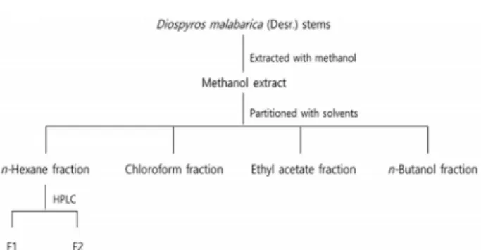
Fig. 1. Isolation procedure of putative antibacterial compound from D. malabarica stems.
물 유래 구강 건강소재의 이러한 문제점을 해결하고 부작용을 최소화할 수 있는 천연물 유래 구강 건강소재에 관한 관심이 고조되고 있으며, 특히 구강 내 세균에 대해 선택적인 항균 효과가 있는 구강 건강소재 개발에 관한 연구와 더불어 구강 내 생물막을 제어할 수 있는 신규 또는 천연 항균소재 개발 등에 관한 연구가 활발히 진행되고 있다[11, 23, 24, 36]. 외국에 서는 치아우식증 억제 물질을 함유한 것으로 양귀비과 식물 (bloodroot), 세이지(sage), 몰약(myrrh), 감초 뿌리(licorice root), 콩과식물의 뿌리혹(quercus infectoria gall), 벌집(vespae nidus), 물레나물과 식물(Cratoxylum formosum), 치커리(chico- ry), 꿀풀(Prunella vulgaris) 등이 보고되었다[25].
인도감나무(Diospyros malabarica)는 인도, 일본, 나이지리 아, 필리핀 등지에 자생하며 아유르베다 의학에서 피부 감염 등으로 인한 작열감, 발열, 설사, 이질, 당뇨, 요로 감염 및 말라 리아에 효과가 있는 것으로 알려져 있다[18, 27]. 인도감나무에 대한 생리활성은 인도감나무 열매의 항산화 활성, 껍질의 항 산화 활성과 항설사 효과 등이 보고되었으나[26, 28] 아직까지 인도감나무가 치아우식증의 원인균인 S. mutans의 생물막에 미치는 영향에 대한 연구는 미흡한 실정이다.
본 연구에서는 천연물 유래 구강 건강소재로써 인도감나무 줄기 추출물의 이용 가능성을 평가하기 위해 TLC, TLC-bio- autography, HPLC, ESI-MS 등을 이용하여 인도감나무 줄기 추출물로부터 S. mutans에 대해 항균활성이 있는 항균물질을 분리하고 주사전자현미경과 real-time PCR을 이용하여 추출 물이 S. mutans의 치태에 미치는 영향을 조사하였다.
재료 및 방법
사용 균주 및 추출물
본 실험에 사용한 균주 Streptococcus mutans KCTC3065는 한국생명공학연구원 생물자원센터(KCTC)에서 분양받아 brain- heart infusion agar (BHIA; BD, Franklin Lakes, USA)에 계대 한 다음 37℃에서 혐기배양하면서 사용하였다. 인도감나무 줄 기 추출물은 메탄올 추출물이며 한국생명공학연구원 해외생 물소재센터(KRIBB, Daejeon, Korea)에서 분양받아 사용하였 다.
인도감나무 줄기 추출물의 유기용매 분획
인도감나무 줄기 추출물로부터 항균물질을 분리하기 위해 추출물은 유기용매의 극성이 낮은 것부터 높은 순서로 Fig.
1과 같이 순차적으로 분획하였다. 인도감나무 줄기 추출물을 멸균증류수에 현탁한 다음 동량의 n-hexane을 가한 후 n-hex- ane 분획과 수용성으로 분획하여 hexane 분획물을 얻었다. 다 시 이 수용성 분획에 chloroform, ethyl acetate, n-butanol을 순차적으로 가하여 chloroform, ethyl acetate, n-butanol, 수용 성 분획물을 얻었다. 모든 분획물은 감압 농축 후 methanol에
용해하였으며 S. mutans KCTC3065에 대한 분획물의 항균활 성은 disc diffusion method [7]에 따라 분획물 농도가 1 mg/
disc인 paper disc (8 mm diameter, ADVANTEC, Tokyo, Japan)를 균주가 도말된 BHI 고체배지에 얹어 배양한 다음 disc 주위에 생성된 clear zone의 크기를 측정하여 확인하였다.
유기용매 분획물의 TLC 및 TLC-bioautography 용매 분획물 중 항균활성을 나타내는 분획물로부터 항균물 질을 분리하기 위해 TLC (Thin layer chromatography)와 TLC-bioautography를 수행하였다. TLC는 TLC plate (Silica gel 60F254, Merck Co.)의 하단으로부터 1 cm 위에 n-hexane 분획물을 점적하였으며 chloroform과 ethyl acetate를 4:1의 비율로 혼합한 전개용매를 이용하여 전개하였다. 전개한 TLC plate는 245 nm와 365 nm에서 UV lamp를 이용하여 spot을 확인하고 spot의 Rf치(Rate of flow value)를 구하였다. S. mu- tans KCTC3065에 대한 각 spot의 항균활성은 Dawanjee 등[8]
의 방법을 이용하여 TLC-bioautography법으로 확인하였다 [4]. 전개가 끝난 TLC plate는 용매를 완전히 제거한 후 S. mu- tans KCTC3065가 도말된 BHI 고체배지에 뒤집어 얹어 배양 한 다음 TLC plate 주위에 생성된 투명환(clear zone)의 크기 를 측정하여 확인하였다.
인도감나무 줄기 추출물의 HPLC와 ESI-MS분석 인도감나무 줄기 추출물로부터 항균물질을 분리하기 위하 여 고성능 액체크로마토그래피(HPLC; High Performance Liquid Chromatography)를 이용하여 n-hexane 분획물로부터 항균물질을 분리하였다. n-Hexane 분획물은 HPLC 이동상에 2 mg/ml의 농도로 용해한 다음 0.2 um membrane filter (ADVANTEC, Tokyo, Japan)로 여과한 후 HPLC에 2 ml를 주입하였다. HPLC는 ODS-AP column (20×500 mm, JAI, Tokyo, Japan)이 장착된 순환분취형 HPLC (recycling prepa- rative HPLC; JAI NEXT, Tokyo, Japan)를 사용하였고 이동상 은 chloroform 과 ethyl acetate (4:1, v/v) 혼합용액을 사용하 였으며 유속은 5.0 ml/min, 검출기는 UV detector (255 nm)를 사용하였다. 분리된 HPLC 분획물의 S. mutans KCTC3065에
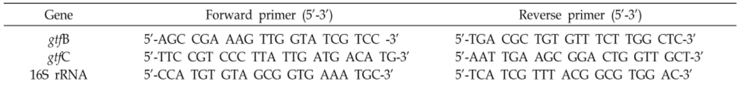
Table 1. Oligonucleotides used for real-time PCR in this study
Gene Forward primer (5’-3’) Reverse primer (5’-3’)
gtfB gtfC 16S rRNA
5’-AGC CGA AAG TTG GTA TCG TCC -3’
5’-TTC CGT CCC TTA TTG ATG ACA TG-3’
5’-CCA TGT GTA GCG GTG AAA TGC-3’
5’-TGA CGC TGT GTT TCT TGG CTC-3’
5’-AAT TGA AGC GGA CTG GTT GCT-3’
5’-TCA TCG TTT ACG GCG TGG AC-3’
대한 항균활성은 disc diffusion method [7]를 이용하여 확인 하였고 항균활성이 확인된 HPLC 분획물의 분자량은 electro- spray ionization–mass spectrometry (ESI-MS; Waters Model 3100, USA)를 이용하여 확인하였다.
주사전자현미경을 이용한 생물막 관찰
인도감나무 줄기 추출물이 S. mutans KCTC3065의 생물막 에 미치는 영향을 알아보기 위해 주사전자현미경(Scanning electron microscope, SEM; JEOL LTD., JSM-6701F, Japan)을 이용하여 추출물 처리에 의한 S. mutans KCTC3065의 바이오 필름 바이오매스(biofilm biomass) 변화를 관찰하였다. Cover- slip (12 mm, SPL Ltd, Korea)이 들어있는 48 well-plate에 OD600=0.4의 밀도로 BHI 액체배지에 현탁한 S. mutans KCTC 3065 배양액을 처리한 후 추출물을 1 mg/ml이 되도록 처리한 다음 37℃에서 33 시간 동안 혐기배양하였다. 배양이 완료된 후 coverslip은 PBS (Phosphate buffer saline, Bioneer, Dae- jeon, Korea) 완충용액에 2회 세척하고 30, 50, 60, 70, 80, 95, 100%의 에탄올을 순차적으로 처리한 다음 100% hexame- thyldisilazane (Sigma Aldrich Co., USA)으로 완전히 탈수한 후 실온에서 건조하였다. 건조된 시료는 platinum으로 코팅한 후 주사전자현미경을 이용하여 추출물을 처리하지 않은 대조 구와 1 mg/ml의 추출물을 처리한 처리구에서 S. mutans KCTC3065의 바이오필름 바이오매스를 각각 5,000배와 10,000 배의 배율로 관찰하였다.
Real-time PCR을 이용한 생물막 생성 관련 유전자 발현 비교 분석
인도감나무 추출물이 S. mutans KCTC3065의 생물막 생성 관련 유전자 발현에 미치는 효과를 검증하기 위해 glucosyl- transferase 유전자를 primer [32]로 사용하여 real-time PCR을 수행하였다. Total RNA는 BHI 액체배지에 현탁한(O.D
600=0.4-0.6) S. mutans KCTC3065에 0.2-1.0 mg/ml 농도의 추 출물을 처리한 다음 37℃에서 배양한 후 배양액을 원심분리하 여 얻은 pellet으로부터 RNeasy Kit (Qiagen, Hillden, Ger- many)를 이용하여 분리하였으며 cDNA는 superscript VILO cDNA synthesis (Thermo Fisher Scientific, USA)를 이용하여 합성하였다. Real-time PCR은 CFX96 touchTM real-time PCR detection system (Bio-Rad, USA)을 이용하여 수행하였으며 실험에 사용된 primer는 Table 1과 같다[32]. PCR 반응은 ssoadvancedTM universal SYBRⓇ green supermix (Bio-Rad,
USA)를 사용하여 95℃에서 30초, 95℃에서 10초, 60℃에서 20 초 씩 35 cycle을 반복하여 수행하였다. PCR 반응 종결 후 melt-curve analysis를 수행하여 유전자 증폭의 정확성을 재확 인하였으며 유전자의 상대적 발현량은 endogenous control로 사용한 S. mutans의 housekeeping gene인 16S rRNA에 대한 gtfB와 gtfC의 상대적 유전자 발현 값으로 나타내었다.
통계처리
모든 실험결과의 평균값과 표준오차는 5회 이상 반복실험 을 수행하여 얻어진 결과를 SAS (Statistical analysis system, USA) program으로 구하였으며 통계적 유의성 검정은 Dun- can’s 다중검정법으로 p<0.05 수준에서 실시하였다.
결과 및 고찰
인도감나무 줄기 추출물의 분획별 항균활성
이전 연구[17]에서 구강 미생물인 P. gingivalis와 S. mutans 에 대해 항균활성이 있음이 확인된 인도감나무 줄기 추출물로 부터 항균물질을 분리하기 위하여 용매의 극성 차이에 따라 n-hexane, chloroform, ethyl acetate, n-butanol, 수용성 분획 물 순으로 단계적으로 분획하여 분획물을 얻었으며 각 분획물 의 용매별 수율은 수용성 분획물(65.8%), n-butanol 분획물 (16.5%), chloroform 분획물(8%), n-hexane 분획물(5.6%), eth- yl acetate 분획물(3.8%) 순으로 나타났다. 각각의 n-hexane, chloroform, ethyl acetate, n-butanol, 수용성 분획물을 감압농 축한 후 disc diffusion method에 따라 S. mutans KCTC3065에 대한 분획물의 항균활성을 확인한 결과, n-hexane 분획물(1 mg/disc)의 항균활성이 가장 우수하였다(Fig. 2). 이상의 결과 로부터 S. mutans KCTC3065에 대해 항균활성이 있는 인도감 나무 추출물의 항균물질은 극성이 낮은 n-hexane에 추출되며 S. mutans KCTC3065에 대하여 비교적 높은 항균활성을 나타 냄을 확인하였다. S. mutans의 생육을 억제하는 오배자 구성물 질의 분리 및 정제에 관한 Shin 등[31]의 연구에서도 S. mutans 에 대해 항균활성이 있는 오배자 추출물은 극성이 낮은 n-hex- ane 분획물이었다.
인도감나무 줄기 추출물로부터 항균물질의 분리 및 정제 S. mutans KCTC3065에 대해 항균활성이 높은 n-hexane 분 획물로부터 항균물질을 분리하기 위해 TLC를 수행한 결과 Rf 치가 0.3, 0.8인 2개 이상의 spot을 확인할 수 있었으며 bio-
Fig. 2. Antibacterial activity of partitioned fraction (1 mg/
disc) with solvents of D. malabarica stems against S. mutans KCTC3065. a: Methanol as a negative control, b: Methanol extracts from D. malabarica stems as a pos- itive control, c: n-Hexane fraction, d: Chloroform frac- tion, e: Ethyl acetate fraction, f: n-Butanol fraction, g:
Water soluble fraction.
Fig. 3. TLC (A) and TLC-bioautography (B) of n-hexane fraction from Diospyros malabarica stems.
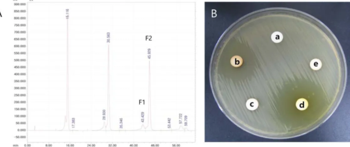
Fig. 4. HPLC chromatogram of hexane fraction from D. malabarica stems (A) and antibacterial activity against S. mutans KCTC3065 of HPLC fraction (1 mg/ml) (B). a: Methanol as a negative control, b: Methanol extracts from D. malabarica stems as a positive control, c: HPLC fraction F1, d: HPLC fraction F2, e: HPLC mobile phase (chloroform ethyl acetate=4:1) a negative control.
autography법을 이용하여 각 spot에 대한 항균활성을 확인한 결과 Rf치가 0.8인 spot에서 항균활성이 확인되었다(Fig. 3).
S. mutans KCTC3065에 대해 항균활성이 우수한 n-hexane 분 획물로부터 recycling preparative HPLC를 이용하여 2개의 분 획물(F1, F2)을 분리하였고 agar diffusion method에 의해 S.
mutans KCTC3065에 대한 항균활성을 측정하였다(Fig. 4). S.
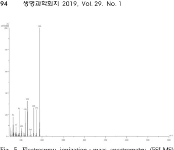
mutans KCTC3065에 대한 HPLC 분획물(1 mg/ml)의 항균활 성은 F2에서만 확인되었으며 F1에서는 항균활성이 나타나지 않았다. 항균활성이 나타난 F2의 구조 분석을 위해 F2를 수집 한 다음 ESI-MS spectrum을 분석한 결과 Fig. 5에서와 같이 molecular ion peak가 m/z 188에서 나타나 분자량이 188로 추정되었다. 그러나 F2 분획물의 순도가 미흡하여 NMR에 의 한 정확한 구조분석은 불가능하였다. 지금까지 인도감나무 줄 기에서 분리된 화합물에 대한 보고는 없으며 이에 대한 추가 적인 연구가 필요할 것으로 사료된다.
주사전자현미경을 이용한 S. mutans KCTC3065의 바이 오필름 바이오매스 관찰
인도감나무 줄기 추출물이 S. mutans KCTC3065의 바이오 필름 바이오매스에 미치는 영향을 알아보기 위해 인도감나무 줄기 추출물을 1 mg/ml의 농도로 처리한 S. mutans KCTC 3065와 추출물을 처리하지 않은 대조구를 주사전자현미경으 로 관찰한 결과는 Fig. 6과 같다. 추출물을 처리하지 않은 대조 구는 추출물 처리구에 비해 세포가 군집을 이루고 모여 있었 으며 세포 주변에 생물막이 형성된 것을 관찰할 수 있었지만 추출물을 1 mg/ml의 농도로 처리한 처리구는 대조구에 비해 세포가 군집을 이루지 않고 독립적으로 존재하고 있었으며 세포 주변에서 생물막을 관찰할 수 없었다(Fig. 6). S. mutans KCTC3065에 의해 형성된 생물막에 대한 glycyrrhetinic acid 의 불안정화 효과에 관한 Yu 등[35]의 보고에 의하면 glycyr- rhetinic acid를 처리하지 않은 대조구의 경우 큰 균 덩어리들 로 구성되어 있는 반면 glycyrrhetinic acid를 15 ug/ml로 처리 한 처리구의 S. mutans KCTC3065는 균 덩어리들이 작게 조각
Fig. 5. Electrospray ionization–mass spectrometry (ESI-MS) spectrum of D. malabarica stems extract.
Fig. 6. Scanning electron microscopy (SEM) micrographs of Streptococcus mutans KCTC3065 biofilms in the absence (A, B) and presence (C, D) of D. malabarica stems extract (1 mg/ml). Magnification is shown by the bar (1 um).
Two different magnifications are shown for each surface;
×5,000 for the upper (A, C) and ×10,000 for the lower images (B, D).
Fig. 7. Effects of Diospyros malabarica stems extract on the genes in relation to the biofilm formation of S. mutans KCTC 3065 by real-time PCR analysis. The genes in relation to the biofilm formation of S. mutans KCTC3065 in the absence and presence of extract of D. malabarica stems.
Results are shown as the SD of five replicates. *p<0.05, as compared with control.
화되어 있으며 생물막의 부피와 두께가 감소한 것을 확인할 수 있었다. 본 연구에서도 추출물을 처리한 처리구의 세포가 군집을 이루지 않고 독립적으로 존재한다는 점에서는 Yu 등 [35]의 보고와 일치하였으며 이는 본 연구자들이 이전의 연구 에서 밝혀낸 인도감나무 줄기 추출물이 생물막 관련 유전자들의 발현을 억제한다는 사실과 연관성이 있는 것으로 보인다[17].
Real-time PCR을 이용한 생물막 생성 관련 유전자 발현 분석
추출물이 S. mutans의 생물막 형성 관련 유전자 발현에 미 치는 영향은 gtfB 유전자와 gtfC유전자를 primer로 이용한 re- al-time PCR로 확인하였으며 그 결과는 Fig. 7에 나타내었다.
Real-time PCR을 이용하여 추출물(0.2, 0.4, 0.6, 0.8, 1.0 mg/
ml) 처리 농도에 따른 gtfB와 gtfC 유전자의 발현 변화를 확인 한 결과, S. mutans KCTC3065의 gtfB 유전자 발현은 추출물의 농도에 따라 큰 변화가 없었지만 gtfC 유전자 발현은 추출물의 농도가 높아질수록 감소하는 경향을 나타내었고 0.8 mg/ml 농도의 추출물과 함께 S. mutans KCTC3065를 배양했을 때 추출물을 처리하지 않고 배양한 S. mutans KCTC3065에 비해 gtfB 유전자 발현이 크게 감소한 것을 확인할 수 있었다. S.
mutans는 다당류 합성과 관련된 3종류의 glucosyltransferase (gtfB, C, D)를 생성하며 gtf 유전자들의 발현은 생물막 형성과 정에서 S. mutans의 치면 부착에 필수적이다. gtfB는 세포 표면 에서 α-1,3 linked liner chain인 비수용성 mutan을 생성하고 gtfC는 주로 치면에서 비수용성/수용성 glucan을 생성하며 gtfD는 α-1,6 linked liner chain인 dextran을 생성한다[9]. Fuji- wara 등[10]의 보고에 의하면 gtfC, gtfD는 초기 부착에 중요하 며 gtfB는 성장 후기에 발현이 증가하면 초기 부착을 강화시키 고 생물막 형성과정에서 S. mutans의 치면 부착에 필수적인 gtfB, C, D 유전자 결손 돌연변이체는 생물막을 형성할 수 없 다. 따라서 인도 감나무 추출물의 처리 농도에 따라 gtf C 유전 자 발현이 감소하였다는 것은 생물막 형성 단계에서 S. mutans 의 초기 치면 부착이 저해됨을 나타내므로 인도감나무 줄기 추출물은 생물막 생성을 억제할 수 있는 천연물유래 구강 건 강소재로써 개발 가능성이 있다고 판단된다.
감사의 글
본 연구는 2017년 경남과학기술대학교 대학회계 연구비 지 원에 의하여 수행된 결과의 일부이며 이에 감사드립니다.
References
1. Bergstorm, J. and Holmberg, B. 1973. The effect of chlorhex- idine emulsion on plaque. An infra individual study of local application. Swed. Dent. J. 66, 461-465.
2. Budtz-Jorgensen, E. and Loe, H. 1972. Chlorhexidine as a denture disinfectant in the treatment of denture stomatitis.
Scand. J. Dent. Res. 80, 457-464.
3. Choi, H. J., Kim, N. J. and Kim, D. H. 2000. Hypoglycemic effect of GE974 isolated from Gyrophora esculenta in normal and diabetic mice. Kor. J. Pharmacogn. 31, 268-272.
4. Choma, I. M. and Grzelak, E. M. 2010. Bioautography de- tection in thin-layer chromatography. J. Chromatogr. A. 1218, 2684-2691.
5. Costerton, J. W., Stewart, P. S. and Greenberg, E. P. 1999.
Bacterial biofilms: A common cause of persistent infections.
Science 60, 756-762.
6. Davies, A. 1973. The mode of action of chlorhexidine. J.
Periodont. Res. 8, 68-75.
7. Davidson, P. M. and Parish, M. E. 1989. Methods for testing the efficacy of food antimicrobials. Food Technol. 43, 148-152.
8. Dewanjee, S., Gangopadhyay, M., Bhattacharya, N., Khanra, R. and Dua, T. K. 2015. Bioautography and its scope in the field of natural product chemistry. J. Pharm. A. 5, 75-84.
9. Ebisu, S., Misaki, A., Kato, K. and Kotani, S. 1974. The struc- ture of water-insoluble glucans of cariogenic Streptococcus mutans, formed in the absence and presence of dextranase.
Carbohydr. Res. 38, 374-381.
10. Fujiwara, T., Hoshino, T., Ooshima, T. and Hamada, S. 2002.
Differential and quantitative analyses of mRNA expression of glucosyltransferases from Streptococcus mutans MT8148.
J. Dent. Res. 81, 109-113.
11. Hassan, S., Danishuddin, M., Adil, M., Singh, K., Verma, P. K. and Khan, A. U. 2012. Efficacy of E. officinalis on the cariogenic properties of Streptococcus mutans a novel and al- ternative approach to suppress quorum sensing mechanism.
PLoS One 7, e40319.
12. Hennessey, T. S. 1973. Some antibacterial properties of chlorhexidine. J. Periodont. Res. 8, 61-67.
13. Huh, M. K. and Kim, H. J. 2014. Antimicrobial effects of sophorae radix extract against oral microorganism. J. Kor.
Soc. Dent. Hyg. 14, 117-122.
14. Iniue, M., Koga, T., Sato, S. and Hamada, S. 1982. Synthesis of adherent insoluble glucan by the concerted action of the two glucosyltransferase components of Streptococcus mutans.
FEBS Lett. 143, 101-104.
15. Kolenbrander, P. E., Palmer, R. J., Periasamy, S. and Jakubovics, N. S. 2010. Oral multispecies biofilm develop- ment and the key role of cell-cell distance. Nat. Rev. Microbiol.
8, 471-480.
16. Kim, H. K., Park, H. W., Shin, I. S., Lee, J. H. and Seo, H.
W. 2008. The antimicrobial effect of horseradish (Armoracia rusticana) root extracts against Streptococcus mutans isolated from human dental plaque. J. Kor. Acad. Pediatr. Dent. 35, 225-234.
17. Kim, H. S., Kwon, H. S., Kim, C. H., Lee, S. W., Sydara, K.
and Cho, S. J. 2018. Effects of methanol extracts from Diospyros malabarica stems on growth and biofilm formation of oral bacteria. J. Life Sci. 28, 110-115.
18. Kumar, S., Shukla, Y. N., Lavania, U. C., Sharma, A. and Singh, A. K. 1997. Medicinal and aromatic plants: prospects for India. J. Med. Arom. Plant Sci. 19, 361-365.
19. Kuramitsu, H. K., He, X., Lux, R., Anderson, M. H. and Shi, W. 2007. Interspecies interactions within oral microbial communities. Microbiol. Mol. Biol. Rev. 71, 653-670.
20. Lee, S. Y., Kim, J. G., Bail, B. J., Yang, Y. M., Lee, K. Y., Lee, Y. H. and Kim, M. A. 2009. Antimicrobial effect of es- sential oils on oral bacteria. J. Kor. Acad. Pediatr. Dent. 36, 1-11.
21. Lim, S. H., Seo, J. S. and Yoon, Y. J. 2003. Effect of leaf- extract from Camellia sinensis and seed-extract from Casia tora on viability of mutans streptococci isolated from the interface between orthodontic brackets and tooth surfaces.
Kor. J. Orthod 33, 381-389.
22. Marsh, P. D. 2001. Dental plaque as a microbial biofilm.
Caries Res. 38, 204-211.
23. Park, S. N., Lim, Y. K., Freire, M. O., Cho, E., Jin, D. and Kook, J. K. 2012. Antimicrobial effect of linalool and α -terpineol against periodontopathic and cariogenic bacteria.
Anaerobe 18, 369-372.
24. Paula, V., Modesto, A., Santos, K. R. and Gleiser, R. 2010.
Antimicrobial effects of the combination of chlorhexidine and xylitol. Br. Dent. J. 209, 19-23.
25. Poureslami, H. 2012. The effects of plant extracts on dental plaque and caries. pp. 396-402. In: Contemporary Approach to Dental Caries, Lim, M.Y. (Ed.). Croatia, In Tech.
26. Pranjal, S. and Debabrat, B. 2014. Phytochemical analysis and antioxidant activity of Gardenia jasminoides ellis and Diospyros malabarica kostel. Int. J. Pharm. Bio. Sci. 5, 199-204.
27. Ramsewak, R. S., Nair, M. G., Stommel, M. and Selanders, L. 2003. In vitro antagonistic activity of monoterpenes and their mixtures against ‘toe nail fungus’ pathogens. Phytother.
Res. 17, 376-379.
28. Rode, M. S., Kalaskar, M. G., Gond, N. Y. and Surana, S.
J. 2013. Evaluation of anti-diarrheal activity of Diospyros ma- labarica bark extract. Bangladesh J. Pharmacol. 8, 49-53.
29. Rosan, B. and Lamont, R. J. 2000. Dental plaque formation.
Microbes Infect. 2, 1599-1607.
30. Schaupp, H. and Wohnaut, H. 1973. Disturbance of taste from oral disinfectants. HNO. 26, 335-341.
31. Shin, A. R., Ohk, S. H., Choi, C. H. and Hong, S. J. 2016.
Identification and partial purification of antibacterial com- pounds against Streptococcus mutans from Galla Rhois. J. Kor.
Acad. Oral. Health 40, 3-8.
32. Takenaka, S., Oda, M., Domon, H., Ohsumi, T., Suzuki, Y., Ohshima, H., Yamamoto, H., Terao, Y. and Noiri, Y. 2016.
Vizantin inhibits bacterial adhesion without affecting bacte- rial growth and causes Streptococcus mutans biofilm to de- tach by altering its internal architecture. Biochem. Biophys.
Res. Commun. 480, 173-179.
33. Wenham, D. G., Davies, R. M. and Cole, J. A. 1981. Insoluble glucan synthesis by mutansucrase as determinant of the car-
초록:Streptococcus mutans에 대한 인도감나무 줄기 추출물의 항균활성 및 생물막 형성 억제 효과
김혜수1․이상우2․콩마니 시다래3․조수정1*
(1경남과학기술대학교 제약공학과, 2한국생명공학연구원, 3라오스 약용식물연구소)
본 연구에서는 천연물 유래 구강 건강소재로써 인도감나무 줄기 추출물의 이용 가능성을 평가하기 위해 TLC, TLC-bioautography, HPLC, ESI-MS 등을 이용하여 인도감나무 줄기 추출물로부터 S. mutans KCTC3065에 대해 항균활성이 있는 항균물질을 분리하고 주사전자현미경과 real-time PCR을 이용하여 추출물이 S. mutans의 생물막 에 미치는 영향을 조사하였다. S. mutans에 대한 인도감나무 줄기 추출물의 항균활성은 극성이 낮은 n-hexane 분 획물에서 확인되었고 TLC, TLC-bioautography, HPLC에 의해 분리된 항균물질의 분자량은 ESI-MS분석 결과 188 로 추정되었다. 인도감나무 줄기 추출물(1 mg/ml) 처리에 따른 S. mutans의 바이오필름 바이오매스 변화는 주사 전자현미경으로 관찰하였으며 추출물을 처리하지 않은 대조구는 추출물 처리구에 비해 세포가 군집을 이루고 모 여 있었으며 세포 주변에서 바이오필름이 관찰되었지만 추출물을 처리한 처리구의 세포 주변에서는 바이오필름 을 관찰할 수 없었다. Real-time PCR을 이용하여 바이오필름 생성 과정에서 치면 부착에 필수적인 GTFs의 발현 양상을 조사한 결과, 인도감나무 추출물이 0.2-1.0 mg/ml의 농도로 처리된 배양액에서 gtfB 유전자 발현은 추출물 의 농도에 따라 큰 변화가 없었지만 gtfC 유전자 발현은 추출물의 농도가 높아질수록 감소하는 경향을 나타내었 다. 이상의 결과를 종합하면 인도감나무 줄기 추출물은 바이오필름 형성 단계에서 치아우식증 원인균인 S. mutans 의 초기 치면 부착을 저해함으로서 바이오필름 생성을 억제할 수 있는 천연물 유래 구강 건강소재로써 이용 가능 성이 높을 것으로 판단된다.
iogenicity of Streptococcus mutans. J. Gen. Microbiol. 127, 407- 415.
34. Whiley, R. A. and Beighton, D. 1998. Current classification of the oral streptococci. Oral Microbiol. Immunol. 13, 195-216.
35. Yu, J. H., Lee, D. M. and Lee, S. H. 2016. Destabilizing effect of glycyrrhetinic acid on pre-formed biofilms of Streptococ- cus mutans. J. Kor. Acad. Oral Health 40, 38-42.
36. Zhiyan, H., Qian, W., Yuejian, H., Jingping, L., Yuntao, J.
and Rui, M. 2012. Use of the quorum sensing inhibitor fur- anone C-30 to interfere with biofilm formation by Streptococ- cus mutans and its luxS mutant strain. Int. J. Antimicrob.
Agents 40, 30-35.
37. Zijnge, V., Leeuwen, M. B., Degener, J. E., Abbas, F., Thurnheer, T., Gmur, R. and Harmsen, H. J. 2010. Oral bio- film architecture on natural teeth. PLoS One 5, e9321.