Gomisin A Ameliorates Endoplasmic Reticulum Stress-induced Hepatic Steatosis
Ye-Rang Yun
1,2and Myeong Ho Jung
1,2*
1
Division of Longevity and Biofunctional Medicine, School of Korean Medicine, Pusan National University, Yangsan 626-870, Korea
2
Healthy Aging Korean Medical Research Center, School of Korean Medicine, Pusan National University, Yangsan 626-870, Korea Received December 29, 2016 /Revised January 12, 2017 /Accepted January 12, 2017
Previously, we have shown that Schisandra chinensis (Turcz.) Baill. (S. chinensis) has a protective effect against endoplasmic reticulum (ER) stress-induced hepatic steatosis. Gomisin A is a bioactive phytoes- trogen derived from S. chinensis. In the present study, the in vitro and in vivo effects of gomisin A on ER stress and hepatic steatosis were investigated. We quantified the expression of markers of ER stress, including glucose regulated protein 78 (GRP78), C/EBP homolog protein (CHOP), and X-box- binding protein-1 (XBP-1), in HepG2 cells treated with tunicamycin or palmitate. Tunicamycin treat- ment in HepG2 cells induced the expression of markers of ER stress, including GRP78, CHOP, and XBP-1c. However, treatment with gomisin A reduced the expression of markers of ER stress. These inhibitory effects were also observed in palmitate-incubated HepG2 cells. The in vivo inhibitory effects of gomisin A were assessed in mice injected with tunicamycin or fed with a high fat diet (HFD). Gomisin A reduced the expression of markers of ER stress and decreased triglyceride levels in the livers of mice after tunicamycin injection or HFD feeding. Furthermore, gomisin A decreased the expression of inflammatory genes in palmitate-incubated HepG2 cells and the liver of HFD-fed obese mice. These results suggest that gomisin A inhibits ER stress and ameliorates hepatic steatosis induced by ER stress.
Key words : Endoplasmic reticulum (ER) stress, gomisin A, hepatic steatosis, high fat diet, inflammation
*Corresponding author
*Tel : +82-51-510-8468, Fax : +82-51-510-8437
*E-mail : [email protected]
This is an Open-Access article distributed under the terms of the Creative Commons Attribution Non-Commercial License (http://creativecommons.org/licenses/by-nc/3.0) which permits unrestricted non-commercial use, distribution, and reproduction in any medium, provided the original work is properly cited.
Journal of Life Science 2017 Vol. 27. No. 2. 233~240 DOI : https://doi.org/10.5352/JLS.2017.27.2.233
Introduction
The endoplasmic reticulum (ER) plays critical roles in the synthesis of secreted and membrane proteins by mediating protein folding, production of lipids and sterols, and the storage of intracellular Ca
2+[22]. However, pathological fac- tors that disrupt ER homeostasis lead to the accumulation of unfolded protein in the ER lumen, provoking ER stress.
Cells usually survive early stress by attenuating protein translation, removing unfolded proteins, and upregulating protein chaperons via the unfolded protein response (UPR) [22]. However, prolonged ER stress can lead to cell death and cause several diseases including ischemia/reperfusion injury, heart disease, and diabetes [1, 3, 23, 26]. Recent stud- ies show that hepatic ER stress is observed in metabolic dis- eases such as obesity and diabetes [1, 3, 23, 26]. ER stress contributes to development of insulin resistance and hepatic steatosis in non-alcoholic fatty liver disease (NAFLD) [2, 17,
18, 20].
NAFLD is a common hepatic disorder that is charac- terized by excessive lipid accumulation in the liver. The first stage consists of hepatic steatosis caused by triglyceride (TG) accumulation in hepatocytes. Symptoms range from simple hepatic steatosis to steatohepatitis, fibrosis, and hep- atocarcinoma[7]. Because the prevalence of NAFLD is in- creasing, it is necessary to develop agents that can prevent hepatic lipid accumulation and treat NAFLD-associated hep- atic disorders. It has been reported that ER stress is an im- portant pathological factor in this pathological process [2, 17, 20]. Thus, an agent that can attenuate ER stress may be a good therapeutic option for the treatment of NAFLD.
The fruit of S. chinensis has been used as a traditional herbal medicine in China, Korea, Japan, and Russia. Several studies have demonstrated the diverse pharmacological ac- tivities of S. chinensis, which include anti-oxidant, anti-tu- mor, anti-obesity, anti-inflammatory, and cardioprotective effects [4-6, 15, 19]. In addition, hepatoprotective activities of S. chinensis have also been reported [8, 9, 14, 25]. S. chi- nensis contains various bioactive constituents, including li- gnans, triterpenoids, polysaccharides, and sterols [10].
Lignans such as deoxyschizandrin, gomisin A, and gomisin N are the main functional constituents of S. chinensis.
Gomisin A was reported to possess hepatoprotective, anti-
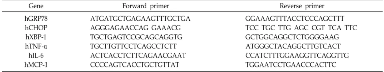
Table 1. List of primers for q-PCR 1. Human
Gene Forward primer Reverse primer
hGRP78 hCHOP hXBP-1 hTNF-α hIL-6 hMCP-1
ATGATGCTGAGAAGTTTGCTGA AGGGAGAACCAG GAAACG TGCTGAGTCCGCAGCAGGTG TGCTTGTTCCTCAGCCTCTT ACTCACCTCTTCAGAACGAAT CCCCAGTCACCTGCTGTTAT
GGAAAGTTTACCTCCCAGCTTT TCC TGC TTG AGC CGT TCA TTC GCTGGCAGGCTCTGGGGAAG ATGGGCTACAGGCTTGTCACT CCATCTTTGGAAGGTTCAGGTTG TGGAATCCTGAACCCACTTC
2. Mouse
Gene Forward primer Reverse primer
mGRP78 mCHOP mXBP-1 mTNF-α mIL-6 mMCP-1
GAAAGGATGGTTAATGATGCTGAG CAGTCATGGCAGCTGAGTCC GAG TCC GCA GCA GGT G CCCTCACACTCAGATCATCTTCT TAGTCCTTCCTACCCCAATTTCC GCATCCACGTGTTGGCTCA
GTCTTCAATGTCCGCATCCTG TAGGTGCCCCCAATTTCATC GTG TCA GAG TCC ATG GGA GCTACGACGTGGGCTACAG TTGGTCCTTAGCCACTCCTTC CTCCAGCCTACTCATTGGGATCA oxidative, and anti-inflammatory effects [11, 13, 16, 24].
In the present study, we investigated the in vitro and in vivo inhibitory effect of gomisin A on ER stress and ER stress-induced hepatic steatosis. The in vitro inhibitory ef- fects were examined in HepG2 cells treated with pharma- ceutical (tunicamycin) or physiological (palmitate) stress inducers. The in vivo protective effects of gomisin A were investigated in tunicamycin-injected mice or high fat diet (HFD) obese mice.
Materials and Methods
Reagents and cell culture
Gomisin A was purchased from ChemFaces (Wuhan, China). Tunicamycin and palmitate were purchased from Sigma-Aldrich (St. Louis, MO, USA). The human hep- atocellular carcinoma cell line HepG2 was obtained from the American Type Culture Collection (Manassas, VA, USA).
HepG2 cells were cultured in Dulbecco’s Minimum Eagle’s Essential Medium (DMEM, Hyclone, South Logan, UT, USA) supplemented with 10% heat-inactivated fetal bovine serum (FBS), 20 U/ml penicillin, and 20 μg/ml streptomycin.
Quantitative PCR (qPCR)
Total RNA was isolated from the liver of the experimental mice and HepG2 cells with TRIzol
TM(Invitrogen, Darmstadt, Germany). cDNA was synthesized using the GoScript Reverse Transcription System (Promega, Madison, Wiscon-
sin, USA) according to the manufacturer’s protocol. The pri- mers (Cosmo Genetech, Seoul, Korea) used in this study are listed in Table 1.
Animal study
C57BL/6 mice (8 weeks of age) were purchased form
Central Lab. Animal Inc. (Seoul, Korea). They were ran-
domly divided into 4 groups (n=5): no treatment group,
treatment with tunicamycin alone, treatment with tunicamy-
cin and a low dose of gomisin A (5 mg/kg body weight),
and treatment with tunicamycin and a high dose of gomisin
A (20 mg/kg body weight). Gomisin A was administered
orally for 4 days. On Day 4, tunicamycin (1 mg/kg body
weight) was administered intraperitoneally for 24 hr via
injection. On Day 5, gomisin A was again administered for
24 hr. To see the protective effects of gomisin A against
HFD-induced hepatic steatosis, C57BL/6 mice were fed a
normal diet (ND) or an HFD for six weeks. Then, the
HFD-fed mice were randomly divided into the following
three groups (n=6 per group): an HFD (distilled water-treat-
ed) group, HFD+low-dose gomisin A (5 mg/kg of body
weight) group, and HFD+high-dose gomisin A (20 mg/kg
of body weight) group. The experimental diets were the
AIN93G-based on the High-fat diet containing 60% kcal fat
and the control diet containing 10% kcal fat. Animal experi-
ments were approved by the Pusan National University
Animal Experiment Ethics Committee and were conducted
in accordance with the institutional guidelines for the care
A
B
Fig. 1. Gomisin A inhibits ER stress in HepG2 cells. (A) q-PCR analysis of markers of ER stress in tunicamycin-treated HepG2 cells. HepG2 cells were pre-incubated in the absence or presence of gomisin A (10, 50, or 100 μM) for 16 hr prior to addition of tunicamycin (2 μg/ml) for 6 hr. TM; tunicamycin, GA; gomisin A. (B) q-PCR analysis of markers of ER stress in palmitate-in- cubated HepG2 cells. HepG2 cells were pre-incubated in the absence or presence of gomisin A (10, 50, or 100 μM) for 16 h prior to adding palmitate (400 μM) for 24 hr. PA; palmitate, GA; gomisin A. Values are expressed as the mean ± SEM (n=3) independent experiments).
#p <0.05,
##p<0.01 vs. untreated control. *p <0.05, **p<0.01 vs. tunicamycin or palmi- tate-treated control.
and use of laboratory animals (ED-PNU2015-0010).
Oil Red O (ORO) staining
Liver specimens were sectioned in blocks and fixed in 10% formalin. After fixation, tissues were dehydrated with a graded series of ethanol and xylene, embedded in paraffin, cut into 3 μm sections. Liver slides were washed with 60%
isopropanol for 5 min and stained with Oil-red O working solution (1.5 mg/ml Oil-red O/60% isopropanol) for 15 min at RT. The slides were washed with distilled water and pho- tographed under a light microscope (Carl Zeiss, DE/Axio Imager A1, Germany).
Biochemical analysis
Serum aspartate aminotransferase (AST) and alanine ami- notransferase (ALT) levels were determined using a com- mercial kit (AM 101-K, Asan Pharmaceutical, Korea).
Hepatic lipids were extracted from the liver according to the following procedure: Briefly, liver tissues were homo- genized in a chloroform-methanol solution (2:1, v/v), and then incubated for 1 hr at room temperature and centrifuged (3,000 rpm, 10 min). The obtained bottom layer (organic phase) was dried overnight. After dissolving in ethanol, hep- atic TG and total cholesterol (TC) were determined using a TG and TC kit (AM 157S-K and AM 202-K, Asan
Pharmaceutical, Korea) and normalized to the protein concentration.
Statistical analysis
Data were expressed as the mean ± SEM. Statistically sig- nificant differences were determined by one-way ANOVA followed by Duncan’s multiple-range tests. For all statistical analyses, p values below 0.05 were considered significant.
Results
Gomisin A inhibits ER stress in HepG2 cells An MTT assay revealed that gomisin A was not cytotoxic to HepG2 cells at a concentration of 100 μM, (data not shown). Then, we investigated the inhibitory effect of gomi- sin A on ER stress in HepG2 cells treated with tunicamycin.
As shown in Fig. 1A, tunicamycin alone increased tran-
scription of markers of ER stress, including GRP78, CHOP,
and XBP-1. In contrast, gomisin A suppressed this induced
transcription in a dose-dependent manner. We then repeated
our investigation of the inhibitory effects of gomisin A
against ER stress in HepG2 cells with palmitate, because tu-
nicamycin is not a physiological inducer of ER stress. As
shown in Fig. 1B, palmitate incubation increased tran-
scription of markers of ER stress such as GRP78, CHOP, and
A
B
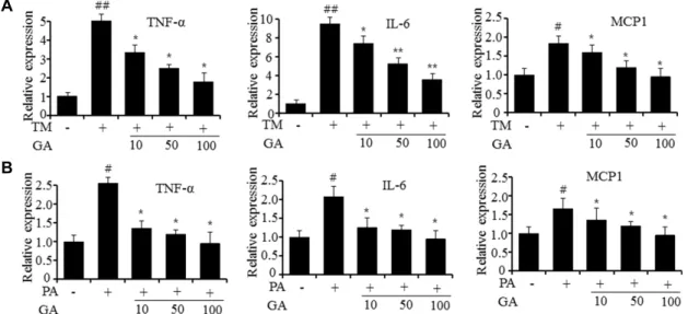
Fig. 2. Gomisin A represses the expression of inflammatory genes in HepG2 cell. (A) qPCR of TNF-α, IL-6 and MCP-1 in tunicamy- cin-treated HepG2 cells. TM; tunicamycin, GA; gomisin A. (B) qPCR of TNF-α, IL-6 and MCP-1 in palmitate-treated HepG2 cells. PA; palmitate, GA; gomosin A. Values are expressed as the mean ± SEM (n=3 independent experiments).
#p<0.05,
##