Received: November 7, 2017 Revised: November 30, 2017 Accepted: December 4, 2017 JOURNAL OF
TRAUMA AND INJURY
CASE REPORT
J Trauma Inj 2017;30(4):231-234 http://doi.org/10.20408/jti.2017.30.4.231
Correspondence to Young Goun Jo, M.D.
Department of Surgery, Chonnam Na- tional University Medical School, 42 Jeb- ong-ro, Dong-gu, Gwangju 61469, Korea Tel: +82-62-220-6456
Fax: +82-62-227-1635 E-mail: [email protected]
http://www.jtraumainj.org eISSN 2287-1683
pISSN 1738-8767
Copyright © 2017 The Korean Society of Trauma
This is an Open Access article distributed under the terms of the Creative Commons Attribution Non-Commercial License (http://creativecommons.org/licenses/by-nc/4.0/) which permits unrestricted noncommercial use, distribution, and reproduction in any medium, provided the original work is properly cited.
Isolated Common Hepatic Duct Injury after Blunt Abdominal Trauma
Yun Chul Park, M.D., Young Goun Jo, M.D., Wu Seong Kang, M.D., Eun Kyu Park, M.D., Hee Jun Kim, M.D., Jung Chul Kim, M.D.
Department of Surgery, Chonnam National University Medical School, Gwangju, Korea
Extrahepatic bile duct injury is commonly associated with hepatic, duodenal, or pan- creatic injuries, and isolated extrahepatic bile duct injury is rare. We report a patient who presented with an isolated extrahepatic bile duct injury after blunt trauma. A 50-year-old man was referred to our hospital after having suffered a fall down injury.
His laboratory findings showed hyperbiliribinemia with elevated aspartate aminotrans- ferase and alanine aminotransferase level. Initial abdominal computed tomography (CT) showed a mild degree of hemoperitoneum without evidence of abdominal solid organ injury. On the 3rd day of hospitalization, the patient complained of dyspnea and severe abdominal discomfort. Follow-up abdominal CT showed no significant inter- val change. Owing to the patient’s condition, Emergency laparotomy revealed a large amount of bile-containing fluid collection and about 1 cm in size laceration on the left lateral side of the common hepatic duct. Primary repair of the injured bile duct with T-tube insertion was performed On postoperative day (POD) 30, endoscopic retro- grade cholangiopancreatography showed minimal bile leakage and endoscopic sphinc- teroplasty and endoscopic retrograde biliary drainage were performed. On POD 61, the T-tube was removed and the patient was discharged.
Keywords: Abdominal injuries; Hepatic duct
INTRODUCTION
Traumatic injuries of the extrahepatic bile ducts are primarily reported in association with a laparoscopic cholecystectomy, and they rarely occur after abdominal trauma [1]. The incidence of extrahepatic bile duct injury is known to be 1-5%. Extrahepatic bile duct injury is commonly associated with hepatic, duodenal, or pancreatic injuries, and isolated extrahepatic bile duct injury is rare [2]. Based on the severity and extent of the injury, multiple treatment modalities such as simple drainage, primary repair
232 http://doi.org/10.20408/jti.2017.30.4.231
Journal of Trauma and Injury Volume 30, Number 4, December 2017
of the injured tissue, and/or enteric anastomosis, are used for the management of this condition [3]. We report a patient who presented with an isolated extrahepatic bile duct injury after a fall and underwent successful repair of the injured bile duct with T-tube drainage.
CASE REPORT
A 50-year-old man was referred to our hospital after having suffered a fall 4 days prior to presentation. He fell from a breakwater over 4 meters high. His bilirubin levels continued to rise while he was hospitalized at an outside clinic, and upon arrival at our hospital, his laboratory findings showed a total bilirubin level of 16.56 mg/dL (reference range [RR] 0.22-1.3 mg/dL), direct bilirubin level of 11.71 mg/dL (RR 0.05-0.3 mg/dL), aspartate aminotransferase level of 152 U/L (RR 10-37 U/L), ala- nine aminotransferase level of 86 U/L (RR 10-37 U/L), as well as hypoalbuminemia. Physical examination showed abdominal distension without any tenderness, and the pa- tient did not complain of abdominal pain. Initial abdom- inal computed tomography (CT), performed at the local hospital, had shown a mild degree of hemoperitoneum;
however, there was no evidence of abdominal solid organ injury. Abdominal CT was repeated at the emergency de-
partment and showed an increased amount of fluid col- lection and edematous thickening of the gallbladder wall (Fig. 1). No other abnormalities were noted in this study.
We planned conservative management, and the patient was admitted to the ward. Although his bilirubin levels decreased after hospitalization, abdominal distension was observed to have slightly increased. On the 3rd day of hospitalization, the patient complained of dyspnea and severe abdominal discomfort. Arterial blood gas analysis showed PO2 level of 64.3 mmHg (RR 75-100 mmHg), PCO2 level of 31.2 mmHg (RR 35-48 mmHg) and bi- carbonate ion level of 23.0 mmol/L (RR 22-26 mmol/L).
Physical examination showed generalized tenderness with rebound tenderness. Follow-up abdominal CT showed the slightly increased amount of fluid collection; owing to the patient’s condition an emergency laparotomy was performed, which revealed a large amount of bile-con- taining fluid collection and inflammatory changes in the gallbladder. We performed a cholecystectomy followed by bile duct exploration, which revealed a longitudinal
Fig. 1. Abdominal computed tomography showed an increased amount of fluid collection and edematous thickening of the gallbladder wall.
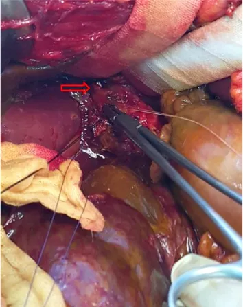
Fig. 2. Longitudinal laceration on the left lateral side of the common hepatic duct was noted after bile duct exploration (red arrow).
233
http://www.jtraumainj.org Yun Chul Park, et al. Isolated Extrahepatic Duct Injury
laceration measuring approximately 1 cm in size on the left lateral side of the common hepatic duct (Fig. 2). We performed primary repair of the injured bile duct with T-tube insertion. We placed two closed suction drains in the sub-hepatic space, followed by closure of the abdom- inal wall. Postoperatively, his bilirubin level gradually decreased. On postoperative day (POD) 7, he complained of abdominal pain, and an abdominal CT examination showed resolution of the fluid collection and an expected level of postoperative changes in the operative bed. Al- though the patient’s condition gradually improved during hospitalization, approximately 200 mL of bile-containing fluid continued to drain through the closed suction drain.
We performed an endoscopic retrograde cholangiopan- creatography (ERCP) on POD 30. Minimal bile leakage was detected, and endoscopic sphincteroplasty and en- doscopic retrograde biliary drainage were performed.
On POD 61, the T-tube was removed after performing a T-tube cholangiography. The drain was removed 10 days later, and the patient was discharged.
DISCUSSION
Isolated extrahepatic bile duct injury following blunt ab- dominal trauma is rare because of the anatomical location and structure of the bile ducts. The exact mechanism of injury is unknown; however, a few hypotheses explain this condition: collision by the spinal column of coronal structure, rupture because of increased internal pressure, and lacerations between the fixed and movable part at the intraductal junction [4-6].
Diagnosis is often delayed because following an injury to the extrahepatic bile ducts, patients usually present with nonspecific symptoms, such as abdominal discom- fort or jaundice. Therefore, this entity should be consid- ered among the differential diagnoses and a thorough clinical examination should be performed in patients presenting with blunt abdominal trauma. A diagnostic peritoneal lavage (DPL) could be performed if a fluid collection without any associated liver injury is detected at the time of the initial abdominal CT. An ERCP or a magnetic resonance cholangiopancreatography could be considered if DPL confirms the presence of bile in the
fluid [7]. It is often difficult to detect injuries to the biliary tract after a laparotomy; however, if bile duct injury is suspected, cholangiography could be a useful tool [8]. In our case, we could not detect the site of injury even after extensive irrigation at first. However, we could find the site after thorough bile duct exploration below the site of the suspected injury.
Similar to the standard approach utilized for the management of other cases of abdominal trauma, ther- apeutic options should be selected based on the patient’s condition. Hemodynamically unstable patients who are unresponsive to initial therapy should undergo urgent surgical intervention and may require damage control surgery. If the patient is stable, surgical treatment can be performed after multiple diagnostic approaches including abdominal CT have been attempted. Surgical treatment of extrahepatic bile duct injury involves the use of various treatment methods based on the location and size of the injured area-an isolated simple drainage procedure, end- to-end anastomosis of the bile ducts, primary repair after debridement with or without T-tube insertion/placement, subsequent enteric diversion after ligation of the bile ducts, and enteric anastomosis with the duodenum or jejunum are the approaches commonly used [3,9]. Re- garding end-to-end anastomosis, there are good results noted, although a high rate of postoperative stricture for- mation is often an area of concern. It is thought to be the result of complex interactions after ischemic changes due to trauma, changes in tissue state after mobilization, and tensions after restoration.
Therefore, biliary-enteric anastomosis should be considered when the high rate of postoperative biliary stricture is expected. After appropriate dissection of the damaged area, if technically good, a biliary-enteric anas- tomosis shows well functional results of more than 85%
of patients in long-term follow-ups [8]. Additionally, a cholecystectomy is often performed at the time of sur- gery, which is often required to treat the inflammatory changes of the gallbladder and formation of gallstones [3,8]. We performed a cholecystectomy in our patient for the management of inflammatory changes of the gallblad- der, and after confirming the injured site, primary repair with T-tube insertion and external drainage was done.
After surgery, to deal with bile leakage, endoscopic biliary
234 http://doi.org/10.20408/jti.2017.30.4.231
Journal of Trauma and Injury Volume 30, Number 4, December 2017
drainage was performed and 2 months later drainage tube was removed.
In conclusion, the extrahepatic biliary injury is a rare entity, which is often difficult to diagnose. Therefore, it is important to perform a thorough evaluation in patients who present with a high index of suspicion for extrahe- patic biliary injury, and appropriate treatment should be instituted based on the extent of the damage.
REFERENCES
1. Ivatury RR, Rohman M, Nallathambi M, Rao PM, Gunduz Y, Stahl WM. The morbidity of injuries of the extra-hepatic biliary system. J Trauma 1985;25:967-73.
2. Parks RW, Diamond T. Non surgical trauma to the extra- hepatic biliary tract. Br J Surg 1995;82:1303-10.
3. Kitahama A, Elliott LF, Overby JL, Webb WR. The extra- hepatic biliary tract injury: perspective in diagnosis and treatment. Ann Surg 1982;196:536-40.
4. Fish JC, Johnson GL. Rupture of duodenum following blunt trauma: report of a case with avulsion of papilla of vater. Ann Surg 1965;162:917-9.
5. Lewis KM. Traumatic rupture of the bile ducts. Ann Surg 1938;108:237-42.
6. Mason LB, Sidbury JB, Guiang S. Rupture of the extrahe- patic bile ducts from nonpenetrating trauma. Ann Surg 1954;140:234-41.
7. Pandey MK, Babu A, Kumar D, Kumar S, Mishra B. Trau- matic common bile duct injury and spontaneous formation of choledochoduodenal fistula managed by cholecystojeju- nostomy: a case report and review of literature. IJSS Case Reports & Reviews 2015;2:12-15.
8. Rodriguez-Montes JA, Rojo E, Martín LG. Complications following repair of extrahepatic bile duct injuries after blunt abdominal trauma. World J Surg 2001;25:1313-6.
9. Bade PG, Thomson SR, Hirshberg A, Robbs JV. Surgical options in traumatic injury to the extrahepatic biliary tract. Br J Surg 1989;76:256-8.