198
©The Korean Society of Food Science and Technology
피부각질형성세포에서 다양한 베리류의 피부노화개선 효과 비교
이수정·최혜란·이진철·박희전·이희권·정종태·이태범*
(재)고창복분자연구소
The Anti-aging Effects of Various Berries in the Human Skin Keratinocyte (HaCaT) Cells
Su Jung Lee, Hye Ran Choi, Jin-Cheol Lee, Hee Jeon Park, Hee Kwon Lee, Jong Tae Jeong, and Tae-Bum Lee*
Gochang Black Raspberry Research Institute
Abstract Ultraviolet B (UV-B) irradiation is a negative factor that induces skin damage, inflammation, and aging. UV- B irradiation induces the inflammatory response through interleukin (IL)-6 and IL-8 expression in keratinocytes. In addition, it induces the production of reactive oxygen species (ROS) and the activation of matrix metalloproteinase-1 (MMP-1), which plays an important role in collagen 1 degradation in the extracellular matrix. We investigated the anti- aging effects of five kinds of berry in human skin keratinocyte (HaCaT) cells using juice of black raspberry (Rubus occidentalis), blueberry wild (Vacciniun angustifolium) and cultivar (Vacciniun corymbosum), black chokeberry (Aronia melanocarpa (Michx.) Elliott), and mulberry (Morus abla). HaCaT cells irradiated with UV-B exhibited increased ROS generation, as well as IL-6, IL-8, and MMP-1 gene expression, when compared to the control cells that were not irradiated with UV-B. However, pre-treatment of berry juice before UV-B irradiation significantly down-regulated the UV-B-induced ROS generation and inflammatory cytokine and MMP-1 expression. The results suggest that all berries have anti-aging effects including lowering inflammatory cytokine levels, ROS generation, and MMP-1 expression in HaCaT cells during UV-B irradiation.
Keywords: berry, ultraviolet B, inflammatory cytokine, reactive oxygen species, matrix metalloproteinase-1
서 론
피부는 다양한 환경적인 요인에 대해 신체를 보호하는 물리적 장벽으로써의 기능을 갖는 가장 첫 번째 기관이며, 다양한 환경 적인 자극은 면역계의 활성화를 일으킨다. 피부노화는 나이가 들 어감에 따라 피부의 생리적 기능감소와 구조적인 변화로 인해 나 타나는 생리적 노화(intrinsic aging)와, 자외선(ultrviolet, UV)과 같 은 외부자극에 의한 화학적 스트레스가 원인이 되는 광노화 (photoaging)로 구분할 수 있다. 피부가 자외선에 노출되면 홍반, 부종, 화상반응, 색소침작, 거칠어짐, 탄력손실 및 주름과 같은 일 련의 피부손상 혹은 노화과정이 촉진된다(1). 자외선은 태양광 스 펙트럼에서 가시광선보다 짧은 파장대인 200-400 nm 영역의 한 부분이다. 자외선은 파장의 길이에 따라 3가지로 분류되는데 장 파장의 UV-A (320-400 nm), 중파장의 UV-B (280-320 nm), 그리 고 단파장의 UV-C (200-280 nm)로 나누어진다(2,3). 그중에서도 특히 UV-B는 염증성 피부질환, 광노화 그리고 암을 유발할 수 있는 가장 중요한 요소로 작용한다(4). UV-B는 피부의 가장 바 깥쪽인 표피층에 주로 작용하여 cytokine과 chemokine의 분비를
통해 염증반응을 일으키며(5-7) 특히 UV-B로 유도된 tumor necro- sis factor-alpha (TNF-α) (8), interleukin-6 (IL-6) 및 interleukin-8 (IL-8)과 같은 pro-inflammatory cytokine이 피부손상에 기여하는 것으로 확인되었다(9). 피부에 UV-B의 노출은 활성산소종(reactive oxygen species, ROS)의 생성을 야기한다(10-12). UV-B 노출에 의 해 케라틴세포에서 지속적으로 생성된 ROS는 효소, 비효소적인 방법에 의해 빠르게 제거되지만, 과생성된 ROS는 피부손상을 일 으킨다. ROS로부터 생성된 free radical은 세포의 구조와 기능의 저하를 초래하며 이는 궁극적으로 세포내 비정상적인 유전자 발 현의 결과를 가져온다. UV-B로 생성된 ROS는 mitogen-activated protein kinase (MAPK) 신호전달 과정에 영향을 미친다. MAPK 는 전사인자인 nuclear factor kappa B (NF-κB)를 활성화 시키는 것으로 잘 알려져 있고, NF-κB는 전사인자인 AP-1 (activation protein 1)의 활성화를 통해 ROS의 생성 그리고 콜라겐 분해를 촉진하여 피부에 손상을 야기한다는 다양한 연구 결과가 보고되 었다(13,14).
Matrix metalloproteinases (MMPs)는 zinc 이온을 포함하는 칼슘 의존적인 endoprotenase로 collagens, elastins, gelatin, matrix gly- coproteins 및 proteoglycan 같은 extracellular matrix (ECM)를 분 해하는 효소로 작용한다(15). MMPs의 활성은 hormones, growth factors, cytokines에 의해 조절이 이루어지며, 최근 연구에 의하면 인간에서 26가지의 MMPs가 존재한다고 보고되었다. 그중에서 특히 MMP-1은 collagenase-1으로 알려져 있으며, MMP-1의 활성 은 collagen 섬유의 분해 원인으로 중요하게 작용한다(5). 자외선 에 의해 MMP-1과 AP-1의 발현이 증가한다는 연구결과가 보고
*Corresponding author: Tae Bum Lee, Gochang Black Raspberry Research Institute, Gochang, Jeonbuk 525-943, Korea
Tel: 82-63-560-5170 Fax: 82-63-563-6680 E-mail: [email protected]
Received August 27, 2013; revised November 25, 2013;
accepted December 9, 2012
된 바 있으며(16), Hwang 등(17)은 UV-B에 의한 MMP-1은 MAPK/NF-κB/AP-1 신호경로를 통해 활성화 된다고 보고하였다.
결국 UV-B로 유도된 inflammatory cytokine의 발현, ROS의 생성 및 MMP-1 mRNA의 발현은 광노화라고 알려진 피부노화에 기 여하며, 심지어 피부암으로도 발전할 수 있다. 베리류는 베리류 특유의 색, 폴리페놀 또는 가장 특별한 안토시아닌과 같은 물질 이 항산화, 항염증 및 항암작용을 일으키는데 중요하게 작용하는 것으로 알려져 있다. 특히 복분자(Rubus occidentalis)는 anthocya- nins (cyanidin 3-sambubioside, cyanidin 3-glucoside, cyanidin 3- xylosylrutinoside, cyanidin 3-rutinoside and pelargonidin 3-rutino- side), flavonoids (flavonols and flavanols), tannins (proanthocya- nidins, ellagitannins and gallotannins), stilbenoids (resveratrol) 및 phenolic acids와 그리고 lignans가 풍부한 것으로 보고되었다(18).
본 실험에서는 인간각질형성세포주인 HaCaT 세포에서 UV-B로 유도된 inflammatory cytokines 발현, ROS 생성 및 MMP-1 mRNA의 유도와 같은 세포내 반응을 베리류 중에서 복분자, 블 루베리(Vaccinium corymbosum, 재배종 및 야생종), 블랙초크베리 (Aronia melanocapa) 및 오디(Morus alba)의 억제활성을 비교해 보고자하였다.
재료 및 방법
세포주 및 세포 배양
인간각질형성세포주인 HaCaT 세포를 56oC에서 30분간 열처리 된 fetal bovine serum (PAA, Etobicoke, ON, Canada) 10%와 항생제(penicillin/streptomycin, Lonza, Walkersville, MD, USA)를 함유한 Dulbecco’s modified Eagle’s media (DMEM, Welgene, Daegu, Korea)를 사용하여 5% CO2, 37oC 세포 배양기에서 배양 하였다.
자외선 B 조사
자외선 조사를 위해 serum free 배지를 세포배양 dish 또는 well 에 채운 후 UV-B lamp (Crosslinker Model CL-1000, UVP, LCC, Upland, CA, USA)를 사용하여 세포에 UV-B를 조사하였다.
HaCaT 세포가 자외선을 받은 뒤 죽지 않고 염증과 산화적 스트 레스를 일으킬 수 있는 적당한 선량을 결정하기 위해 0, 25, 50, 100 mJ/cm2의 선량별로 조사하였다.
베리류 착즙액 제조와 처리
영하 40oC에 보관한 복분자(BRB)를 비롯한 블루베리(재배종, BB (cv.)), 블루베리(야생종, BB (WT)), 블랙초크베리(BCB) 및 오 디(MB)를 각각 10 kg을 24시간 해동 후 착즙포에 담고 손으로 으깨었다. 착즙포에 공기가 들어가지 않도록 준비한 다음 유압식 착즙기(KR-70, Koryeo, Incheon, Korea)를 이용하여 하강버튼을 천천히 누르면서 압력이 2000 psi가 될 때까지 착즙하였다. 복분 자, 블루베리(재배종), 블루베리(야생종), 블랙초크베리 및 오디 착 즙액의 처리는 다음과 같이 실시하였다. Serum free 배지로 dish 또는 well을 한번 washing한 뒤 serum free 배지를 채워 넣고 베 리류 착즙액을 처리하였다.
세포독성 검사
다양한 베리류가 HaCaT 세포에 독성을 미치는지 확인하기 위 해 96 well plate에 well당 4×10⁴ cell/mL로 분주한 후 24시간 동 안 배양하고, 복분자, 블루베리(재배종), 블루베리(야생종), 블랙초 크베리 및 오디 착즙액을 처리한 뒤 24시간 뒤에 methyl thiazolyl
diphenyl tetrazolium (MTT) assay를 실행하였다. MTT assay에는 cell viability assay kit (EZ-Cytox, Dogen, Seoul, Korea)를 이용 하였고, 모든 well에 MTT 용액 10 µL를 가해주고 다시 37oC, 5%
CO2에서 4시간 배양한 뒤 흡광도 측정을 위해 1분 정도 부드럽 게 shaking을 한 뒤 microplate reader (Synergy HT, Bio-Tek, Winooski, VT, USA)를 이용하여 450 nm에서 흡광도를 측정하였다.
유전자 발현 분석을 위한 RNA 분리 및 real-time RT-PCR 베리류가 피부각질형성 세포인 HaCaT 세포에서 inflammatory cytokines mRNA (TNF-α, IL-6, IL-8)와 MMP-1 mRNA의 발현 에 미치는 영향을 확인하기 35 mm culture dish에 3×10⁵ cell/mL 로 분주하고 24시간 배양하였다. 24시간 뒤에 serum free 배지로 한번 washing 한 후 serum free 배지를 2 mL씩 채운 뒤 베리류 착즙액을 500 µg/mL의 농도로 1시간 동안 전처리하였다. 1시간 뒤 배지를 없애고, serum free 배지를 500 µL씩 분주하고 나서, 100 mJ/cm2 세기로 UV-B를 세포에 조사하였다. 이후 serum free 배지에 베리류 착즙액의 농도가 500 µg/mL이 되도록 하여 1.5 mL씩 추가로 분주한 뒤 24시간 배양하였다. 베리류를 처리한 세 포에서 RNA를 추출하기 위해 total RNA extraction reagent인 tri reagent (RNAiso PLUS, Takara, Otsu, Japan)를 이용하였다. Tri reagent 1 mL을 세포에 넣은 후 15초 동안 vortex하였다. 이어 15,000 rpm에서 15분 동안 원심분리한 후 chloroform 200 µL를 넣고 20초간 vortex한 후 원심 분리한 다음 상층액을 새로운 튜 브에 옮겼다. 차가운 isopropanol 동량을 넣고 상온에서 10분 동 안 방치하였다. 이를 다시 15,000 rpm에서 20분간 원심 분리하여 RNA 침전물을 얻었다. RNA 침전물을 75% ethanol로 씻은 후, 침전물을 수 분간 실온에서 건조시켜 0.1% diethyl pyrocarbonate (DEPC) water에 녹였다. RNA 농도(1 OD=40 µg/mL)는 DUR730 spectrophotometer (Beckman, Fullerton, CA, USA)를 이용하여 260 nm에서 측정하였다.
First strand cDNA을 50 mM Tris-HCl (pH 8.3), 75 mM KCl, 3 mM MgCl2, 10 mM DTT (Invitrogen, Carlsbad, CA, USA), 1 U/µL RNasin (Invitrogen), 1 mM each dNTP, oligo (dT)20 100 ng과 MMLV reverse transcriptase (Invitrogen) 200U가 함유된 20 µL의 용액에서 총 RNA 2 µg으로부터 합성하였다. 실시간 PCR 은 Fast Start DNA Master SYBR Green I kit (Roche, Man- nheim, Germany)을 이용하여 Light Cycler 2.0에서 증폭하였다. 사 용한 primer와 PCR 조건은 Table 1, 2와 같으며 유전자의 정량 분석은 Light Cycler Software 4.0 (Roche)을 이용하였다.
Reactive oxygen species (ROS) 측정
피부각질형성 세포인 HaCaT에서 ROS를 유도하기 위해 96 well black plate에 1×10⁵ cell/mL로 분주하고 24시간 배양하였다. 24시 간 뒤에 serum free 배지로 한번 washing 한 후 serum free 배지 를 200 µL씩 채운 뒤 베리류 착즙액을 500 µg/mL의 농도로 1시 간 동안 전처리하였다. 1시간 뒤 배지를 없애고, serum free 배지 를 20 µL씩 분주하고 나서, UV-B를 100 mJ/cm2의 선량으로 세 포에 조사한 뒤 serum free 배지를 180 µL씩 추가로 분주하였다.
이때 배지에 베리류 착즙액의 처리 농도가 500 µg/mL이 되도록 하여 24시간 동안 배양하였다. 이후 ROS 생성량을 측정하기 위 해 dichlorofluorescein diacetate (DCFH-DA) 염색 방법을 사용하 였다(19). 본 실험에서 DCFH-DA의 농도는 2.5 µM로 처리 하였 고, DCFH-DA 처리 후 1시간 뒤에 형광분석 ELISA reader기 (Bio-Tek)를 통해 excitation wavelength 485 nm, emission wave- length 530 nm를 이용해 분석하였다. 실험에 사용된 양성대조군
은 N-acetyl-L-cysteine (NAC, Sigma aldrich, St. Louis, MO, USA)을 사용하였다.
통계분석
모든 분석 자료는 평균±표준오차(mean±SE)로 나타내었으며 실 험결과는 SPSS 12.0K (SPSS Inc., Chicago, IL, USA)를 이용하 여 p<0.05 수준에서 통계처리 하였고 t-test와 Duncan’s multiple range test로 검증하였다.
결과 및 고찰
세포독성 검사
다양한 베리류, 즉 복분자를 비롯한 블루베리(재배종), 블루베 리(야생종), 블랙초크베리 및 오디 추출물의 세포독성여부를 확 인한 결과 최고농도인 500 µg/mL에서 독성을 보이지 않았고(Fig.
1), 이러한 결과를 바탕으로 베리류 착즙액이 HaCaT 세포에 독 성이 없는 것으로 사료되어 500 µg/mL의 농도에서 실험을 진행 하였다.
베리류의 inflammatory cytokines mRNA 발현억제 비교 UV-B가 염증을 유도하는지 확인 해보고자 UV-B의 선량이 증 가할 때 inflammatory cytokines의 발현을 확인해 보았다. 먼저 HaCaT 세포에 다양한 선량별로 UV-B를 조사하여 HaCaT 세포 가 죽지 않는 최대 선량을 확인한 결과 100 mJ/cm2임을 알 수 있 었다(data not shown). 이후 UV-B를 HaCaT 세포에 0, 25, 50, 100 mJ/cm2의 선량별로 조사하여 TNF-α, IL-6 및 IL-8 mRNA의 발현을 확인해 보았다. 그 결과 UV-B의 선량이 증가함에 따라 TNF-α, IL-6 및 IL-8 mRNA의 발현이 증가함을 확인하였다(Fig.
2). 마지막으로 UV-B 선량은 HaCaT 세포가 죽지 않는 최대 선 량인 100 mJ/cm2로 하여 다양한 베리류 착즙액이 UV-B로 유도
된 inflammatory cytokines의 발현에 어떠한 영향을 미치는지 확 인해 보았다. 그 결과 UV-B 100 mJ/cm2 그룹에서 inflammatory cytokines의 발현이 현저히 증가됨을 확인하였고, UV-B 100 mJ/
cm2를 조사한 후베리류 착즙액을 처리한 그룹들에서 증가되었던 inflammatory cytokines의 발현이 현저히 감소되었다(Fig. 3). 특히 복분자와 블루베리(재배종)의 TNF-α 억제활성이 가장 뛰어났다.
IL-6의 경우 블랙초크베리, 블루베리, 복분자 순으로 inflammatory cytokines의 발현 억제 효과를 보여주었고 블루베리 재배종과 야 생종의 IL-6 억제 활성은 비슷한 것으로 나타났다. 또한 블랙초 크베리, 블루베리(야생종), 복분자가 IL-8의 발현을 눈에 띄게 억 제 하였다. Torri 등(20)은 블루베리의 염증억제에 대한 효과를 보 고한 바 있고, 복분자 또한 TNF-α와 IL-6의 억제효과에 대한 연 구에서는 복분자가 염증반응의 매개물질인 nitric oxide (NO), prostaglandin E2 (PGE2) 그리고 inflammatory cytokine 등의 생성 을 억제함으로 염증반응을 완화시켜주는 것으로 판단하였다(21).
항염증에 대한 블랙초크베리의 다양한 연구보고 중에서 Ohgami 등(22)의 in vitro 연구결과에 따르면 블랙초크베리가 NO 생성효 소와 cyclooxygenase-2 (COX-2) 효소를 억제함으로써 염증 유발 에 관여하는 NO, PGE2그리고 TNF-α 생성을 억제하는 것으로 보고하였다. 오디는 한국에서 재배되는 베리류 중 산업화 및 상 품화를 하는데 있어서 가장 우수한 가격경쟁력을 가지고 있다.
따라서 피부노화 연구결과를 바탕으로 기능성 음료로서 복분자 제품을 개발시 오디와의 혼합을 통해 복분자 단독음료와 동일한 피부노화 억제 효과를 증명할 수 있다면 제품의 가격경쟁력에서 우위를 가질 수 있을 것으로 사료된다. 복분자와 오디 복합처리 시의 inflammatory cytokines 발현 억제에 대해 시너지 효과가 있 는지 여부를 확인하기 위해 복분자와 오디를 각각 250 µg/mL씩 함께 처리하였다. 그 결과 복분자 250 µg/mL과 오디 250 µg/mL 의 농도로 동시에 처리할 경우, 복분자와 오디를 각각 500 µg/mL 로 처리한 그룹에 비해 시너지 효과를 보이지 않았지만 오디만 을 처리했을 때보다는 염증성 사이토카인의 발현이 감소됨을 확 인하였다(Fig. 3).
베리류의 MMP-1 mRNA 발현 비교
MMP-1의 발현에 베리류 착즙액이 어떠한 영향을 미치는지 확 Fig. 1. Effect of various berries on the survival of HaCat cell line.
The cells were cultured in the presence of various concentrations of samples for 24 h. Values are shown as means±SE from three independent experiments.
Table 1. PCR primers for real-time RT-PCR Gene Amplicon
(bp) Primer
TNF-α 324 S TCAGCCTCTTCTCCTTCCTG AS TGAAGAGGACCTGGGAGTAG IL-6 124 S AAATGCCAGCCTGCTGACGAAG
AS AACAACAATCTGAGGTGCCCATGCTAC IL-8 179 S ATGACTTCCAAGCTGGCCGT
AS TGTGGTCCCTCTCAATCACTC MMP-1 160 S AGTGACTGGGAAACCAGATGCTGA
AS GCTCTTGGCAAATCTGGCCTGTAA β-Actin 157 S CACTGTGCCCATCTACG
AS CTTAATGTCACGCACGATTTC Table 2. Real-time RT-PCR conditions
Gene Hot start Denaturation Annealing Extension TNF-α
95oC 10 min
95oC 15 s
60oC, 10 s
72oC, 15 s
IL-6 57oC, 10 s
IL-8 60oC, 10 s
MMP-1 62oC, 10 s
β-Actin 60oC, 10 s
인한 결과, UV-B를 100 mJ/cm2로조사한 그룹에서 MMP-1의 발 현이 현저히 증가되었고, UV-B를 조사한 후 다양한 베리류 착즙 액을 500 µg/mL의 농도로 처리 하였을 경우, 베리류 착즙액을 처 리한 모든 그룹에서 MMP-1 mRNA의 발현이 현저하게 감소되 었다(Fig. 4). 모든 베리류 처리 그룹에서 MMP-1 mRNA의 발현 억제효과를 보여주었지만, 그중에서도 특히 블루베리(야생종)의 Fig. 2. Expression of inflammatory cytokines mRNA in a dose- dependent manner by UV-B exposure in the HaCaT cell line.
Values are expressed as mean±SE. * indicates significant difference with the control group (p<0.05).
Fig. 3. Effect of various berries on mRNA levels of inflammatory cytokines by UV-B exposure in the HaCaT cell line. a-g indicates significant differences between groups at p<0.05 by Duncan’s multiple comparison test. BRB: black raspberry juice 500 µg/mL, BB (WT): blueberry (wild type) juice 500 µg/mL, BB (cv.):
blueberry (cultivar) juice 500 µg/mL, BCB: black chokeberry juice 500µg/mL, MB: mulberry juice 500 µg/mL, BRB+MB: black raspberry juice 250µg/mL+mulberry juice 250 µg/mL.
효과가 가장 탁월 하였다. 복분자의 MMP-1 저해활성은 보고된 바 있다(23). 복분자(250 µg/mL)와 오디(250 µg/mL)를 동시에 처 리한 그룹과 복분자 또는 오디를 각각 500 µg/mL로 처리한 그룹 을 비교해본 결과, 복분자와 오디를 동시에 처리한 그룹이 복분 자와 오디만을 각각 처리한 그룹보다 MMP-1 mRNA의 발현이 좀 더 감소하는 효과를 보여주었다.
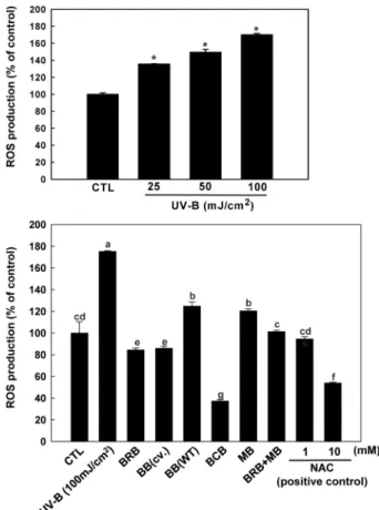
베리류의 ROS 억제활성 비교
UV-B의 조사를 통해 세포손상과 노화의 원인이 되는 ROS를 유도한 뒤, 다양한 베리류의 항산화활성을 비교해보았다. UV-B 선량이 0, 25, 50, 100 mJ/cm2로 증가함에 따라 ROS의 생성량도 눈에 띄게 증가되었고, 특히 100 mJ/cm2의 선량을 조사한 그룹은 대조군에 비해 ROS의 생성량이 약 1.8배 증가 되었다(Fig. 5).
UV-B 조사 후 베리류 착즙액을 처리한 결과 모든 베리류 처리 그룹에서 ROS의 생성이 눈에 띄게 감소되었고, 그중에서도 특히 블랙초크베리의 활성이 가장 뛰어난 것을 확인하였다(Fig. 5).
NAC은 광범위하게 연구가 진행된 ROS 제거제이다(24). NAC은 glutathione의 전구체로 작용하며 직접적으로 H2O2 또는 hydroxyl radical (OH)과 같은 ROS 제거제로 작용하여 ROS를 감소시키거 나 ROS에 의한 산화스트레스 및 염증성 반응을 감소시킨다(25- 27). 양성대조군인 NAC 10 nM보다 블랙초크베리의 효과가 더 높 은 것으로 나타났다. 블랙초크베리에 함유된 안토시아닌 중 cyanidin 3-galactoside, cyanidin 3-arabinoside, cyanidin 3-glucoside, cyani- din-3-O-xyloside 4종이 주요 성분으로 이루어져 있다(28,29). 이런 안토시아닌이 블랙초크베리의 강력한 항산화능력을 나타내는 것 으로 생각되며, 이미 블랙초크베리의 항산화와 항염증에 관한 연 구가 보고된바 있다(30-32). 그 다음으로 높은 ROS 생성 저해 효 과를 보인 그룹은 복분자, 블루베리(재배종)의 순으로 두 착즙액 의 ROS 생성 저해 효과가 비슷한 것으로 나타났다. 복분자의 항 산화활성에 대해 많은 연구결과가 존재하며(33), 복분자의 항산 화 활성은 복분자에 포함된 페놀성 화합물 및 flavonoids류가 관 여할 것으로 추정한다(34-36). 블루베리의 경우 다른 채소와 과일
과 비교했을 때 페놀성 화합물중 anthocyanins의 함량이 높다고 보고되었고(37,38), 이러한 anthocyanins가 ROS 생성억제와 같은 항산화 능력과 관계가 있다는 연구결과가 보고된 바 있다(39,40).
그러나 복분자 250 µg/mL과 오디 250 µg/mL의 농도로 동시에 처 리그룹의 경우 ROS 생성 억제에 대한 시너지 효과를 보이지 않 았다.
요 약
다양한 베리류인 복분자, 블루베리(재배종), 블루베리(야생종), 블랙초크베리 및 오디의 inflammatory cytokines 발현 억제효과, MMP-1 발현 억제활성 그리고 ROS 생성 저해효과를 비교해보았 다. Inflammatory cytokines 발현 억제효과 비교에서 복분자와 블 루베리(재배종)의 TNF-α 억제활성이 가장 뛰어났고, IL-6의 경우 블랙초크베리, 블루베리, 복분자 순으로 inflammatory cytokines의 발현 억제 효과를 보여주었다. 또한 블랙초크베리, 블루베리(야생 종), 복분자가 IL-8의 발현을 눈에 띄게 억제 하였다. 더불어 복 분자와 오디의 inflammatory cytokines 발현 억제에 대해 시너지 효과가 있는지 여부를 확인하기 위해 복분자와 오디를 각각 250 µg/mL씩 함께 처리한 결과 복분자 250 µg/mL과 오디 250 µg/mL Fig. 4. Effect of various berries on expression of MMP-1 mRNA
in HaCaT cell line. a-e indicates significant differences between groups at p<0.05 by Duncan’s multiple comparison test. BRB: black raspberry juice 500 µg/mL, BB (WT): blueberry (wild type) juice 500 µg/mL, BB (cv.): blueberry (cultivar) juice 500 µg/mL, BCB:
black chokeberry juice 500 µg/mL, MB: mulberry juice 500 µg/mL, BRB+MB: black raspberry juice 250 µg/mL+mulberry juice 250 µg/
mL.
Fig. 5. ROS generation in a concentration dependent manner and effects of different berries and NAC on production of ROS by UV-B irradiation in the HaCaT cell line. NAC: N-Acetyl-L- Cysteine. * indicates significant difference with the control group (p<0.05). a-g indicates significant differences between groups at p<0.05 by Duncan’s multiple comparison test. BRB: black raspberry juice 500 µg/mL, BB (WT): blueberry (wild type) juice 500 µg/mL, BB (cv.): blueberry (cultivar) juice 500µg/mL, BCB: black chokeberry juice 500 µg/mL, MB: mulberry juice 500 µg/mL, BRB+MB: black raspberry juice 250 µg/mL+mulberry juice 250 µg/mL.
의 농도로 동시에 처리할 경우, 복분자와 오디를 각각 500 µg/mL 로 처리한 그룹에 비해 시너지 효과를 보이지 않았지만 오디만 을 처리했을 때보다는 염증성 사이토카인의 발현이 감소됨을 확 인하였다. 또한 베리류 착즙액을 처리한 모든 그룹에서 MMP-1 mRNA의 발현이 현저하게 감소되었고, 그중에서도 특히 블루베 리(야생종)의 효과가 가장 탁월하였다. 복분자(250 µg/mL)와 오디 (250µg/mL)를 동시에 처리한 그룹과 복분자 또는 오디를 각각 500µg/mL로 처리한 그룹을 비교해본 결과, 복분자와 오디를 동 시에 처리한 그룹이 복분자와 오디만을 각각 처리한 그룹보다 MMP-1 mRNA의 발현이 좀 더 감소하는 효과를 보여주었다.
ROS 생성저해 효과를 비교한 결과, 블랙초크베리가 가장 뛰어났 고 그 다음으로 복분자, 블루베리(재배종)의 순으로 두 착즙액의 ROS 생성 저해 효과가 비슷한 것으로 나타났다. 그러나 복분자 250µg/mL과 오디 250 µg/mL의 농도로 동시에 처리그룹의 경우 ROS 생성에 대한 시너지 억제효과를 보이지 않았다. 이상의 결 과들로부터 다섯 가지 베리류 즉, 복분자, 블루베리(재배종), 블 루베리(야생종), 블랙초크베리 및 오디의 inflammatory cytokines 과 MMP-1 mRNA 발현 및 ROS 생성에 대한 저해 효과는 각각 의 베리류에서 차이를 보였으나, 다섯가지 베리류 모두 항염증, 항산화 작용을 통해 피부노화 개선에 기여할 것으로 사료된다.
감사의 글
본 연구는 2012년 전라북도 고부가가치식품 가공기술개발 지 원사업에 의한 연구비로 수행된 것으로 이에 감사드립니다.
References
1. Chung JH. Photoaging in Asians. Photodermatol. Photo. 19: 109- 121 (2003)
2. Afaq F, Adhami VM, Ahmad N, Mukhtar H. Botanical antioxi- dants for chemoprevention of photocarcinogenesis. Front. Biosci.
7: 784-792 (2002)
3. Clydesdale GJ, Dandie GW, Muller HK. Ultraviolet light induced injury: Immunological and inflammatory effests. Immunol. Cell Biol. 79: 547-568 (2001)
4. Katiyar SK, Matsui MS, Elmets CA, Mukhtar H. Polyphenolic antioxidant(-)-epigallocatechin-3-gallate from green tea reduces UVB-induced inflammatory responses and infiltration of leuko- cytes in human skin. Photochem. Photobiol. 69: 148-153 (1999) 5. Fisher GJ, Wang ZQ, Datta SC, Varani J, Kang S, Voorhees JJ.
Pathophysiology of premature skin aging induced by ultraviolet light. New Engl. J. Med. 337: 1419-1428 (1997)
6. Di Girolamo N, Wakefield D, Coroneo MT. UVB mediated induction of cytokines and growth factors in pterygium epithelial cells involves cell surface receptors and intracellular signaling.
Invest. Ophth. Vis. Sci. 47: 2430-2437 (2006)
7. Shimauchi T, Sugita K, Nishio D, Isoda H, Abe S, Yamada Y, Hino R, Ogata M, Kabashima K, Tokura Y. Alterations of serum Th1 and Th2 chemokines by combination therapy of interferon-γ and narrowband UVB in patients with mycosis fungoides. J. Der- matol. Sci. 50: 217-225 (2008)
8. Tanaka K, Hasegawa J, Asamitsu K, Okamoto T. Prevention of the ultraviolet B-mediated skin photoaging by a nuclear factor κB inhibitor, parthenolide. J. Pharmacol. Exp. Ther. 315: 624-630 (2005)
9. Yarosh D, Both D, Kibitel J, Anderson C, Elmets C, Brash D, Brown D. Regulation of TNFα production and release in human and mouse keratinocytes and mouse skin after UV-B irradiation.
Photodermatol. Photo. 16: 263-270 (2000)
10. Yoshizumi M, Nakamura T, Kato M, Ishioka T, Kozawa K, Wakamatsu K, Kimura H. Release of cytokines/chemokines and cell death in UVB-irradiated human keratinocytes, HaCaT. Cell
Biol. Int. 32: 1405-1411 (2008)
11. Erden Inal M, Kahraman A, Kken T. Beneficial effects of querce- tin on oxidative stress induced by ultraviolet A. Clin. Exp. Der- matol. 26: 536-539 (2001)
12. Thiele J, Elsner, P. Oxidants and Antioxidants in Cutaneous Biol- ogy. Karger Medical and Scientific Publishers, Basel, Switzer- land. pp. 1-17 (2001)
13. Miyachi Y. Photoaging from an oxidative standpoint. J. Dermatol.
Sci. 9: 79-86 (1995)
14. Kligman LH, Kligman AM. The nature of photoaging: its preven- tion and repair. Photodermatology 3: 215-227 (1986)
15. Verma RP, Hansch C. Matrix metalloproteinases (MMPs): Chemi- cal-biological functions and (Q)SARs. Bioorg. Med. Chem. 15:
2223-2268 (2007)
16. Watanabe H, Shimizu T, Nishihira J, Abe R, Nakayama T, Tan- iguchi M, Sabe H, Ishibashi T, Shimizu H. Ultraviolet A-induced production of matrix metalloproteinase-1 is mediated by macroph- age migration inhibitory factor (MIF) in human dermal fibro- blasts. J. Biol. Chem. 279: 1676-1683 (2003)
17. Hwang YP, Choi JH, Kim HG, Choi JM, Hwang SK, Chung YC, Jeong HG. Cultivated ginseng suppresses ultraviolet B-induced collagenase activation via mitogen-activated protein kinases and nuclear factor NF-κB/activator protein-1-dependent signaling in human dermal fibroblasts. Nutr. Res. 32: 428-438 (2012)
18. Boivin D, Blanchette M, Barrette S, Moghrabi A, Beliveau R.
Inhibition of cancer cell proliferation and suppression of TNF- induced activation of NFκB by edible berry juice. Anticancer Res. 27: 937-948 (2007)
19. Rosenkranz AR, Schmaldienst S, Stuhlmeier KM, Chen W, Knapp W, Zlabinger GJ. A microplate assay for the detection of oxidative products using 2',7'-dichlorofluorescin-diacetate. J.
Immunol. Methods 156: 39-45 (1992)
20. Torri E, Lemos M, Caliari V, Kassuya CA, L. Bastos JK, Andrade SF. Anti-inflammatory and antinociceptive properties of blueberry extract (Vaccinium corymbosum). J. Pharm. Pharmacol.
59: 591-596 (2007)
21. Yang HM, Lim SS, Lee YS, Shin HK, Oh YS, Kim JK. Compar- ison of the anti-inflammatory effects of the extracts from Rubus coreanus and Rubus occidentalis. Korean J. Food Sci. Technol.
Soc. 39: 342-347 (2007)
22. Ohgami K, Ilieva I, Shiratori K, Koyama Y, Jin XH, Yoshida K, Kase S, Kitaichi N, Suzuki Y, Tanaka T, Ohno S. Anti-inflamma- tory effects of aronia extract on rat endotoxin-induced uveitis.
Invest. Ophth. Vis. Sci. 46: 275-281 (2005)
23. Bae JY, Lim SS, Choi JS, Kang YH. Protective actions of Rubus coreanus ethanol extract on collagenous extracellular matrix in ultraviolet-B irradiation-induced human dermal fibroblasts. Nutr.
Res. Pract. 1: 279-284 (2007)
24. Zafarullah M, Li WQ, Sylvester J, Ahmad M. Molecular mecha- nisms of N-acetylcysteine actions. Cell Mol. Life Sci. 60: 6-20 (2003)
25. Tsuji F, Miyake Y, Aono H, Kawashima Y, Mita S. Effects of bucillamine and N-acetyl-L-cysteine on cytokine production and collogen-induced arthritis (CIA). Clin. Exp. Immunol. 115: 26-31 (1999)
26. Rahman A, Kefer J, Bando M, Niles WD, Malik AB. E-selectin expression in human endothelial cells by TNF α-induced oxidant generation and NF-κB activation. Am. J. Physiol. 275: 533-544 (1998)
27. Verhasselt V, Vanden Berghe W, Vanderheyde N, Willems F, Hae- geman G, Goldman M. N-acetyl-L-cysteine inhibits primary human T cell responses at the dentritic cell level: association with NF-kB inhibition. J. Immunol. 162: 2569-2574 (1999) 28. Oszmianski J, Sapis JC. Anthocyanins in fruits of Aronia melano-
carpa (chokeberry). J. Food Sci. 53: 1241-1242 (1988)
29. Wu X, Gu L, Prior RL, McKay S. Characterization of anthocya- nins and proanthocyanidins in some cultivars of ribes, aronia, and sambucus and their antioxidant capacity. J. Agr. Food Chem. 52:
7846-7856 (2004)
30. Matsumoto M, Hara H, Chiji H, Kasai T. Gastroprotective effect of red pigments in black chokeberry fruit (Aronia melanocarpa Elliot) on acute gastric hemorrhagic lesions in rats. J. Agr. Food
Chem. 52: 2226-2229 (2004)
31. Jeong JM. Antioxidative and antiallergic effects of aronia (Aronia melanocarpa) extract. J. Korea Soc. Food Sci. Nutr. 37: 1109- 1113 (2008)
32. Kokotkiewicz A, Jaremicz Z, Luczkiewicz M. Aronia plants: a review of traditional use, biological activities, and perspectives for modern medicine. J. Med. Food 13: 255-69 (2010)
33. Tulio AZ Jr, Reese RN, Wyzgoski FJ, Rinaldi PL, Fu R, Scheer- ens JC, Miller AR. Cyanidin 3-rutinoside and cyanidin 3-xylosyl- rutinoside as primary phenolic antioxidants in black raspberry. J.
Agr. Food Chem. 56: 1880-1888 (2008)
34. Sun P, Ye W, Zheng G, Wang Z, Chen Y, Ogihara Y, Takeda T.
A new flavonol glycoside, epimedin K, from Epimedium kore- anum. Chem. Pharm. Bull. 44: 446-447 (1996)
35. Lee JW, Do JH. Determination of total phenolic compounds from the fruit of Rubus coreanum and antioxidative activity. J. Korean
Soc. Food Sci. Nutr. 29: 943-947 (2000)
36. Wang SY, Lin HS. Antioxidant activity in fruits and leaves of blackberry, raspberry, and strawberry varies with cultivar and developmental stage. J. Agr. Food Chem. 48: 140-146 (2000) 37. Wang H, Cao G, Prior R. Total antioxidant capacity of fruits. J.
Agr. Food Chem. 44: 701-705 (1996)
38. Mazza G, Oomah BD. Herbs, Botanicals and Teas. CRC Press, Boca Raton, FL, USA. pp. 289-318 (2000)
39. Prior RL, Cao G, Martin A, Sofic E, McEwan J, O’Brein C, Lischner N, Ehlenfeldt M, Kalt W, Krewer G, Mainland CM.
Antioxidant capacity as influenced by total phenolic and anthocy- anin content, maturity and variety of Vaccinium species. J. Agr.
Food Chem. 46: 2686-2693 (1998)
40. Kalt W, Forney CF, Martin A, Prior RL. Antioxidant capacity, vitamin C, phenolics, and anthocyanins after fresh storage of small fruits. J. Agr. Food Chem. 47: 4638-4644 (1999)