Hydrogen peroxide로 산화적 스트레스가
유도된 HaCaT keratinocyte에서 금은화의 세포 보호 효과
서승희#, 최미옥*
광주여자대학교 미용과학과
Protectvie effects of Lonicerae Japonicae Flos against hydrogen peroxidase-induced oxidative stress on Human keratinocyte, HaCaT cells.
Seung-Hee Seo
#, Mee-Ok Choi
*Dept. of Beauty Science Graduate School, Kwangju Women's University.
ABSTRACT
Objectives : Lonicerae Japonicae Flos (LJF) has been shown anti-oxidant, anti-inflammatory, anti-viral, anti-rheumatoid properties. However, it is still largely unknown whether LJF inhibits skin injury against oxidative stress in human keratinocyte, HaCaT cells. The purpose of this study was to evaluate the protective effects of LJF against hydrogen peroxide(H2O2)-induced oxidative stress in human keratinocytes, HaCaT cells.
Methods : To evaluate out the protective effects of LJF on oxidative injury in HaCaT cells, an oxidative stress model of HaCaT cells was established under a suitable concentration (500 µM) hydrogen peroxide. HaCaT keratinocyte cells were pre-treated with LJF (0.1, 0.25 or 0.5 mg/ml), and then stimulated with H2O2. Then, the cells were harvested to measure the cell viability, DNA damage, and release of reactive oxygen species (ROS).
Results : LJF (0.1, 0.25 or 0.5 mg/ml) itself did not show any significant toxicity in HaCaT cells. The treatment of H2O2 caused the oxidative stress, leading to the cell death, and DNA injury. However, pretreatment with LJF reduced cell death, and DNA injury. The stimulation of H2O2 on HaCaT cells resulted in excessive release of ROS, which is the main factor of oxidative stress. The excessive release of ROS was inhibited by LJF treatment significantly.
Conclusions : These results could suggest that LJF exhibited the protective effects of HaCaT cells against H2O2-induced oxidative stress by inhibiting ROS release. It could be explained that LJF inhibit skin damages against oxidative stress. Thus, LJF would be useful for the development of drug or cosmetics treating skin troubles.
Key words : Keratinocyte, HaCaT cell, Lonicerae Japonicae Flos, hydrogen peroxide (H2O2), reactive oxygen species (ROS)
서 론1)
금은화(金銀花, Lonicera japonica Flos, LJF)의 기원식물 은 대한약전과 중국약전에 의하면 다양하다. 대한약전에서는 인동과(Caprifoliaceae)에 속한 인동덩굴 Lonicera japonica
Thunb.의 꽃봉오리를 기원으로 하지만 중국약전에서는 인동 덩굴은 물론, 화남인동 (華南忍冬) Lonicera confusa (Sweet) DC, 고선인동 (菰腺忍冬) Lonicera hypoglauca Miq. 황갈모인동 (黄褐毛忍冬) Lonicera fulvotomentosa
*교신저자 : 최미옥, 광주광역시 광산구 여대길 201번지 광주여자대학교 미용과학과 ·Tel : 063-850-6837 ·E-mail : [email protected]
#제1저자 : 서승희, 광주광역시 광산구 여대길 201번지 광주여자대학교 미용과학과 ·Tel : 063-850-6837 ·E-mail : [email protected]
·접수:2013년 6월 19일 ·수정:2013년 7월 3일 ·채택:2013년 7월 19일
Hsu et S. C. Cheng 등을 기원으로 사용한다. 본 연구에서 는 인동덩굴 Lonicera japonica Thunb.을 이용하여 실험을 진행하였다. 한방에서는 性味가 寒하고 甘하며, 心, 肺, 胃經 에 歸經하여 涼散風熱, 擁腫精瘡, 喉痹, 丹毒, 熱血毒痢, 風 血感冒, 溫炳發熱의 效能이 있어서 癰腫疔瘡, 喉痺, 丹毒, 熱 毒血痢, 風熱感冒, 溫病發熱等의 證狀에 常用되고 있다1,2). 또 한, 임상적으로 감염성 질환, 외이도염, 화농증, 중이염과 질 환뿐만 아니라 바이러스성 결막염, 인플루엔자, 폐렴 등의 염 증성 질병에도 사용되고 있다3,4). 이외의 알려진 효능으로 in vitro 모델에서의 연구에서 항균, 항바이러스 작용으로 그람 양성균과 그람음성균에 대해 억제작용이 있으며5), 항산화작용
6), 간보호작용7), 항암작용8), 면역증강9) 등이 연구 보고 되어 있다.
인체 피부는 크게 표피, 진피, 피하조직으로 구성되어 있 다. 이 중에서 각질형성세포(keratinocytes)는 표피층에 존재 하고 있으며, 외부환경으로부터 신체 내부를 보호하거나 보습, 체온조절 등 중요한 역할을 한다10). 하지만 피부 표 피에 해당하는 각질형성세포(keratinocytes)는 바깥층에서 태양광선(자외선)을 비롯한 외부 자극에 직접적으로 노출 되게 되는데 이러한 지속적인 외부 자극의 노출을 통해 산 화적 스트레스(oxidative stress)가 발생하게 된다. 산화적 스트레스(oxidative stress)는 과산화수소 (hydrogen peroxide, H2O2), 수산기(hydroxyl radicals, OH-), 페록실 기(peroxyl radicals, ROO-), 퍼옥시니트라이트 (peroxynitrite, ONOO-) 그리고 과산화물음이온 (superoxide anion, O2-)을 비롯한 다양한 활성산소 (reactive oxygen species, ROS)를 생성하여 각질형성세 포의 탄력 및 보습기능의 저하, 노화의 촉진, 그리고 피부 세포의 염증과 괴사 등을 유발 하는 것으로 알려졌다11,12). 따라서 산화적 스트레스(oxidative stress)로부터 각질형성 세포를 보호하고 노화를 지연시키기 위해서는 체내 항산화 방어 시스템을 향상시키는 것이 매우 중요하다13). 선행연 구들에서 금은화의 항산화, 항염증과 같은 다양한 연구가 진행 되었으나, 피부에서 금은화의 세부적인 효능 연구는 보고된 바가 없었다. 이에 본 연구에서는 각질형성세포 HaCaT keratinocyte에서 과산화수소(hydrogen peroxide, H2O2)로 유발되는 산화적 스트레스에 대한 금은화 물추출 물의 효과를 알아보고자 하였다.
재료 및 방법
1. 재료
1) 약재실험에 사용한 금은화(Lonicerae Japonicae Flos)는 옴니 허브(경북 영천, 대한민국)에서 구입하여 정선해서 사용하였다.
2) 세포주 및 세포 배양
본 실험에 사용된 HaCaT 세포는 Addexobio (San Diego, Califonia, USA)로부터 분양 받아 실험에 사용하였다. HaCaT 세포는 형질 전환된 각질형성세포로 사람 피부의 정상 각질형 성세포와 형태 및 반응 양식이 동일하면서 계대배양이 제한되
지 않아 유지하기가 편한 장점이 있다. 각질형성세포주인 HaCaT 세포주를 10% fetal bovine serum(FBS)과 1%
penicillin/streptomycine을 첨가한 Dulbecco’s modified Eagle’s medium (DMEM) (GibcoBRL, Braunscheig, Germany) 배지를 사용하여 5% CO2, 37℃ 세포 배양기에서 배양하면서 실험에 사용하였다.
2. 방법
1) 약물 추출금은화 100g을 1L에 약탕기(대웅, 한국)로 3시간 가열 추 출한 다음 여과한 후, 동결 건조하여 건조분말 (15.3g)을 얻 었으며 실험을 위하여 4℃에 보관했다 (수율:15.3%). 생리식 염수 녹여 stock solution (100 mg/ml)을 제조하였다. 제조 된 stock solution은 -20℃에 보관하면서 분석방법에 적합 하도록 희석하여 사용하였다.
2) 세포 독성 분석
HaCaT cell의 생존율은 밀집세포의 미토콘드리아 탈수소 효소에 의해 자줏빛 formazan 생성물로 변하는 MTT 환원을 바탕으로 MTT 분석법으로 측정했다. 세포들은 DMEM 배지 에서 5☓104 cells/well의 밀도로 현탁하여 0.1, 0.25, 0.5 mg/ml의 농도로 금은화 물 추출물을 처리 하였다. 24시간 배양 후 5 mg/ml의 농도로 MTT 용액을 첨가하고 다시 30 분 동안 배양하였다. MTT-formazan 생성물은 DMSO 200 µl를 첨가 함 으로써 용해했다. formazan의 양은 용해액을 96well plate에 loading 한 후, 540 nm에 흡수되는 양을 측정함으로서 결정하였다.
3) DNA 응축 분석 (4‘,6-diamidino-2-phenylindole, DAPI) DAPI (Sigma, MO, USA)는 intact cell membrane을 투 과하여 DNA와 결합하게 되고, 이때 358nm(Max.) 정도 파 장대의 빛을 흡수하여 blue-cyan 계통의 형광을 발한다. 그 렇게 결합한 DAPI는 DNA를 염색함으로서, 핵과 chromatin 의 형태를 관찰 할 수 있게 해 준다. HaCaT 세포에 H2O2 를 처리한 후 1시간 동안 배양하였다. PBS로 2회 세척 후 4% paraformaldehyde 용액으로 30분 동안 실온에서 고정하 고 PBS로 3회 세척하였다. 세포의 핵은 DAPI (10 mg/ml) 사용하여 5분간 염색하고 PBS로 3회 세척하였고, prolong gold anti-fading mount solution (invitrogen, CA, USA) 를 이용하여 마운팅하였다. 그 후 Flourscence microscopy (Olympus☓ 70, Tokyo, Japan)으로 관찰하였다.
4) DNA 분절 측정 (1) 세포 DNA분절 샘플 준비
Cellular DNA fragmentation Kit (Millipore, MA, USA) 를 이용하여 DNA의 분절 정도를 측정하였다. 먼저 HaCaT 세포를 2☓105 cells/ml로 현탁하고, bradU labeling solution 을 10 µM을 첨가하여 5% CO2, 37℃ 조건하에서 2시간 배 양 하였다. 그 후 250 g에서 10분간 원심 분리하여 상층액을 제거한 다음, 세포에 DMEM 배지를 각 200 µl씩 첨가하여 96well plate에 넣어 주었다. 실험군에 LJ를 0.1, 0.25, 0.5
mg/ml 농도로 한 후 1시간 뒤, H2O2 500 µM을 처리하고 5% CO2, 37℃ 조건하에서 1시간 배양하였다. 1시간 후에 96well plate를 250 g에서 10분간 원심 분리 후, 상층액을 제거 하였다. 96well plate 각 well에 1☓ incubation solution 200 µl를 첨가한 후, 상온에서 30분간 배양하였다.
그 후 250 g에서 10분간 원심분리하여 나온 상층액을 100 µl 취하여 실험에 사용하였다.
(2) 세포 DNA 분절 분석
96 well plate에 DNA coating solution 100 µl를 첨가한 뒤, 4℃에서 overnight하였다. 현탁액을 털어내어 버린 뒤, 1☓ incubation solution 200 µl를 첨가하여 상온에서 30분 간 blocking 하였다. 그 후, washing buffer 200 µl를 첨가 하여 3회 씻어낸 다음, exonuclease Ⅲ solution 100 µl를 첨가하여 37℃에서 30분간 denaturing 하였다. washing buffer 200 µl를 첨가하여 3회 씻어 낸 다음, anti-Brad-POD conjugate solution을 각 well에 100 µl씩 첨가하여 4℃에 서 overnight하였다. 그 후, washing buffer 200 µl를 첨가 하여 3회 씻어낸 다음, substrate solution 100 µl를 첨가해 DNA가 분절된 정도를 370 nm에서 spectrometer를 이용하 여 흡광도를 측정함으로서 결정하였다.
5) ROS 생성도 분석 (Dichlorofluorescein diacetate, DCF-DA)
세포 내 활성산소의 생성을 측정하기 위하여 형광 probe는 DCF-DA를 이용하였다. 비형광물질인 DCF-DA (Sigma, MO, USA)는 세포내 H2O2와 관련된 peroxide 존재시 ROS 에 의해 산화되어진다. 2,7-dichlorofluorescein로 탈 에스테 르화 된 DCF-DA는 형광의 DCF로 변환되어 녹색의 형광을 띄게 된다. HaCaT 세포에 H2O2를 처리 한 후 1시간 동안 배 양하였다. 세포를 수확하기 전에 2 µM DCF-DA를 처리하여 37℃에서 10분 동안 배양하였다. 배양한 세포는 PBS로 세척 하여 1% trypsine-EDTA 용액을 처리하여 세포를 수확하고, 다시 PBS로 세척하여 flow cytometry (FACS Calibur, BD Biosciences)로 형광을 측정하였다. 정보의 분석은 Cell Quest software( Becton Dickinson)를 이용하였다.
6) 통계 처리
실험 결과에 대한 통계 처리는 SPSS V10.0을 이용하여 one way ANOVA로 검정하여 P값이 0.05 미만일 때 통계적 으로 유의한 차이가 있는 것으로 판정하였다.
결 과
1. 금은화 물추출물이 HaCaT 세포 독성에 미치 는 영향
금은화 물추출물이 사람 각질형성세포인 HaCaT 세포의 대 사 및 독성에 영향을 주어 세포 사멸 및 산화적 스트레스 발 생에 관여할 수 있음을 배제하기 위하여, HaCaT 세포에 농 도별로 금은화를 0.1, 0.25, 0.5, 혹은 1 mg/ml의 농도로
처리하여 3시간 동안 배양한 후 세포 독성 분석법을 실시해 생존율을 관찰하였다. 그 결과, 아래 Fig. 1과 같이 금은화는 HaCaT 세포에서 최고 농도 0.5 mg/ml까지는 유의성있는 세포 독성을 보이지 않았으나, 1 mg/ml의 농도에서는 유의 성있는 세포 독성을 보여, 실험의 농도를 0.1, 0.25, 0.5 mg/ml로 설정하였다 (Fig. 1).
Fig 1. The effects of Lonicerae Japonicae Flos (LJF) extracts on cell viability in human keratinocyte HaCaT cells. HaCaT cells were incubated with or without LJF indicated doses (0.1, 0.25, 0.5 or 1 mg/ml) for 3 h. The cell viability was measured by MTT assay.
The results sere similar in 3 additional experiments. *p < 0.05 significant as compared to untreated normal.
2. 금은화 물추출물이 H
2O
2로 유도한 HaCaT 세포의 독성에 미치는 영향
활성산소의 한 종류로 잘 알려 진 H2O2는 직접적으로 사 람의 각질형성세포 내에서 산화적 스트레스를 유발한다고 알 려져 있다12,13). 따라서, 먼저 H2O2처리 후 사람 각질형성세 포인 HaCaT 세포에 어떠한 영향을 미치는지 알아보고, 금은 화의 효과를 살펴보고자 하였다. HaCaT 세포에 금은화를 0.1, 0.25, 0.5 mg/ml 농도별로 처리하여 1시간 동안 배양 한 후 H2O2 500 µM 을 2시간 처리 한 뒤 세포 생존률을 살 펴보았다. 그 결과 H2O2 500 µM을 처리하였을때 감소하던 세포 생존률이 금은화 처리 군에서 농도 의존적으로 증가한 것을 확인 할 수 있었다 (Fig. 2).
Fig 2. The effects of LJF extracts on the H2O2-induced cytotoxicity in human keratinocyte HaCaT cells. HaCaT cells were pre-treated with or without LJF (0.1, 0.25, or 0.5 mg/ml) for 1h, then induced with H2O2 (500 µM). After 2 h, the cell viability was measured by MTT assay. The results were similar in 3 additional experiments. *p < 0.05 significant as compared to untreated normal, †p < 0.05 : significant as compared to H2O2 alone.
3. 금은화 물추출물이 H
2O
2로 유도된 산화적 스 트레스로 인한 세포 내 DNA 손상에 미치는 영향
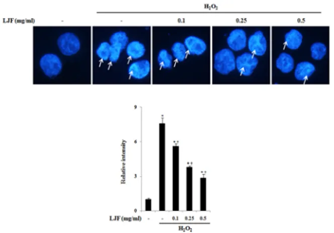
H2O2 처리 시 나타나는 세포 DNA 손상에 금은화가 주는 영향을 알아보기 위하여, 금은화를 농도별(0.1, 0.25, 0.5
mg/ml)로 1시간 전 처리 하고, H2O2 (500 µM)을 1시간 처 리하였다. Fig. 3에서와 보는 바와 같이 정상군에 비하여 대 조군인 H2O2 처리 군에서 핵의 응축이 일어나는 것을 확인 할 수 있었다. 하지만 금은화를 0.1, 0.25, 0.5 mg/ml을 처 리 하였을 때 H2O2 (500 µM)처리로 인한 핵의 응축을 유의 하게 감소되는 것을 볼 수 있었다 (Fig 3).
Fig 3. The effects of LJF on the H2O2-induced cell death in human keratinocyte HaCaT cells. The cells were pre-treated with or without LJF (0.1, 0.25, or 0.5 mg/ml) for 1h, and then induced with or with out H2O2 (500 µM). After 1 h, the cells were harvested for detection of DAPI. To find our the DNA fragment, the cells were stained with DAPI. Detail method were described Materials and Method. The similar results were obtained from three additional experiment. *p < 0.05 significant as compared to untreated normal,
†p < 0.05 : significant as compared to H2O2 alone.
4. 금은화 물추출물이 H
2O
2로 유도된 산화적 스 트레스로 인한 DNA 분절에 미치는 영향
H2O2 처리 시 나타나는 세포 DNA 분절에 금은화가 주는 영향을 알아보기 위하여, 금은화를 0.1, 0.25, 0.5 mg/ml을 H2O2 처리 1시간 전에 전 처리 하고, 1시간 후 H2O2 (500 µM)을 1시간 처리하였다. 그 결과 정상군에 비하여 대조군인 H2O2 처리 군에서 DNA분절이 일어나는 것을 확인 할 수 있 었다 (Fig 4). 하지만 금은화를 0.1, 0.25, 0.5 mg/ml을 처 리 하였을 때 H2O2 (500 µM) 처리로 인한 DNA 분절을 유 의하게 감소시킬 수 있었다 (Fig. 4).
Fig 4. The effects of LJF on the H2O2-induced DNA fragmentation in human keratinocyte HaCaT cells. The cells were pre-treated with or without LJF (0.1, 0.25, or 0.5 mg/ml) for 1h, and then induced with or without H2O2 (500 µM). After 1 h, the cells were harvested for detection of DNA fragmentation. The cells were visualized by DNA fragmentation assay. Detail method were described Materials and Method. The similar results were obtained from three additional experiment. *p < 0.05 significant as compared
to untreated normal, †p < 0.05 : significant as compared to H2O2
alone.
5. 금은화 물추출물이 H
2O
2로 유도된 산화적 스 트레스로 인한 ROS 생성에 미치는 영향
H2O2 처리 시 ROS 생성이 활성화 되고, ROS 분비가 사 람 각질 형성세포의 사멸 및 염증 반응을 촉진 한다는 보고에
따라서14,15), 금은화가 ROS의 생성에 어떤 영향을 주는지 알
아보기 위하여, 금은화를 농도 별(0.1, 0.25, 0.5 mg/ml)로 1시간 전에 전처리 하고, H2O2 500 µM을 처리 하였다.
H2O2 처리 1시간 후 세포에 DCF-DA를 염색하여, ROS의 생성을 관찰 하였다. 그 결과 정상군 보다 H2O2 (500 µM) 처리군에서 ROS 생성이 크게 증가하였다. 하지만 금은화를 처리 하였을 때 H2O2 처리로 인한 ROS 생성이 농도 의존적 으로 유의하게 억제됨을 확인 할 수 있었다 (Fig. 5).
Fig 5. The effects of LJF on the hydrogen peroxide-induced ROS production in human keratinocyte HaCaT cells. The cells were pre-treated with or without LJF (0.1, 0.25, or 0.5 mg/ml) for 1 h, and then induced with or without H2O2 (500 µM). After 1 h, the cells were harvested for detection of ROS. Then the ROS production were measured by DCFDA positive cell percent. Detail methods were described Materials and Methods. The similar results were obtained from three additional experiments. *p < 0.05 significant as compared to untreated normal, †p < 0.05 : significant as compared to H2O2 alone.
고 찰
최근 삶의 질 개선과 함께 피부 관련 분야에 대한 관심이 높아지면서 외부 환경적 요인에 의한 피부 노화를 개선시키기 위해 화장품, 의약품 등을 비롯한 다양한 분야에서 항노화, 항산화에 대한 연구가 활발하게 이루어지고 있다. 자외선을 비롯한 지속적인 외부자극에 피부가 노출될 경우 산화적인 피 부손상은 물론 광노화를 유발한다고 알려져 있다16). 외부 자 극에 의한 피부 노화는 피부의 각질형성세포와 섬유아세포를 활성화시켜 유해한 활성산소(ROS)를 생성하여 산화적 스트레 스를 유발하며 결국에는 피부세포의 손상은 물론 나아가서 피 부의 퇴화나 사멸을 초래하게 된다고 알려져 있다17,18). 각질 형성세포라 불리는 Keratinocytes는 피부에 있어 다양한 외 부환경과 자극으로부터 피부를 보호해 주고, 수분 등의 내부 물질이 피부 밖으로 소실되지 않도록 방어하는 중요한 역할을 한다. 그러므로 피부 최외곽에서 다양한 스트레스에 노출되는 keratinocytes에서 산화적 스트레스에 대한 방어 시스템은 중요한 의미를 가진다. 하지만 과도한 산화적 스트레스에 의
해 keratinocyte가 손상되면, 방어 시스템의 붕괴가 와서 피 부에 손상을 입게 된다. 따라서, 본 연구에서는 항산화, 항염 증에 유용하게 사용되어온 금은화를 이용하여 HaCaT keratinocytes에서 항산화 활성을 평가하고 세포 손상을 억 제하는 효과를 탐색하였다.
금은화는 오래전부터 한의학에서는 호흡기 질환, 관절염 치료, 해독 및 해열제로 사용되어 왔으며19), 이러한 주요 효 능은 금은화에 함유되어 있는 항산화 물질에 의한 것임이 밝 혀져, 최근에는 금은화의 함유 성분들과 그 생리활성에 대한 연구가 진행되고 있다20). 또한, 최근 진행된 연구로는 금은화 에틸아세테이트 분획물의 돌연변이 생성억제21) 및 항산화효과
22)에 관한 내용과 금은화 주요 함유 성분들의 HIV-1 RT 저 해23), phospholipase A2 저해24) 등이 보고되었다. 그러나 피부세포에서의 금은화의 효과에 관한 국내외 연구는 미미한 실정이며, 특히 각질형성세포에서의 항산화 효과에 관련된 연 구는 진행된 바가 없었다. 본 실험에서는 금은화 물추출물의 항산화 활성을 평가하기 위해서 HaCaT keratinocytes에 활 성산소의 한 종류로 잘 알려 진 H2O2 (500 µM)를 이용하여 산화적 스트레스를 유도하였다.
H2O2는 산화적 스트레스 뿐만 아니라 산소 radical의 모든 부분에 관여하는 것으로, 대부분의 세포와 조직에 자유롭게 분포한다25). 또한 H2O2는 다양한 신호기전에 관여하여 생리 활성에 주요한 작용을 한다26,27). 최근 많은 연구에서는 H2O2
를 통한 세포 사멸에 초점을 맞추고 있다28,29). H2O2는 세포 사멸 과정에서 형태학적인 세포 사멸, 세포 수축, DNA 분절 등 다양한 변화를 일으킨다30). 이러한 변화에는 다양한 원인 이 존재하지만, ROS가 주요하게 작용한다고 알려져 있다
31,32)
. ROS는 DNA의 구조를 붕괴시키면서 DNA를 손상시키 고, 세포 소기관들을 다양하게 공격하면서 세포 사멸 및 염증 을 유도하는 것으로 알려져 있다33-35). 고로 ROS를 통한 세 포 손상 및 사멸을 억제하는 것은 세포 보호에 있어서 아주 중요한 의미를 가진다. 본 연구에서는 기존의 연구와 일치하 게 H2O2가 세포 사멸을 증가함을 알 수 있었다. 또한 H2O2
는 DNA chromatin 응축 및 DNA 분절 등을 유도하여 세포 손상을 유도하였다. 이에 금은화 물 추출물을 전 처리한 후, H2O2를 통하여 과도한 산화적 스트레스를 주었을 때, 이러한 세포 사멸 및 손상을 농도 의존적으로 억제함을 알 수 있었다.
또한 H2O2에 의한 과도한 ROS생성도 금은화에 의해 억제됨 을 보았을 때, 금은화는 H2O2에 의한 ROS 생성을 억제하여서 추가적인 세포 사멸 및 손상을 억제함을 유추할 수 있다.
본 연구에서는 금은화 물 추출물의 사람 각질형성 세포에 서의 항산화 효과를 통한 세포 보호 효과를 살펴보고자 하였 으며, 그 결과 금은화 물 추출물은 각질형성세포에서의 항산 화, 세포보호 효과가 우수 하였다. 따라서, 본 연구 결과는 추후에 금은화를 이용한 피부에서의 다양한 효능 및 기전 연 구를 하는데 기초적인 자료가 될 것으로 판단된다. 또한, 추 후 연구에서 금은화 물 추출물의 피부에 대한 세부 기전 연구 및 활성 성분 분석에 대한 추가적인 실험이 필요할 것으로 사 료된다.
결 론
금은화 물추출물을 이용하여 사람각질형성세포인 HaCaT keratinocyte에서 H2O2로 유발한 산화적인 손상에 대한 효 과를 관찰한 결과 다음과 같은 결론을 얻었다.
1. 금은화 물추출물은 HaCaT keratinocyte에서 0.5 mg/ml에서까지 유의성 있는 세포 독성을 보이지 않았 으며, H2O2로 유발한 산화적 손상에 대해 농도 의존적 으로 세포 생존률을 증가 시켰다.
2. 금은화 물추출물은 HaCaT keratinocyte에서 H2O2로 유발한 DNA chromatin의 응축 및 DNA 분절을 감소 시켰다.
3. 금은화 물추출물은 HaCaT keratinocyte에서 H2O2로 유발한 ROS의 생성을 농도 의존적으로 억제 하였다.
이상의 결과, 금은화 물추출물은 사람각질형성세포인 HaCaT keratinocyte에서 H2O2로 유발한 산화적인 스트레스에서 세 포 보호 및 ROS 생성 억제 효과가 우수하게 나타났으며, 이 와 같은 결과를 바탕으로 금은화 물추출물이 피부의 산화적 손상을 개선하는데 주요하게 작용할 수 있을 것으로 추측된 다. 추후에 금은화 물추출물의 피부에서 산화적인 손상을 억 제하는 구체적인 기전 연구가 추가적으로 필요할 것으로 사료 된다.
Reference
1. Schools of Korean Medicine Textbook complolation committee. Bonchohak. Seoul : Yeonglimsa. 2007 : 240-1.
2. Ahn SH, Kim HH. Lonicerae Flos inhibited COX-2 and MMP-9 in LPS induced Arthritis of mouse through regulation of MIF. Kor J Ori Med Phywiol Pathol. 2010 ; 24(2) : 242-8.
3. Dan Bensky, Andrew Gamble. Chinese herbal medicins. Materia Media. WA : Eastland Press.
1986 : 85-6.
4. Huang KC. The Pharmacology of Chinese herbs (2nd Ed.). FL : CRC. 1998 : 8-9.
5. Bae JH, Kim MS, Kang EH. Antimicrobial Effect of Lonicerae Flos Extracts on Food-borne Pathogens.
Food Sci Biotechnol. 2005 ; 37(4) : 642-7.
6. Joo JS, Kim JS, Jeong JG, Kim BK. Study of efficacy of Foeniculi fructus and Lonicerae flos extract on acute pancreatitis. Kor J Herbology.
2010 ; 25(4) : 39-45.
7. Ohata S, Sato N, Tu SH, Shinoda M, Protective effects of Taiwan crude drugs on experimental liver injuries. Yakugaku Zasshi. 1993 ; 113(12) : 870-80.
8. Rim BM, Rim CW, Choi JY. Chung YS, Jeong HG.
Effects of Lonicera japonica extract as a biological
response modifier. Environ Mut Car. 1992 ; 12(1) : 45-54.
9. Luo ZH. The combined modulating effects of cerium nitrate with certain Chinese traditional drugs on altered cell-mediated immunities in scald mice. Zhonghua Wai Ke Za Zhi. 1990 ; 28(9) : 562-5, 574-5.
10. Halliwell B, Gutteridge JM. The importance of free radicals and catalytic ions in human diseases.
Mol Aspects Med. 1985 ; 8(2) : 89-193.
11. Kim ES, Kim JS, Kim GN. Anti-oxidant Function of Genistein against H2O2-induced Oxidative Stress in HaCaT Keratinocytes. Kor J Aesthet Cosmetol.
2012 ; 10(3) : 541-7.
12. Cross CE, Halliwell B, Borish ET, Pryor WA, Ames BN, Saul RL, McCord JM, Harman D.
Oxygen radicals and human disease. Ann Intern Med. 1987 ; 107(4) : 526-45.
13. Applegate LA, Noel A, Vile G, Frenk E, Tyrrell RM. Two genes contribute to different extents to the heme oxygenase enzyme activity measured in cultured human skin fibroblasts and keratinocytes:
implications for protection against oxidant stress.
Photochem Photobiol. 1995 ; 61(3) : 285-91.
14. Frank RG, de Gruijl. Photocarcinogenesis: UVA vs.
UVB radiation. Skin Pharmacol Appl Skin Physiol.
2002 ; 15(5) : 316-20.
15. Heck DE, Vetrano AM, Mariano TM, Laskin JD.
UVB light stimulates production of reactive oxygen species: unexpected role for catalase. J Biol Chem, 2003 ; 278(25) : 22432-6.
16. Dimri GP, A Testori, M Acosta, J Campisi. Replicative senescene, aging and growth-regulatory transcription factors. Biol Signals. 1996 ; 5(3) : 154-62.
17. Chiba K, Sone T, Kawakami K, Onoue M. Skin roughness and wrinkle formation induced by repeated application of squalene monohydroperoxide to the hairless mouse, Exp Dermatol. 1999 ; 8(6) : 471-9.
18. Leonard S, S Wang, L Zang, V Castranova, V Vallyathan, X Shi. Role of molecular oxygen in the generation of hydroxyl and superoxide anion radicals during enzymatic Cr(VI) reduction and its implication to Cr(VI)-induced carcinogenesis carcinogenesis. J Environ pathol toxicol oncol. 2000 ; 19(1-2) : 49-60.
19. Lee J, Jeong SI, Jang SI. Effects of aqueous extract from Lonicera japonica flower on trimellitic anhydride-induced contact hypersensitivity in BALB/c mice. Kor J Herbology. 2008 ; 23(2) : 51-8.
20. Yoo JM, Baik IH, Kim JH, Kim SR, Rhyu MR.
Screening of the antioxidant activity of some medicinal plants. Korean J Food Sci Technol. 2005
; 36(2) : 333-8.
21. Chung KC, Kwon DY, Baek SH, Kim SH, Chang HW. Effect of Lonicera Flos's ethyl acetate fraction on mutagenicity. Yakhak Hoeji. 1988 ; 32 : 328-33.
22. Choi CW, Jung HA, Kang SS, Choi JS. Antioxidant constituents and a new triterpenoid glycoside from Flos Lonicerae. Arch Pharm Res. 2007 ; 30(1) : 1-7.
23. Chang CW, Lin MT, Lee SS, Liu KC, Hsu FL. Lin JY. Differential inhibition of reverse transcriptase and cellular DNA polymerase-alpha activities by lignans isolated from Chinese herbs, Phyllanthus myrtifolius Moon, and tannins from Lonicera japonica Thunb and Castanopsis hystrix. Antiviral Res. 1995
; 27(4) : 367-74.
24. Chang HW, Baek SH, Chung KW, Son KH, Kim HP, Kang SS. Inactivation of phospholipase A2 by naturally occurring biflavonoid, ochnaflavone. Biochem Biophys Res Commun. 1994 ; 205(1) : 843-9.
25. Barbouti A, Doulias PT, Nousis L, Tenopoulou M, Galaris D. DNA damage and apoptosis in hydrogen peroxide-exposed Jurkatcells: bolus addition versus continuous generation of H2O2. Free Radic Biol Med. 2002 ; 33 : 691– 702.
26. Sundaresan M, Yu ZX, Ferrans VJ, Irani K, Finkel T. Requirement for generation of H2O2 for platelet-derived growth factor signal transduction.
Science 1995 ; 270 : 296– 9.
27. Forman HJ, Torres M. Redox signaling in macrophages.
Mol Aspects Med. 2001 ; 2 : 189–216.
28. Lee YJ, Shacter E. Hydrogen peroxide inhibits activation, not activity, of cellular caspase-3 in vivo. Free Radic Biol Med. 2000 ; 29 : 684–92.
29. Chandra J, Samali A, Orrenius S. Triggering and modulation of apoptosis by oxidative stress. Free Radic Biol Med. 2000 ; 29 : 323– 33.
30. Hengartner MO. The biochemistry of apoptosis.
Nature. 2000 ; 407 : 770–6.
31. Henselei U, Rosenbach T, Kolde G. Induction of apoptosis in human HaCaT keratinocytes. Arch Dermatol Res. 1996 ; 288 : 676– 83.
32. Mathiasen IS, Jaattela M. Triggering caspase-independent cell death to combat cancer.
Trends Mol Med. 2002 ; 8 : 212–20.
33. Halliwell B. The wanderings of a free radical.
Free Radic Biol Med. 2009 ; 46 : 531-42.
34. Droge W. Free radicals in the physiological control of cell function. Physiol Rev. 2002 ; 82 : 47-95.
35. Thannickal VJ, Fanburg BL. Reactive oxygen species in cell signalling. Am J Physiol Lung Cell Mol Physiol. 2000 ; 279 : L1005-L1028.