58 서 론
플라보노이드(flavonoid)는 페놀 복합체(phenolic compound) 이며 디페닐프로판(diphenylpropane: C6C3C6) 골격(skeleton)
에 의해 특성화된 구조를 가진다. 이 구조는 단량체 플라 바놀(monomeric flavanol), 플라본(flavone), 플라바놀(flava- nol), 플라바논(flavanon) 등에서 나타나고 있는데, 이러한 모든 화합물은 암의 억제(cancer prevention)을 포함한 여 러 인간의 건강 증진에 잠재적인 역할을 알아내기 위한
인간 Keratinocyte HaCaT 세포에서
에토포사이드 또는 과산화수소에 의해 유도되는 아폽토시스에 미치는 플라보노이드들의 영향
건국대학교 동물생명공학과
이응룡․안재연․강용진․최태원․조쌍구1
Effects of Flavonoids on the Etoposide- or Hydrogen Peroxide-Induced Apoptosis in Human Keratinocyte HaCaT Cells
Eung-Ryoung Lee, Jae Yeon Ahn, Yong-Jin Kang, Tae-Won Choi and Ssang-Goo Cho1 Department of Animal Biotechnology and RCTCP, Konkuk Unitersity, Seoul 143-701, Korea
Apoptosis is a fundamental cellular activity which allows for the maintenance of physiological balance and a protective mechanism against carcinogenesis. Recently, Flavonoids, a broadly distributed class of plant pigments, were known to regulate apoptosis and considerable scientific and therapeutic interest has focused on the structure and functions of these flavonoids in cancer chemotherapy. Despite of the many studies, the pro-oxidant/anti-oxidant properties of flavonoids remain debatable, and the detailed molecular mechanisms of their effects remain largely unknown. The objective, then, of the present work was to assess the apoptosis-modulating effects of several flavonoids in human keratinocyte HaCaT cells. In this study, we treated several flavonoids to human HaCaT keratinocytes and found that 3,4’-dihydroxy flavone and eriodictyol slightly increse cell viability, although other flavonoids including keamferol, quercetin, taxifolin, apigenin, naringenin and isohamnetin showed cell growth inhibition. 3,4’-dihydroxy flavone showed anti-apoptotic effect on etoposide or H2O2-induced cell death. Treatment of cells with 3,4’- dihydroxy flavone has decreased the nuclear fragmentation, PARP or pro-caspase 3 cleavage induced by etoposide or hydrogen peroxide in HaCaT keratinocytes. Taken together, our data strongly suggest that phytochemicals such as flavonoids and etoposide differentially regulate apoptosis in human HaCaT keratinocytes. (Cancer Prev Res 11, 58-64, 2006)
ꠏꠏꠏꠏꠏꠏꠏꠏꠏꠏꠏꠏꠏꠏꠏꠏꠏꠏꠏꠏꠏꠏꠏꠏꠏꠏꠏꠏꠏꠏꠏꠏꠏꠏꠏꠏꠏꠏꠏꠏꠏꠏꠏꠏꠏꠏꠏꠏꠏꠏꠏꠏꠏꠏꠏꠏꠏꠏꠏꠏꠏꠏꠏꠏꠏꠏꠏꠏꠏꠏꠏꠏꠏꠏꠏꠏꠏꠏꠏꠏꠏꠏꠏꠏꠏꠏꠏꠏꠏꠏꠏꠏꠏꠏꠏꠏꠏꠏꠏꠏꠏꠏ Key Words: Keratinocyte, HaCaT, Apoptosis, Flavonoid, 3,4'-dihydroxy flavone, DAPI
책임저자:조쌍구, ꂕ 143-701, 서울시 광진구 화양동 1 건국대학교 동물생명공학과
Tel: 02-450-4207, Fax: 02-455-1044 E-mail: [email protected]
접수일:2005년 10월 28일, 게재승인일:2005년 11월 30일
Correspondence to:Ssang-Goo Cho
Department of Animal Biotechnology, Konkuk University, 1 Hwayang- dong, Gwangjin-gu, Seoul 143-701, Korea
Tel: +82-2-450-4207, Fax: +82-2-455-1044 E-mail: [email protected]
중요한 연구를 하는데 필요하다. 예전의 몇몇 연구에서 어떤 종류의 플라보노이드들은 항종양(anti-tumor) 성질 을 나타내기도 하고, 이들의 항산화적 성질을 이용하여 아폽토시스(apoptosis)와 분화, 그리고 세포주기(cell cycle) 을 조절하는 것으로 보고되었다.1) 그러나 in vitro에서는 아주 적은 경우에서만 그들의 항산화적 활성이 나타나 며 이들은 또한 농도에 따라 pro-oxidant나 anti-oxidant compound로서의 기능에 대한 다른 결과들이 도출되고 있는 실정이다.2) 즉, 플라보노이드의 기능에 대한 많은 연구에도 불구하고 이들의 pro-oxidant/anti-oxidant 성질은 논쟁으로 남아 있으며, 또한 이들 효과에 대한 자세한 분자 기작(molecular mechanism)은 잘 알려지지 않았다.
플라보노이드는 유전자 발현 억제뿐만 아니라 auxin indolyl acetic acid의 exocytosis를 통하여 식물 성장의 많은 조절 기능에 영향을 끼치고 다른 생물학적 기능도 조절 하는 것으로 알려져 있다. 게다가 플라보노이드는 reverse transcriptase와 protease와 같은 viral enzyme은 물론이고 특 정 bacterial strain의 성장을 억제하거나 사멸시키는 기능 도 가지고 있다. 이것은 또한 특정 병원성 원생동물을 사멸시킨다고도 알려져 있으나, 동물 세포에 대해 독성 은 매우 낮다고 알려져 있다. 플라보노이드는 특정 효소 를 특이적으로 저해하는 성질로 인하여 많은 중요한 일 반 병의 치료에서 특정 호르몬이나 신경전달물질을 자 극시키거나 free radical을 제거하는데 이용되어 왔다.3) 플 라보노이드는 광범위하게 퍼져 있는 식물 색소의 한 부 류이고 대부분은 phenylalanine에서 합성되어질 수 있다.
최근 과학적인 자연 치료 방법에 대한 관심이 증가함에 따라, 이들 플라보노이드의 구조와 기능에 초점이 맞춰 지고 있다.4,5) 이들은 녹색식물의 세포 어디에나 산재되 어 있고, 자연에서 식물의 색깔에 많이 관련되어 있다.
대부분은 자외선(UV light)에 의해서 자극되어 밝은 형광 을 뿜는 것으로 알려져 있다.
아폽토시스는 생리적인 균형(balance)를 유지하는데 적 용되는 기본적인 세포 활동일 뿐만 아니라 손상된 세포 나 비정상적인 세포의 제거를 통하여 carcinogenesis에 대 항하는 protective mechanism으로 알려져 있다.6) 이것은 복 잡한 면역 방어 기작과 positive와 negative cell survival reg- ulatory factor의 불균형과 연관된 많은 질병의 발생에도 관련되어 있다. 아폽토시스 과정(Apoptotic process)은 많 은 pro-와 anti-apoptotic molecules의 정밀한 조절에 의해 이루어진다. 그리고 이는 주로 cysteine protease인 caspase family에 의해서 일어난다.7)
신체와 외부의 경계인 epidermal keratinocyte은 정기적, 연속적으로 radiation과 chemical을 포함한 다양한 환경적
및 생리적 스트레스(environmental, physiological stress)에 노출되어 있다. Keratinocyte는 때때로 이러한 유해한 자 극에 아폽토시스가 일어나거나, 그렇지 않으면 생존 할 수 있는 신호전달을 개시한다.8) 본 연구에서 keampferol, quercetin, isohamnetin을 포함한 다양한 종류의 플라보노 이드를 HaCaT keratinocyte에 처리하여 보았다. Keamp- ferol, quercetin, 및 isohamnetin 등은 명확하게 pro-apoptotic effect를 가지는 것으로 조사되었으나 3,4’-dihydroxy flavone 를 포함한 몇몇의 flavonoid는 anti-apoptotic한 영향을 나타 내었다. 본 연구에서 아폽토시스는 여러 플라보노이드 들에 의해 특이적으로 조절되고, 이것은 그들의 diphe- nylpropane (C6C3C6) skeleton 안에서의 특이적인 구조적 차이에 의해 좌우된다는 것을 알 수 있었다.
재료 및 방법
1. 재료
N-acetylcysteine, DAPI, catalase와 propidium iodide는 Sigma- Aldrich (St. Louis, USA)사에서 구입하였다. Etoposide, PD98059, SB203580, SP106203은 BioMol (Plymouth Meet- ing, PA)사, dichlorofluorosteine diacetate (DCFHDA)는 Mole- cular Probes (Eugene, USA)사, 그리고 electrophoresis reagents 와 Bio-Rad protein assay kit는 Bio-Rad Laboratories (Hercules, USA)사에서 각각 구입하였다. 실험에 사용한 항체인 anti- Actin, anti-PARP, anti-Caspase-3는 Santa Cruz Biotech Inc.
(Santa Cruz, USA)사에서 그리고 anti-phospho ERK는 Cell Signaling Technology Inc. (Beverly, MA)사에서 각각 구입하 였다. Enhanced chemiluminescence (ECL) Western blot detec- tion kit는 Amersham Phamacia Biotech. (Oxford, UK)사에서 구입하였고, Lipofectamine, Lipofectamine Plus reagent, 그리 고 G418은 Gibco BRL (Gaithersberg, USA)사에서 구입하였 고, 세포배양 용기는 NUNC (New York, USA)사에서 구입 하였다.
2. 방법
1) 세포배양: 인간 keratinocyte인 HaCaT 세포주는 10%
fetal bovine serum과 항생제(50μg/ml streptomycin과 50 U/ml penicillin)가 첨가된 DMEM으로 37oC, 5% CO2 배양 기에서 100 mm 배양조에 배양하였고 2일에서 3일에 한 번 씩 분주함과 동시에 배양액을 갈아주었다.
2) DAPI 염색: HaCaT 세포를 PBS로 세번 수세한 후, 세포을 acetone/methanol (50:50, vol/vol)로 5분간 고정시 킨 후 PBS로 3번 정도 수세 후 형광을 띠며 DNA에 결합 하는 염색액인 4, 6-diamidino-2-phenylinodole (DAPI, 2μg/
ml) 용액으로 5분간 염색하였다. 그리고 PBS로 2∼3번 정도 수세 후 염색된 핵의 모양은 형광현미경(Zeiss) 하에 서 ultraviolet excitation filter (300∼500 nm)를 통해 관찰하 였다.
3) MTT assay: Etoposide 및 과산화 수소에 의한 세포독 성을 알아보기 위해 세포를(1×10⁶ cells/ml) 37oC, 5% CO2
에서 24시간 배양하였다. 배양된 세포는(5×10⁴ cells/ml) 96 well에 100μl씩 분주한 후 24시간 배양 후 각 시료들 을 처리해 주었다. 시료 처리 48시간 후 MTT (2 mg/ml) 시약을 50μl 가하고 4시간을 배양하였다. 그 후 상층액 을 제거 한 후 DMSO를 150μl씩 넣어 주었다. Microplate (ELISA) reader로 450 nm의 파장에서 흡광도를 측정하였 다.
4) Western Blotting: 전기영동을 위한 단백질 시료의
추출은 처리 시간별로 세포를 ice-cold Tris buffered saline (TBS; 20 mM Tris-HCl, pH 8.0, 137 mM NaCl)으로 3회 수 세한 후, lysis buffer (TBS, 1% NP-40, 1 mM sodium orthova- nadata, 10μg/ml leupeptin 및 1 mM PMSF)를 넣어 4oC에서 20분간 반응시키고 12,000×g에서 15분간 원심분리하여 상층액을 모았다. 동일한 양의 단백질을 sodium dodecyl sulfate-polyacrylamide gel electrophoresis (SDS-PAGE)로 분리 시킨 후, 단백질을 nitrocellulose membrane에 transfer하였 다. 이 membrane을 항체의 비특이적 결합을 차단하기 위 하여 blocking buffer (5% non-fat milk와 0.1% Tween 20을 함유한 TBS용액)에서 1시간 동안 반응시킨 후, 각 검증 단백질에 대한 항체를 가하여 1∼2시간 동안 반응시켰 다. 이어서 0.1% Tween 20을 함유한 TBS-T 용액으로 10 분씩 3차례 수세한 다음, peroxidase-conjugated anti-mouse IgG 혹은 anti-rabbit IgG를 secondary antibody로 반응시켰 다. 이어서 ECL system으로 반응시킨 후 X-ray film상에서 단백질을 검증하였다. 각 시료의 단백질 정량은 BCA protein assay kit를 사용하여 565 nm에서 흡광도를 측정하 여 실시하였다.
결 과
여러 플라보노이드들이 HaCaT keratinocyte의 세포 활 성(cell viability)에 미치는 영향 - 인체의 첫 방어선은 주로 외피와 내피로 구성된 피부이다. Keratinocyte는 각질세포 또는 각질형성세포로서, 표피 세포(epidermis cell)의 95%
을 차지하며 각질을 형성하게 된다. 멜라닌 세포와 함께 세포성경계를 형성하고 있으며, 여러 가지 연속성 분화 과정에서, 바닥세포, 가시세포, 과립세포로서 알려져 있 다. 인간 HaCaT keratinocyte cell line은 발생 때 우연히 생
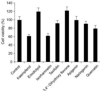
겨난 불멸의 인간 epithelial cell line이며,9) p53의 특정 잔 기가 mutation된 cell line이다. 하지만, HaCaT 세포는 쥐에 게 접종시켜도 암화 되지는 않는 것으로 보고되었다.10) 인간 HaCaT 세포에 미치는 다양한 플라보노이드들의 영 향을 알아보기 위해서 isohamnetin, kaempferol, 3,4’-dihydroxy flavone, eriodictyol, taxifolin, apigenin, narningenin 그리고 quercetin을 48시간 동안 처리하였다. MTT assay를 통해 세포활성(cell viability)를 조사하였는데, MTT assay는 살아 있는 세포의 미토콘드리아의 물질대사 능력을 측정하는 것으로 세포내 산화 환원반응을 보여줌으로써 세포의 활성도를 나타내어 준다. 대부분의 플라보노이드들은 HaCaT keratinocyte의 세포 활성을 감소시키는 것으로 조 사되었는데, kaempferol과 isohamnetin이 가장 좋은 효과를 보여주었다(Fig. 1). 그러나 몇몇 플라보노이드는 세포 활 성에 다른 결과를 보여주었는데, 흥미롭게도 3,4’-dihyd- roxy flavone과 eriodictyol은 HaCaT keratinocyte에서 세포 활 성을 증가시키는 결과를 보였다.
1. 3,4‘-dihydroxy flavone, eriodictyol은 etopo- side, H2O2에 의한 세포사멸를 억제시킨다 좀 더 정확하게 플라보노이드의 세포 활성 증가 효과 를 알아보기 위해서, 본 연구에서는 phenolic phytochemical 이고 아폽토시스 유도 물질 중 하나로 잘 알려진 etopo-
Fig. 1. Effects of flavonoids on cell viability of HaCaT cells.
Human keratinocyte HaCaT cells were treated with the indicated amounts of flavonoids and cell viability was determined by MTT assay. HaCaT cells were exposed to 10μM of the indicated flavonoids for 48 hr. As a control, cells were treated with the same amount of DMSO without any flavonoids. Data are the means±S.E. of values from three independent experiments.
side와 산화제인 과산화 수소(hydrogen peroxide: H2O2)를 이용해서 HaCaT 세포에서 아폽토시스를 유도한 상태에 서 이들 플라보노이드들의 효과를 조사하였다. 예상된 결과와 같이 etoposide와 H2O2는 HaCaT 세포의 세포활성 를 감소시켰다(Fig. 2). 그리고, etoposide와 H2O2에 의해 유도된 세포사멸에 미치는 플라보노이드의 영향을 보기 위해 본 실험에서 사용되어진 플라보노이드들을 1시간 전처리 한 후, etoposide와 H2O2를 처리하고 24시간 동안 배양하였다. 이 농도에서 etoposide와 H2O2는 아폽토시스 를 유도시키며, kaempferol, quercetin, taxifolin, apigenin, narningenin 그리고 isohamamnetin을 같이 처리하였을 경 우, etoposide만을 처리했을 경우보다 세포활성을 더욱 감 소시키거나, 큰 영향을 주지 않았다. 반면, 3,4’-dihydroxy flavone과 eriodictyol은 etoposide에 의해 유도되는 세포활 성의 감소를 억제시키는 것으로 조사되었다(Fig. 2B). 또 한, 사람 피부에 영향을 주는 환경적 요소 중 하나로 알 려진 산화제에 의한 스트레스에 대한 저항성을 알아보 고자 H2O2를 각각의 플라보노이드와 함께 처리해 보았 다. Etoposide 처리에서와 마찬가지로 H2O2에 의한 세포 사멸을 3,4’-dihydroxy flavone과 eriodictyol이 저해시키는 결과를 얻을 수 있었다(Fig. 2A). 또한, 본 실험 결과에서 는 이러한 세포사멸 억제능력 면에서 eriodictyol 보다 3,4’-dihydroxy flavone이 더 뛰어난 것으로 조사되었다 (Fig. 2).
2. 3,4‘-dihydroxy flavone에 의한 아폽토시스의 억 제 작용
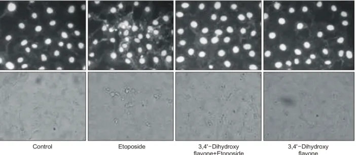
본 연구에서는 또한 10μM의 etoposide와 3,4’- dihy- droxy flavone에 세포를 노출시킨 후, DAPI 염색을 통해 세포 내에서 아폽토시스가 일어나고 있는 핵(apoptotic nuclei)의 형태학적 변화를 관찰하였다. 세포는 아폽토시 스가 일어나면 먼저 핵의 응축이 일어나고 그 후 핵은 fragmentation을 일으키는 형태학적 특성을 나타낸다. 본 결과에서도 etoposide는 HaCaT 세포에서 핵의 fragmen- tation을 유발시키지만, 3,4’-dihydroxy flavone은 etoposide에 의한 핵의 fragmentation을 억제하는 것으로 조사되었다 (Fig. 3).
3. 3,4‘-dihydroxy flavone의 세포 내 신호전달 기작 에 따른 아폽토시스 억제
일반적으로 세포사멸이 유도되면 caspase-3가 활성화 되어, 그 기질인 poly (ADP-ribose) polymerase (PARP)가 절 단되어 85,000 dalton (85 kDa)의 단편이 생성된다.1) Eto- poside를 처리한 세포에서도 시간이 경과함에 따라 caspase-3의 활성화가 증가하고 PARP의 절단이 점차 증 가하는 것으로 나타났다. 반면에, 3,4’-dihydroxy flavone를 전 처리한 세포에서는 etoposide에 의해 유도되는 caspase-3 의 활성화와 PARP의 절단이 현저하게 감소하는 것을 확 인 할 수 있었다(Fig. 4). 지금까지의 결과를 보면 다양한 Fig. 2. Effects of flavonoids, etoposide and H2O2 on cell viability of HaCaT cells. Cell viability was determined by MTT assay. (A) Cells were pre-incubated with the indicated flavonoids (10μM) for 1 h followed by the stimulation with H2O2 (100μM) for 48 h. (B) Cells were pre-incubated with the indicated flavonoids (10μM) for 1 h followed by the stimulation with etoposide (10μM) for 48 h. Data are the means±S.E. of values from three independent experiments.
플라보노이드가 아폽토시스에 서로 다른 영향을 준다는 것을 알 수 있었으며, 또한 아직 완전히 연구되지 않은 3,4’-dihydroxy flavone이 인간 HaCaT keratinocyte에서 etpop- side 또는 산화제인 H2O2에 의한 아폽토시스를 억제할 수 있음을 확인하였다.
고 찰
최근 항암 약품의 개발에 있어서 자연계에 존재하는 물질의 사용은 과학적, 산업적으로나 아주 중요한 주제
가 되고 있다. 특히 taxol과 etoposide와 같은 여러 phyto- chemicals은 이미 치료용 항암 약품으로 이용되고 있다.12) 플라보노이드는 최근 큰 관심을 받고 있는 천연물질 집단 중에 하나인데, kaempferol과 quercetin를 포함한 몇 몇 플라보노이드가 항산화 효과에 영향을 주고 또한 발 암을 억제할 수 있다는 여러 연구가 발표되었다.4,5) 종양 의 활성을 억제하는 것 말고도 몇몇 플라보노이드들은 항과민성 반응, 항바이러스, 그리고 항염증 반응의 특징 을 포함한 유익한 생물학적 활동을 나타내는 것으로 보 고되었으며, 또한, 몇몇 플라보노이드는 anti-apoptotic 특 Fig. 3. 3,4’-Dihydroxy flavone suppresses etoposide-induced apoptosis in HaCaT cells. Cells were pre-incubated with 3,4’-dihydroxy flavone (10μM) for 1 h and then exposed to etoposide (10μM) for 24 h. Cells were fixed and permeablized with methanol/acetic acid (1:1, vol/vol) and then loaded with 0.8μg/ml DAPI for 5 min. Fluorescence images were obtained under fluorescence microscope.
Data are the means±S.E. of values from three independent experiments.
Control Etoposide 3,4' Dihydroxy
flavone+Etoposide
- 3,4' Dihydroxy
flavone -
Fig. 4. 3,4’-Dihydroxy flavone suppresses etoposide-induced apoptotic caspase activation in HaCaT cells. Cells were pre-incubated with 3,4’-dihydroxy flavone (10μM) for 1 h and then exposed to etoposide (10μM) for the indicated time periods. Proteins were separated on 10% SDS-polyacrylamide gels (30μg/lane) and transferred to nitrocellulose membrane. Cleavage of PARP and pro- caspase-3 was analyzed by Western blotting with mouse monoclonal antibodies. The blots were re-probed with anti-Actin antibody to confirm equal amount of protein loading.
0 2 4 8 12 24
Etoposide
4 8 12 24
3,4' Dihydroxy flavone+Etoposide -
hr PARP
Pro caspase 3- -
Actin
성이 있는 것으로 발표되었다.13) 플라보노이드와 관련된 다양한 생물학적 기능을 알기 위해 수행된 많은 연구에 도 불구하고, 세포 효과에 기본이 되는 명확한 분자 메카 니즘은 아직 알려지지 않고 있다. 본 연구에서 우리는 플라보노이드가 인간 HaCaT keratinocytes의 세포활성에 특정한 영향을 준다는 것을 증명하였다. Kaempferol, isorhamnetin, naringenin, taxifolin, apigenin 그리고 quercetin 을 포함한 여러 플라보노이드는 세포 성장을 억제하는 영향을 보여주었으나 3,4’-dihydroxy flavone과 eriodictyol이 세포성장을 유도하고 외부 스트레스에 대한 방어 기작 을 증가시킨다는 결과를 얻었다. 또한 3,4’-dihydroxy flav- one은 etoposide에 의해 유도된 세포사멸을 억제한다는 것이 증명되었다(Fig. 1).
최근 여러 플라보노이드가 세포주기 정지와 다양한 암세포에서 아폽토시스를 유도한다는 것이 보고되었
다.14,15) 그러나 플라보노이드의 anti-apoptotic 효과에 대
해서는 아직까지 많은 연구가 되고 있지 않으며, 특히 본 연구에 사용된 3,4’-dihydroxy flavone의 anti-apoptotic 효 과에 대해서는 전혀 알려진 바가 없다. 3,4’-dihydroxy flavone이 apoptotic cell damage로부터 세포를 보호한다는 본 연구 결과는 표피 세포에 있어서는 최초의 보고인데, 이와 같은 연구 결과를 활용함으로써 각종 스트레스 환 경으로부터 인체의 첫 방어선인 피부 keratinocyte의 보호 에 기여될 수 있을 것으로 기대된다.
플라보노이드가 특정 효소를 억제하고 신경전달물질 을 자극하고 산화제를 제거하는 등의 능력을 지니고 있 기 때문에 여러 질병의 치료에 사용되어지고 있다.4,5) 비 록 대다수의 연구들이 플라보노이드의 항산화 효과에 중점을 두었지만 플라보노이드의 농도와 구조에 있어서 항산화 그리고 pro-oxidant한 면에서의 기능은 상대적으 로 많이 보고되고 있지 않다.7,16) 또한 여러 보고들이 다 양한 플라보노이드가 특정세포의 외부 스트레스에 의한 아폽토시스를 특징적으로 조절하는 것을 증명하였다.
이러한 특징적 효과는 적어도 플라보노이드가 자체적으 로 가진 structure-activity relationship (SAR) 부분에 있어서 의 구조 때문으로 생각된다. 이전 논문들은 플라보노이 드에 있는 다수의 hydroxyl (OH) 치환기들이 활성화 산호 (reactive oxygen species: ROS)를 제거하는 능력에 있어 중 요한 요소라고 예상하였다. OH 그룹을 더 많이 가진 플 라보노이드들은 더 잠재적인 항산화 특성을 가지며 또 한 항염증 효과를 강화시키는데 더 효과가 있다고 알려
졌다.17,18) 그러나 OH 치환기의 효과는 아직 자세히 결론
지어지지 않았고, 특정 플라보노이드의 diphenylpropane (C6C3C6) skeletons에 있는 특정 부위에서 각각의 OH가
주는 영향을 특정화 짓기 위해 앞으로 더 많은 연구가 필요하다. 그리고 이전 연구들은 methoxyl (OCH3) 그룹 이 플라보노이드의 항염증과 항산화 효과에 부정적인 영향을 준다고 보여주었다. 3-OH 플라본이 표피 성장 요소에 의한 분열을 억제하고 glycoside의 처리가 일반적 으로 플라보노이드와 관련된 아폽토시스 유도 효과를 억제하는 것으로 나타나고 있다.16,19)
본 연구에서, kaempferol, isorhamnetin, naringenin, taxi- folin, apigenin 그리고 quercetin이 HaCaT keratinocytes에 부 정적인 영향을 주었으나 3,4’-dihydroxy flavone는 세포에 긍정적인 영향을 주었다(Fig. 1). 이들 결과들은 3,4’-dihyd- roxy flavone과 kaempferol을 포함한 여러 플라보노이드에 서 타고난 특정 structure-activity relationship (SAR)가 있음 을 분명히 나타낸다. 본 연구의 결론을 바탕으로 플라보 노이드 diphenylpropane (C6C3C6) 골격의 5 또는/그리고 7 탄소의 특정 OH 치환기들이 아폽토시스 조절 능에 영향 을 미칠 것으로 예상된다. 앞으로 최근 연구 결과를 바탕 으로 아폽토시스 조절에 있어 structure-activity relationship (SAR)의 역할을 명확히 밝히기 위해 자연에서 얻을 수 있는 플라보노이드와 화학적으로 합성 가능한 여러 플 라보노이드의 아폽토시스 조절능을 비교해 볼 계획이 다. 결론적으로, 본 연구는 다양한 플라보노이드에 의거 한 pro-apoptosis/anti-apoptosis의 특성을 좀 더 연구하는데 도움이 되는 정보를 제공할 것으로 생각된다.
결 론
3,4’-dihydroxy flavone은 etoposide와 H2O2에 의한 인간 keratinocyte HaCaT 세포의 아폽토시스를 저해하였다. 따 라서 본 플라보노이드의 활성이 인간 keratinocyte의 외부 자극에 대한 세포 방어에 활용될 것으로 기대된다.
감사의 글
This work was supported by the Korea Research Foundation Grant funded by the Korean Government (KRF-2005-070- C00095) and a Grant from the ERC program of the Korea Science & Engineering Foundation (No. R11-2002-100-01000-0).
참 고 문 헌
1) Hengartner MO. The biochemistry of apoptosis. Nature 407, 770-776, 2000.
2) Igney FH, Krammer PH. Death and anti-death: tumour resistance to apoptosis. Nat Rev Cancer 2, 277-288, 2002.
3) Cody V. Crystal and molecular structures of flavonoids. Prog Clin Biol Res 280, 29-44, 1988.
4) Hertog MG, Hollman PC, Katan MB, Kromhout D. Intake of potentially anticarcinogenic flavonoids and their deter- minants in adults in The Netherlands. Nutr Cancer 20, 21-29, 1993.
5) Havsteen BH. The biochemistry and medical significance of the flavonoids. Pharmacol Ther 96, 67-202, 2002.
6) Fu Y, Hsieh TC, Guo J, Kunicki J, Lee MY, Darzynkiewicz Z, Wu JM. Licochalcone-A, a novel flavonoid isolated from licorice root (Glycyrrhiza glabra), causes G2 and late-G1 arrests in androgen-independent PC-3 prostate cancer cells.
Biochem Biophys Res Commun 322, 263-270, 2004.
7) Cemeli E, Schmid TE, Anderson D. Modulation by flavonoids of DNA damage induced by estrogen-like compounds. Environ Mol Mutagen 44, 420-426, 2004.
8) Diker-Cohen T, Koren R, Liberman UA, Ravid A. Vitamin D protects keratinocytes from apoptosis induced by osmotic shock, oxidative stress, and tumor necrosis factor. Ann N Y Acad Sci 1010, 350-353, 2003.
9) Boukamp P, Petrussevska RT, Breitkreutz D, Hornung J, Markham A, Fusenig NE. Normal keratinization in a spontaneously immortalized aneuploid human keratinocyte cell line. J Cell Biol 106, 761-771, 1998.
10) Lehman TA, Modali R, Boukamp P, Stanek J, Bennett WP, Welsh JA, Metcalf RA, Stampfer MR, Fusenig N, Rogan EM.
p53 mutations in human immortalized epithelial cell lines.
Carcinogenesis 14, 833-839, 1993.
11) Yang YL, Li XM. The IAP family: endogenous caspase inhibitors with multiple biological activities. Cell Res 10, 169-177, 2000.
12) Sheppard BC, Rutten MJ, Meichsner CL, Bacon KD, Leonetti PO, Land J, Crass RC, Trunkey DD, Deveney KE, Deveney
CW. Effects of paclitaxel on the growth of normal, polyposis, and cancerous human colonic epithelial cells. Cancer 85, 1454-1464, 1999.
13) Chen YC, Shen SC, Lee WR, Lin HY, Ko CH, Shin CM, Yang LL. Wogonin and fisetin induction of apoptosis through ac- tivation of caspase 3 cascade and alternative expression of p21 protein in hepatocellular carcinoma cells SK-HEP-1. Arch Toxicol 76, 351-359, 2002.
14) Hsu YL, Kuo PL, Lin CC. Acacetin inhibits the proliferation of Hep G2 by blocking cell cycle progression and inducing apoptosis. Biochem Pharmacol 67, 823-829, 2004.
15) Nguyen TT, Tran E, Ong CK, Lee SK, Do PT, Huynh TT, Nguyen TH, Lee JJ, Tan Y, Ong CS, Huynh H.
Kaempferol-induced growth inhibition and apoptosis in A549 lung cancer cells is mediated by activation of MEK-MAPK.
J Cell Physiol 197, 110-121, 2003.
16) Shen SC, Ko CH, Hsu KC, Chen YC. 3-OH flavone in- hibition of epidermal growth factor-induced proliferaton th- rough blocking prostaglandin E2 production. Int J Cancer 108, 502-510, 2004.
17) Areias FM, Rego AC, Oliveira CR, Seabra RM. Antioxidant effect of flavonoids after ascorbate/Fe (2+)-induced oxidative stress in cultured retinal cells. Biochem Pharmacol 62, 111-118, 2001.
18) Hendriks JJ, de Vries HE, van der Pol SM, van den Berg TK, van Tol EA, Dijkstra CD. Flavonoids inhibit myelin phagocytosis by macrophages; a structure-activity relationship study. Biochem Pharmacol 65, 877-885, 2003.
19) Shen SC, Chen YC, Hsu FL, Lee WR. Differential apoptosis- inducing effect of quercetin and its glycosides in human promyeloleukemic HL-60 cells by alternative activation of the caspase 3 cascade. J Cell Biochem 89:1044-1055, 2003.