Received: 23 October, 2015 Revised: 28 October, 2015 Accepted: 2 November, 2015 Corresponding author: Jong-Duk Choi
Departmet of Physical Therapy, College of Health and Medical Science, Daejeon University, 62 Daehak-ro, Dong-gu, Daejeon 34520, Republic of Korea Tel: 82-42-280-2293 Fax: 82-42-280-2295 E-mail: [email protected]
This is an Open-Access article distributed under the terms of the Creative Commons Attribution Non-Commercial License (http://creativecommons.org/licens es/by-nc/4.0) which permits unrestricted non-commercial use, distribution, and reproduction in any medium, provided the original work is properly cited.
Copyright © 2015 Korean Academy of Physical Therapy Rehabilitation Science
http://dx.doi.org/10.14474/ptrs.2015.4.2.66 pISSN 2287-7576
eISSN 2287-7584
Phys Ther Rehabil Sci 2015, 4 (2), 66-72 www.jptrs.org
The effect of balance training with plantar flexor stretching on range of motion, balance, and gait in stroke patients: a
randomized controlled pilot trial
Ki-Suk Park
a, Jong-Duk Choi
baDepartment of Physical Therapy, Graduate School of Health and Medicine, Daejeon University, Daejeon, Republic of Korea
bDepartmet of Physical Therapy, College of Health and Medical Science, Daejeon University, Daejeon, Republic of Korea
Objective: The aim of this study was to investigate the effect of balance training with plantar flexor stretching on ankle dorsi flex- ion range of motion (ROM), balance, and gait ability in stroke patients.
Design: A randomized controlled pilot trial.
Methods: Thirty stroke patients volunteered to participate in this study. The subjects were randomly allocated to two groups: the experimental group (n=15) received the neurodevelopment therapy plus balance training with plantar flexor stretching for 20 mi- nutes in one session. The control group (n=15) received the same neurodevelopment therapy plus plantar flexor static stretching for 20 minutes in one session. Both groups underwent sessions four times a week, for a total of 4 weeks. Measurements included passive range of motion (PROM), active range of motion (AROM) of ankle dorsiflexion using a goniometer, timed up and go (TUG), the functional reaching test (FRT), and the 10 m walk test (10 MWT).
Results: There were significant improvements in AROM and PROM of ankle dorsiflexion, TUG, and FRT scores after the inter- vention in the experimental group (p<0.05). However, the control group showed no statistically significant differences except for PROM of ankle dorsiflexion. The experimental group showed a significant improvement in PROM, TUG, and FRT scores com- pared to the control group (p<0.05).
Conclusions: Balance training with plantar flexor stretching improves ankle dorsiflexion ROM and balance ability in patients with stroke. Therefore, this therapeutic intervention will be effective for rehabilitation of stroke patients in the clinical setting.
Key Words: Muscle stretching exercises, Postural balance, Stroke
Introduction
A stroke is an illness inducing disability by blockage of the blood flow toward the brain or bleeding in the brain tis- sue from various causes. It is the second most common cause of death in Korea after cancer [1,2]. More than 80% of pa- tients surviving after a stroke show weakening of functions and hemiparesis, and damage to their gait ability threatens their independent life [3,4]. Gait ability is closely related to balance, and, in particular, reduced ankle joint mobility by ankle contracture hampers standing and balancing, a sig-
nificant factor in diminishing the effects of rehabilitation
[5]. It is reported that 84% of patients with brain damage
show contracture, and in 76% of them this is accompanied
by contracture of the ankle joints [6]. Given et al. [7] re-
ported that the muscles around the ankle joints easily devel-
op changes in their mechanical property because they have
more type I muscle fibers and connective tissue than other
muscles. Owing to the characteristics of the ankle joints,
stroke patients are known to be more likely to have increased
spasticity and shortened fascia of the gastrocnemius on the
paretic side, producing a reduced range of motion in dorsi-
flexion in the ankle joints [8]. Such changes induce incorrect transmission of somatesthesia from the joint or the muscle receptors or motor response, accompanied by inappropriate ankle strategy, causing difficulty in balance control [9,10].
All the motion in the ankles including dorsiflexion controls the interactions between the feet and the ground, serving as an essential factor in gait and balance. Performing func- tional activities requires at least 10 degrees of range of mo- tion in dorsiflexion. In this context, efforts to increase the range of motion in the ankle are required for stroke patients before planning a program of gait training [11,12].
Various methods of stretching the plantar flexor have been clinically used in order to increase the range of motion and to stimulate somatesthesia in the ankle joints; static stretching is regarded as an intervention that can be per- formed easily. Many patients with damage to the central nervous system also undergo static stretching of the plantar flexor [13]. However, the effects of stretching on stroke pa- tients have been reported differently. Kim [14] reported that a short-term application of passive stretching to the plantar flexor of subjects with stroke reduced the excitability of the α-motor neuron and increased the degree of dorsiflexion.
Lee et al. [15] identified that static stretching of the plantar flexor of stroke patients by using the Q-board for six weeks increased gait velocity in the 10 m walk test (10MWT) and the timed up-and-go test. However, in Bressel and McNair [16]’s research, consistent static stretching of the plantar flexor of stroke patients for 30 minutes reduced the stiffness of the ankle joints for a short period, but gait velocity was not significantly increased in the 10MWT, owing to reduced muscular strength. Yuk [13] reported that standing balance ability temporarily declined immediately after consistent static stretching of the plantar flexor for 15 minutes, because of the sway of the center of gravity by dynamic and neuro- logical changes, suggesting that patients with damage to the central nervous system may be subject to the risk of falling after consistent stretching because of their lessened balance ability. The stretching of the plantar flexor may contribute to an increase in joint mobility by changing muscle length and improving stiffness in the tissues around the joints [10,17], while inducing a temporary decline in balance ability due to changes in tension and somatesthesia [16,18-20].
Changes in ankle degree in the sagittal plane, in particular, affect static balance more [21]; balancing against the back- and-forth posture sway is performed by alternative activa- tion of the tibialis anterior and the gastrocnemius [22]. Thus, for stroke patients to maintain balance with dorsiflexion
may be a considerable challenge, because of the compli- cated process in which the sensory system perceives body motions and the central nervous system integrates and modi- fies the motions to make the musculoskeletal system react appropriately to environmental changes [23,24]. However, most of the previous researches on stretching of the plantar flexor have focused on passive, static stretching, whereas few deal with balance training with plantar flexor stretching.
Also, researches into the effects of consistent stretching have been insufficient to date, and consequently the effects have not been identified clearly.
Thus, this study was devised to investigate the effects of balance training with plantar flexor stretching for four weeks on range of motion in dorsiflexion in the ankle joints, bal- ance, and gait ability of stroke patients. The hypothesis of this study is as follows. The experimental group undergoing balance training with plantar flexor stretching may show im- provement in the range of motion in dorsiflexion in the ankle joints, balance, and gait velocity, when compared to the con- trol group undergoing plantar flexor static stretching.
Methods Subjects
The stroke patients who entered a rehabilitation hospital in Cheongju-si, 30 were selected to be the subjects of this study, after they had received an explanation of the contents and purpose of this experiment and given their consent to participation. All of the protocols used in this study were ap- proved by the Daejeon University. The subjects’ rights were protected according to the guidelines of the Daejeon University.
The inclusion criteria were as follows: patients who were di- agnosed with hemiplegia by stroke at least six months ago, showed 10 or less degrees in the active range of motion (AROM) of dorsiflexion and 20 or less degrees in the pas- sive range of motion (PROM), G2 or under of the grades in the modified Ashworth scale of the plantar flexor, Mini- Mental State Examination Korean version score higher than 24 and those who could walk at least 15 m with or without gait-assist devices. The exclusion criteria were as follows:
patients who had problems in standing and gait caused by or- thopedic surgery or disability in the lower limbs.
Procedures
The 30 subjects were randomly and equally assigned to an
experimental group (n=15) and a control group (n=15). Both
groups underwent neurodevelopmental treatment for 30 mi-
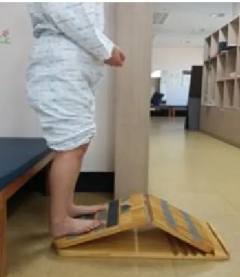
Figure 1. Balance training with Plantar flexor stretching training.
nutes and then plantar flexor stretching for 20 minutes; the experimental group performed balance training with plantar flexor stretching, while the control group performed general plantar flexor static stretching. Both groups underwent the procedure four times a week for four weeks.
The Q-board used in this experiment was an inclined wooden board (40×70 cm), which was angle-adjustable. the tilt angle of the Q-board for plantar flexor stretching was set between 5 and 20 degrees so that subjects would not feel pain or discomfort. When a subject expressed discomfort during the stretching, the angle was adjusted to avoid tissue damage [13]. The control group underwent static stretching leaning against a wall on the Q-board in dorsiflexion. The experi- mental group stood on the Q-board, balancing indepen- dently without leaning against a wall (Figure 1). In order for the subjects to maintain balance using angle strategy as much as possible, their movements and alignment in the hip joints and the knee joints were intentionally limited; if necessary, the therapist issued verbal instruction. Patients who found proper weight bearing and balancing difficult underwent the process after they had been accustomed to the motions by repetition with the help of a therapist. Therapists observed the patients throughout the process to ensure their safety.
Before intervention, PROM and AROM in dorsiflexion was measured in both groups using a goniometer [25], the functional reach test (FRT) and the The timed up and go test (TUG) being administered to assess their balance ability and the 10MWT to measure their gait ability. After the four- week intervention, the same investigators measured the val- ues again.
In the FRT for assessing balance ability, the subjects in the standing position were asked to stretch an arm forward with shoulder flexion of 90 degrees. We measured the distance between the tip of the middle finger in the starting position and the same tip when a subject stretched his/her arm as for- ward as possible. The intrarater reliability was 0.92, and the interrater reliability was 0.98 [26]. TUG is a test to quickly assess a person’s basic mobility and balance; it measures with a stopwatch the time that a subject takes to rise from an armchair, walk 3 meters, turn around, and walk back to the char to sit down. We conducted this measurement three times to obtain mean values. The intrarater reliability was 0.99, and the interrater reliability was 0.98 [27]. To assess gait ability, we used the 10MWT, a tool whose reliability and val- idity have been proven in various studies. Subjects were asked to walk 14 meters of straight-line distance; the 2 meters add- ed to the starting and the finishing point were set to be the distance for acceleration and deceleration. The walking time for the 10 meters was measured with a stopwatch [28].
Data analysis
IBM SPSS ver. 19.0 for Windows (IBM Co., Armonk, NY, USA) was used for statistical processing. The Shapiro-Wilk test was used for the test of normality and result values showed normality. The chi-square test and independent t-test were conducted to test the homogeneity of the two groups.
Before the balance training with plantar flexor stretching for the experimental group and the plantar flexor static stretch- ing for the control group, a paired t-test was conducted to compare the difference within a group and an independent t-test to compare between the groups. The statistical sig- nificance was set at α=0.05.
Results
General characteristics of the subjects
Table 1 shows subjects general characteristics. No stat- istically significant difference was found between the two groups.
Changes in the range of motion in dorsiflexion of the two groups before and after training
In both groups, PROM of dorsiflexion showed a sig- nificant increase in after the training (p<0.05). the rate of change between the groups showed a significant difference (p<0.05).
AROM of dorsiflexion in the experimental group showed
Table 1. General characteristics of the subjects (N=30)
Characteristic Experimental group (n=15) Control group (n=15) χ2/t p
Age (y) 58.90 (13.37) 60.18 (13.01) −0.223 0.826
Gender 1.292 0.450
Male 11 (73.3) 8 (53.3)
Female 4 (26.7) 7 (46.7)
Height (cm) 166.20 (9.73) 162.36 (6.74) 1.059 0.303
Weight (kg) 64.20 (9.73) 59.52 (8.04) 0.922 0.368
Onset (mo) 24.10 (11.71) 22.55 (12.82) 0.289 0.776
Type of lesion 0.536 0.464
Hemorrhage 8 (53.3) 6 (40.0)
Infartion 7 (46.7) 9 (60.0)
Paretic side 0.682 0.682
Right 3 (20.0) 5 (33.3)
Left 12 (80.0) 10 (66.7)
Values are presented as number (%) or mean (SD).
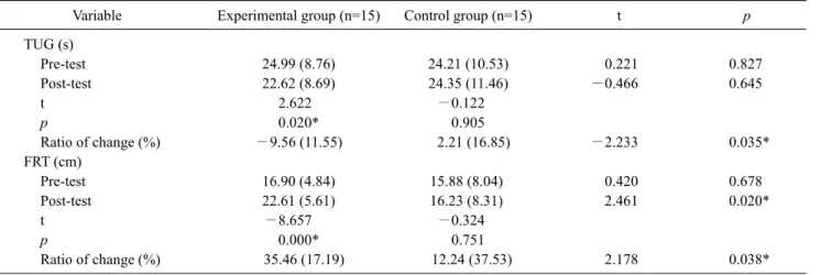
Table 3. Comparison of balance ability within group and between group (N=30)
Variable Experimental group (n=15) Control group (n=15) t p
TUG (s)
Pre-test 24.99 (8.76) 24.21 (10.53) 0.221 0.827
Post-test 22.62 (8.69) 24.35 (11.46) −0.466 0.645
t 2.622 −0.122
p 0.020* 0.905
Ratio of change (%) −9.56 (11.55) 2.21 (16.85) −2.233 0.035*
FRT (cm)
Pre-test 16.90 (4.84) 15.88 (8.04) 0.420 0.678
Post-test 22.61 (5.61) 16.23 (8.31) 2.461 0.020*
t −8.657 −0.324
p 0.000* 0.751
Ratio of change (%) 35.46 (17.19) 12.24 (37.53) 2.178 0.038*
Values are presented as mean (SD).
TUG: timed up and go test, FRT: functional reach test.
*p<0.05.
Table 2. Comparison of ankle joint range of motion within group and between group (N=30)
Variable Experimental group (n=15) Control group (n=15) t p
PROM
Pre-test 13.07 (2.49) 12.73 (2.05) 0.400 0.692
Post-test 15.73 (2.02) 14.00 (1.46) 2.694 0.012*
t −6.162 −5.104
p 0.000* 0.000*
Ratio of change (%) 22.84 (18.07) 11.13 (9.62) 2.215 0.038*
AROM
Pre-test 5.80 (3.14) 5.20 (3.67) 0.481 0.634
Post-test 6.67 (3.66) 5.53 (3.80) 0.833 0.412
t −3.166 −2.092
p 0.007* 0.055
Ratio of change (%) 13.22 (17.06) 5.86 (10.46) 1.426 0.167
Values are presented as mean (SD).
PROM: passive range of motion, AROM: active range of motion.
*p<0.05.
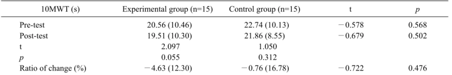
Table 4. Comparison of the gait ability within groups and between groups (N=30)
10MWT (s) Experimental group (n=15) Control group (n=15) t p
Pre-test 20.56 (10.46) 22.74 (10.13) −0.578 0.568
Post-test 19.51 (10.30) 21.86 (8.55) −0.679 0.502
t 2.097 1.050
p 0.055 0.312
Ratio of change (%) −4.63 (12.30) −0.76 (16.78) −0.722 0.476
Values are presented as mean (SD).
10MWT: 10 m walk test.
an increase from 5.80±3.14 degrees in pre-training to 6.67±
3.66 degrees in post-training (p<0.05). There was no sig- nificant difference in the rate of change between the two groups (Table 2).
Changes in balance ability of the two groups before and after training
With regard to comparison of balance ability within a group, the time taken in the TUG significantly declined in post-training in the experimental group (p<0.05).
In the FRT, the distance in the experimental group sig- nificantly increased (p<0.05). The rate of change in the FRT, TUG between the groups showed a statistically significant difference (p<0.05) (Table 3).
Gait ability in the two groups before and after training In the 10MWT, the time in the experimental group de- clined, without a statistically significant difference (Table 4).
Discussion
The purpose of this study was to identify the effects of plantar flexor stretching applied to patients with hemiplegia caused by stroke to improve their range of motion in the an- kle joints, balance, and gait ability. The results of this study showed that the experimental group undergoing balance training with plantar flexor stretching increased their PROM in dorsiflexion and experienced a significant improvement in balance ability when compared to the control group un- dergoing plantar flexor static stretching.
Body movements enabling functional activities depend not only on active neuromuscular control but also on the mo- bility of appropriate soft tissue. The proper range of motion in dorsiflexion in the ankle joints, in particular, is essential for performing functional activities such as walking, run- ning, climbing, and going down stairs normally [29]. After a
stroke, the range of motion in the ankle joints is restricted by a decrease in the size of muscular fibers, a decline in the number of recruitment motor units, a failure to produce proper muscular strength owing to abnormal muscle tone, and weakness or contracture of the dorsi flexor [8], a re- striction that poses the risk of declining balance ability [30].
Lee et al. [31] reported that sensitivity to the phasic reflex
declined when patients with hemiparesis underwent plantar
flexor stretching for 30 minutes. Although short-term stret-
ching is insufficient to develop structural changes in mus-
cles, the mechanoreceptor around the ankles and the Golgi
tendon organ mainly in muscles decrease the excitability of
the gastrocnemius and the α-motor neuron to increase mus-
cular flexibility. After 48 hours of the stretching, however,
the excitability of the α-motor neuron reverts to its extent
before the stretching, and consequently the flexibility of the
ankle joints declines [14,32]. There have been various stud-
ies identifying improvement in the range of motion in the an-
kle joints due to short-term plantar flexor stretching, but few
researchers have focused on changes in the joints brought
about by long-term stretching. In this study, both the ex-
perimental group and the control group showed a significant
increase in AROM of their ankle joints after plantar flexor
stretching was applied for four weeks. However, the rate of
change between the groups showed a significant difference,
perhaps because the experimental group, unlike the control
group, was not restricted in the range of movement of their
body, including the ankles. The results may be due to the fact
that, in the experimental group, the angle by which the an-
kles were stretched may have been larger, as the body moved
back and forth more. It is well known that, even while a per-
son is standing quietly, the muscles contract against small
sways in order to maintain balance [33]. It is suggested that
the AROM of the experimental group also improved be-
cause, when they underwent balance training in dorsiflexion,
they could perceive the sense of stretching while their body
was swayed back and forth and then repeatedly experienced alternative induction of appropriate muscle contraction to maintain their balance.
Lee et al. [15] reported that the time taken in the 10MWT and the TUG was significantly decreased in stroke patients when they underwent plantar flexor stretching for six weeks, perhaps because of an increase in the range of motion in the ankle joints and a decrease in spasticity. In this study, how- ever, the control group undergoing static stretching did not show a significant difference between pre- and post-training in the results of the TUG, the FRT, or the 10MWT. This in- dicates that it was insufficient for them to experience an im- provement in functional ability because of improved PROM, since they were not experiencing an improved AROM, which is related to muscular strength. Song et al. [21] identi- fied the effects of ankle posture of healthy adults on static balance, reporting that changes in the sagittal plane rather than in the frontal plane of the ankle had greater effects.
Such results may indicate that it can be a considerable chal- lenge for stroke patients to maintain balance in dorsiflexion.
Posture control to maintain balance is performed by a com- plicated interaction between the sensory and the muscu- loskeletal system and is integrated and modified within the central nervous system by reacting to environmental changes [23,34]. In this context, the experimental group undergoing balance training with plantar flexor stretching might have experienced an improvement in proprioception and balance ability by transmission of somatesthesia from the joint or muscle receptors in the environment in which their bodies swayed and repeated movements were applied by an appro- priate ankle strategy in order to maintain balance. However, there was no significant difference in the time taken in the 10MWT before and after the training, despite improvement in balance ability, perhaps because even the increased angle of the AROM did not reach the functional angle (10 degrees) that is needed for walking. Also, other functions related to walking other than joint angle may contribute to the insigni- ficance.
The results of this study may not be capable of general- ization to all stroke patients with hemiparesis, because the subjects were chosen from among those who were treated in a rehabilitation hospital and those who met the conditions for inclusion. Also, we did not assess other factors that may affect improvement in balance and gait ability, including muscular strength and proprioception sense. The results of this study may suggest that stroke patients can improve their range of motion in dorsiflexion in the ankle joints and bal-
ance ability more effectively by undertaking balance train- ing with plantar flexor stretching (although this is a consid- erable challenge for them), rather than static, passive plantar flexor stretching.
Conflict of Interest
The authors declared no potential conflicts of interest with respect to the authorship and/or publication of this article.
References
1. Park CH, Chung BI. Effects of treadmill training on hyperexten- sion of the knee and cadence in patients with hemiplegia. Phys Ther Korea 2001;8:89-96.
2. Korea National Statistical Office. Cause of death statistics 2011.
Daejeon: Korea National Statistical Office; 2011.
3. Wall JC, Charteris J, Turnbull GI. Two steps equals one stride equals what?: the applicability of normal gait nomenclature to abnormal walking patterns. Clin Biomech (Bristol, Avon) 1987;
2:119-25.
4. Wade DT, Wood VA, Heller A, Maggs J, Langton Hewer R.
Walking after stroke. Measurement and recovery over the first 3 months. Scand J Rehabil Med 1987;19:25-30.
5. Bohannon RW, Larkin PA. Lower extremity weight bearing un- der various standing conditions in independently ambulatory pa- tients with hemiparesis. Phys Ther 1985;65:1323-5.
6. Yarkony GM, Roth EJ, Heinemann AW, Wu YC, Katz RT, Lovell L. Benefits of rehabilitation for traumatic spinal cord injury.
Multivariate analysis in 711 patients. Arch Neurol 1987;44:93-6.
7. Given JD, Dewald JP, Rymer WZ. Joint dependent passive stiff- ness in paretic and contralateral limbs of spastic patients with hemiparetic stroke. J Neurol Neurosurg Psychiatry 1995;59:271-9.
8. Gao F, Ren Y, Roth EJ, Harvey R, Zhang LQ. Effects of repeated ankle stretching on calf muscle-tendon and ankle biomechanical properties in stroke survivors. Clin Biomech (Bristol, Avon) 2011;26:516-22.
9. Lee KB, Park YH, Song EK, Yoon TR, Jung KI. Static and dy- namic postural balance after successful mobile-bearing total an- kle arthroplasty. Arch Phys Med Rehabil 2010;91:519-22.
10. McHugh MP, Cosgrave CH. To stretch or not to stretch: the role of stretching in injury prevention and performance. Scand J Med Sci Sports 2010;20:169-81.
11. Wolfson L, Whipple R, Judge J, Amerman P, Derby C, King M.
Training balance and strength in the elderly to improve function.
J Am Geriatr Soc 1993;41:341-3.
12. Won JI, An CM. Knee strength and ankle range of motion Influencing gait velocity and gait asymmetry in patients with chronic stroke. Phys Ther Korea 2015;22:1-10.
13. Yuk GC. The acute effects of 15 minutes Plantarflexor static stretch in quite stance. J Korean Soc Phys Med 2012;7:191-7.
14. Kim JS. Effects of gastrocnemius stretching on α-motor neuron excitability and ankle joint active dorsiflexion range of motion. J Korean Content Assoc 2009;9:278-86.
15. Lee JH, Lee JH, Kwon WA, Kim JS. The effect of ankle joint muscle strengthening training and static muscle stretching train- ing on stroke patients plantar pressure and gait. J Korean Acad Ind Coop Soc 2012;13:1153-60.
16. Bressel E, McNair PJ. The effect of prolonged static and cyclic stretching on ankle joint stiffness, torque relaxation, and gait in people with stroke. Phys Ther 2002;82:880-7.
17. Avela J, Finni T, Liikavainio T, Niemelä E, Komi PV. Neural and mechanical responses of the triceps surae muscle group after 1 h of repeated fast passive stretches. J Appl Physiol (1985) 2004;
96:2325-32.
18. Avela J, Kyröläinen H, Komi PV. Altered reflex sensitivity after repeated and prolonged passive muscle stretching. J Appl Physiol (1985) 1999;86:1283-91.
19. Babault N, Kouassi BY, Desbrosses K. Acute effects of 15min static or contract-relax stretching modalities on plantar flexors neuromuscular properties. J Sci Med Sport 2010;13:247-52.
20. Behm DG, Bambury A, Cahill F, Power K. Effect of acute static stretching on force, balance, reaction time, and movement time.
Med Sci Sports Exerc 2004;36:1397-402.
21. Song CH, Kim KR, Lee KJ, Shin DC, Lee YD. The effects of an- kle position on static balance in healthy adult. J Spec Educ Rehabil Sci 2012;51:403-17.
22. Almeida GL, Carvalho RL, Talis VL. Postural strategy to keep balance on the seesaw. Gait Posture 2006;23:17-21.
23. Kisner C, Colby LA. Therapeutic exercise: foundations and tech- niques. 6th ed. Philadelphia: FA Davis; 2012.
24. Carr JH, Shepherd RB. Neurological rehabilitation: optimizing motor performance. 2nd ed. Edinburgh: Churchill Livingstone;
2010.
25. Lee WC, Park HS, Rha JD, Han YK, Cho JJ, Chang BC, et al.
Measurement of the dorsiflexion range in the ankle. Korean J Sport Med 1998;16:29-34.
26. Duncan PW, Weiner DK, Chandler J, Studenski S. Functional reach: a new clinical measure of balance. J Gerontol 1990;45:
M192-7.
27. Podsiadlo D, Richardson S. The timed "Up & Go": a test of basic functional mobility for frail elderly persons. J Am Geriatr Soc 1991;39:142-8.
28. Dean CM, Richards CL, Malouin F. Task-related circuit training improves performance of locomotor tasks in chronic stroke: a randomized, controlled pilot trial. Arch Phys Med Rehabil 2000;81:409-17.
29. Rabin A, Kozol Z, Spitzer E, Finestone AS. Weight-bearing an- kle dorsiflexion range of motion-can side-to-side symmetry be assumed? J Athl Train 2015;50:30-5.
30. Vandervoort AA, Chesworth BM, Cunningham DA, Rechnitzer PA, Paterson DH, Koval JJ. An outcome measure to quantify passive stiffness of the ankle. Can J Public Health 1992;83 Suppl 2:S19-23.
31. Lee SJ, Kwon BS, Park CH. The effect of passive stretching on the spasticity of ankle plantar flexor muscles. J Korean Acad Rehabil Med 2001;25:987-92.
32. Gossman MR, Sahrmann SA, Rose SJ. Review of length-asso- ciated changes in muscle. Experimental evidence and clinical implications. Phys Ther 1982;62:1799-808.
33. Nagano A, Yoshioka S, Hay DC, Himeno R, Fukashiro S.
Influence of vision and static stretch of the calf muscles on pos- tural sway during quiet standing. Hum Mov Sci 2006;25:422-34.
34. Kim SG. Effect of treadmill gradient training on lower limb mus- cle activity in chronic stroke patient. J Korean Acad Ind Coop Soc 2012;13:220-6.