대한외과학회지:제 63 권 제 6 호
□ 원 저 □
Vol. 63, No. 6, December, 2002
458
서 론
유방촬영술의 선별검사(screening)는 유방암 환자의 사망 률을 감소시켰으나 유방촬영술의 정확도 및 진단적 가치에 대한 논란도 많다. 중간암(interval cancer)이란 선별검사 간 격사이에 발생하는 암 혹은 유방암 진단 전의 유방촬영상 에서 정상 소견을 보이는 유방암으로 정의되며, 선별검사 프로그램의 성공은 중간암의 수를 최소화하는 데 있다.(1) 중간암 발생률(interval cancer rate)은 선별검사의 효율성에 대한 중요한 표지자로서 유방촬영술의 정확도를 반영할 뿐 만 아니라 검사의 간격에도 영향을 받는다.(2) 대개 검사간 격을 1년에서 2년으로 시행하는 경우가 대부분이나 국가나 지역에 따라 달라 3년의 간격으로 시행되는 프로그램도 있 으므로, 이 경우 interval cancer의 비율은 증가한다고 볼 수 있다. 또한 치밀유방, 호르몬대체요법 등은 유방촬영술의 정확도에 영향을 주어 중간암 발생에 기여하게 된다.(3) 대 개의 중간암은 증상으로 인해 진단되기 때문에 선별검사로 진단된 유방암과는 다른 특성을 가지리라 예측할 수 있다.
이에 저자들은 중간암 및 선별검사로 진단된 유방암 환자 의 방사선학적 특징과 병리학적 특징을 비교해 보고자 하 였다.
선별검사로 진단된 유방암과 중간암(Interval Cancer)의 비교
연세대학교 의과대학 외과학교실, 성균관대학교 의과대학 삼성제일병원 1외과, 2방사선과
김준영․조백현1․허민희1․강성수1․이지현1․이성공1․조병제2․이경상2․이해경1
Interval Breast Cancers: Comparisons with Screen Detected Cancers
Jun Young Kim, M.D., Back Hyun Cho, M.D.1, Min Hee Hur, M.D.1, Sung Soo Kang, M.D.1, Jee Hyun Lee, M.D.1, Sung Kong Lee, M.D.1, Byung Jae Cho, M.D.2, Kyung Sang Lee, M.D.2 and Hae Kyung Lee, M.D.1
Purpose: Although the screening with a mammography has been shown to reduce breast cancer mortality, it has limitations relating to its sensitivity and efficacy. Interval cancers are those that become symptomatic, and are detected between screening examinations. The success of a screening program in reducing the rate of mortality due to breast cancer relies on keeping the number of interval cancers at a minimum. This study was performed to review the mammographic features of interval cancers, and to compare their clinicopathological factors with those cancers detected by screening.
Methods: Of the 881 women who had operations for breast cancer performed between 1995 and 1999, we retrospec- tively analyzed the medical records and mammograms of 57 who received at least a mammogram before the diagnosis of their breast cancer. These patients were divided into an interval cancer group, who had symptoms, and a screen detected cancer group, who had not. The factors compared included the clinical, radiographic, histopathological, and immunohistochemical features.
Results: Interval cancers were more likely to have masses, than microcalcifications, in their mammographic features, and were more likely to be invasive and at a higher stage according to their histopathological features. The false ne- gative rate was 48% for the screen detected cancers, and 35% for the interval cancers (P=0.414). HRT users had the higher false negative rate of 51.6% than the 26.9% for the nonuser (P=0.103).
책임저자:이해경, 서울특별시 중구 묵정동 1-19
ꂕ 100-380, 성균관대학교 의과대학 삼성제일병원 외과 Tel: 02-2000-7280, Fax: 02-2000-7791
E-mail: [email protected]
접수일:2002년 10월 21일, 게재승인일:2002년 11월 18일 이 내용은 2001년 추계 외과학회에서 포스터 발표되었음.
Conclusion: The interval cancers were found to be different from the screen detected cancers in terms of their radiolo- gical and pathological features. The standardization of screen interval, and additional magnification mammography, or ul- trasonography may contribute to reduce false negative rates of mammography. (J Korean Surg Soc 2002;63:458-461) Key Words: Interval cancers, Screen detected breast can-
cers, False negative rate
중심 단어: 중간암, 선별검사로 진단된 유방암, 위음 성률
ꠏꠏꠏꠏꠏꠏꠏꠏꠏꠏꠏꠏꠏꠏꠏꠏꠏꠏꠏꠏꠏꠏꠏꠏꠏꠏꠏꠏꠏꠏꠏꠏꠏꠏꠏꠏꠏꠏꠏꠏꠏꠏꠏꠏꠏꠏꠏꠏꠏꠏ Department of Surgery, Yonsei University College of Medi- cine, Departments of 1Surgery and 2Radiology, Samsung Cheil Hospital, Sungkyunkwan University School of Medicine, Seoul, Korea
김준영 외:선별검사로 진단된 유방암과 중간암(Interval Cancer)의 비교 459 ꠏꠏꠏꠏꠏꠏꠏꠏꠏꠏꠏꠏꠏꠏꠏꠏꠏꠏꠏꠏꠏꠏꠏꠏꠏꠏꠏꠏꠏꠏꠏꠏꠏꠏꠏꠏꠏꠏꠏꠏꠏꠏꠏꠏꠏꠏꠏꠏꠏꠏꠏꠏꠏꠏꠏꠏꠏꠏꠏꠏꠏꠏꠏꠏꠏꠏꠏꠏꠏꠏꠏꠏꠏꠏꠏꠏꠏꠏꠏꠏꠏꠏꠏꠏꠏꠏꠏꠏꠏꠏꠏꠏꠏꠏꠏꠏꠏꠏꠏꠏꠏꠏꠏꠏꠏꠏꠏꠏꠏꠏꠏꠏꠏꠏꠏ
방 법
1995년 1월부터 1999년 12월까지 삼성제일병원에서 유방 암으로 수술 받은 881명의 환자 중 수술 전 본원에서 한번 이상의 유방촬영술을 시행하여 유방촬영상의 재검토가 가능 한 57명의 환자를 대상으로 23명의 증상 없이 선별검사로 진 단된 환자군과 34명의 증상이 있었던 중간암 환자군으로 분 류하여 후향적 비교 연구를 시행하였다. 대상 환자군의 평균 나이는 51.4세였으며, 이전 검사와의 평균 검사 간격은 14.6 개월이었다(n=57). 선별검사로는 양쪽 유방에 대해 Craniocau- dal view와 lateral oblique view를 시행하였다. 방사선과 의사 가 유방암 진단전의 유방촬영술을 재검토하여 유방암 진단 시의 유방촬영술과 같은 위치에서 병변을 발견할 경우 양성 의 소견이거나 비특이적 소견이더라도 위음성으로 분류하였 다. 비교인자로는 나이, 검사간격, 유방촬영상 소견, 위음성 률, 병리조직학적 유형, 침윤성 유방암의 원발종양 크기, 림 프절 전이 유무, 병기, 호르몬 수용체 유무, c-erbB2, p53 양성 여부, 호르몬치료(HRT) 시행 여부 등이었다. 림프절에 관해 서는 단순히 전이 유무만으로 분류하였으며, 전이된 림프절 의 개수, 위치 또는 특성은 고려하지 않았다. 병리 조직학적 유형은 관상피내암과 침윤성 유관암으로 분류하였으며, 핵 분화등급은 Reverse Nuclear Grade I∼III으로 분류하였다. 유 방암의 병기는 American Joint Committes on Cancer (AJCC)에 따른 TNM 등급으로 분류하였다. 호르몬 치료의 시행 여부는 이전 검사가 시행된 시점에 6개월 이상 시행한 경우를 기준 으로 하였다. 진단당시의 통계학적 분석은 SPSS 10.0을 이용 하여 chi-square test, student t test를 시행하였고, P값이 0.05 미만인 경우 유의한 것으로 판정하였다.
결 과
1) 임상적 특징(Table 1)
임상적 증상 없이 선별검사로 진단된 유방암 환자는 23명
으로 평균 연령은 52세(39∼61세)였고, 마지막 유방촬영술 과의 간격은 14개월(5∼46개월)이었다. 중간암 환자는 34명 으로 평균 연령은 51세(35∼69세), 검사 간격은 15개월(7∼
31개월)로 선별검사군과 차이가 없었다. 선별검사로 진단 된 유방암 환자군에서 호르몬대체요법을 시행 받았던 환자 가 많았다(P=0.029).
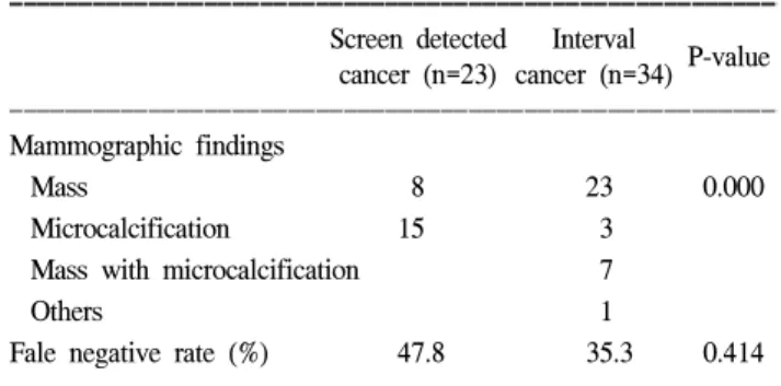
2) 방사선학적 특징(Table 2)
유방촬영상 소견으로 선별검사군에서 미세석회가 많았 고 중간암에서는 결절이 많았다(P=0000). 또한 두 군에서 진단 전의 마지막 유방촬영상의 위음성률을 비교하면 선별 검사군에서 48%, 중간암에서 35%로 선별검사군에서 오히 려 높았으나 통계적 의의는 없었고(P=0.414), 위음성을 제 외한 엄밀한 의미의 중간암은 65%를 차지하였다. 전체 환 자에서 유방촬영상의 위음성률은 40.4%였으며 HRT를 시 행받은 환자에서 52%로 HRT를 시행받지 않은 환자의 26.9%보다 높았으나 통계적 의의는 없었다(P=0.103). 이전 의 유방촬영상에서 위음성 소견을 보였던 환자들의 이전 검사와의 간격을 비교해 볼 때 선별검사군은 평균 12.3개 월, 중간암환자군의 경우 17.5개월이었다. 또한 결절의 경 우 양성으로 간주하였거나 비특이적 소견이어서 더 이상의 검사를 시행하지 않은 경우가 대부분이었고 미세석회의 경 우 기존의 양성으로 간주하였던 것이 범위가 확대되거나 수가 증가한 것이 대부분이었다(표에 나타나 있지 않음).
3) 병리학적 특징(Table 3)
중간암의 경우 병리조직학적 유형으로 침윤성 유관암 (Invasive Ductal Carcinoma)의 비율이 높았고(P=0.015), 병기 가 높았다(P=0.019). 선별검사군에 속하는 12명의 침윤성 유 관암 환자의 경우 평균 종양의 크기는 1.5 cm이었고, 중간암 의 경우 29명의 침윤성 유관암 환자의 평균 종양 크기는 2.4 cm으로 선별검사군보다 컸으나 통계학적 유의 수준은 아니 었다(P=0.083). 그 외에 림프절 전이유무, 호르몬 수용체 유 무, c-erbB2 및 p53 양성여부에는 두 군 간의 차이가 없었다.
Table 1. Clinical characteristics of screen detected cancer and interval cancer of the breast
ꠚꠚꠚꠚꠚꠚꠚꠚꠚꠚꠚꠚꠚꠚꠚꠚꠚꠚꠚꠚꠚꠚꠚꠚꠚꠚꠚꠚꠚꠚꠚꠚꠚꠚꠚꠚꠚꠚꠚꠚꠚꠚꠚꠚꠚꠚꠚꠚꠚꠚꠚꠚꠚꠚꠚ Screen detected Interval
P-value cancer (n=23) cancer (n=34)
ꠏꠏꠏꠏꠏꠏꠏꠏꠏꠏꠏꠏꠏꠏꠏꠏꠏꠏꠏꠏꠏꠏꠏꠏꠏꠏꠏꠏꠏꠏꠏꠏꠏꠏꠏꠏꠏꠏꠏꠏꠏꠏꠏꠏꠏꠏꠏꠏꠏꠏꠏꠏꠏꠏꠏ Mean age, years 51.9±6.0 51.0±8.2 0.659 Interval, months* 14.0±8.1 15.0±7.2 0.616 Hormone replacement therapy
Done 17 14 0.029
Not done 6 20
ꠏꠏꠏꠏꠏꠏꠏꠏꠏꠏꠏꠏꠏꠏꠏꠏꠏꠏꠏꠏꠏꠏꠏꠏꠏꠏꠏꠏꠏꠏꠏꠏꠏꠏꠏꠏꠏꠏꠏꠏꠏꠏꠏꠏꠏꠏꠏꠏꠏꠏꠏꠏꠏꠏꠏ
*interval between two mammograms.
Table 2. Radiological characteristics of screen detected cancer and interval cancer of the breast
ꠚꠚꠚꠚꠚꠚꠚꠚꠚꠚꠚꠚꠚꠚꠚꠚꠚꠚꠚꠚꠚꠚꠚꠚꠚꠚꠚꠚꠚꠚꠚꠚꠚꠚꠚꠚꠚꠚꠚꠚꠚꠚꠚꠚꠚꠚꠚꠚꠚꠚꠚꠚꠚꠚꠚ Screen detected Interval
P-value cancer (n=23) cancer (n=34) ꠏꠏꠏꠏꠏꠏꠏꠏꠏꠏꠏꠏꠏꠏꠏꠏꠏꠏꠏꠏꠏꠏꠏꠏꠏꠏꠏꠏꠏꠏꠏꠏꠏꠏꠏꠏꠏꠏꠏꠏꠏꠏꠏꠏꠏꠏꠏꠏꠏꠏꠏꠏꠏꠏꠏ Mammographic findings
Mass 8 23 0.000
Microcalcification 15 3
Mass with microcalcification 7
Others 1
Fale negative rate (%) 47.8 35.3 0.414
ꠏꠏꠏꠏꠏꠏꠏꠏꠏꠏꠏꠏꠏꠏꠏꠏꠏꠏꠏꠏꠏꠏꠏꠏꠏꠏꠏꠏꠏꠏꠏꠏꠏꠏꠏꠏꠏꠏꠏꠏꠏꠏꠏꠏꠏꠏꠏꠏꠏꠏꠏꠏꠏꠏꠏ
460 대한외과학회지:제 63 권 제 6 호 2002
ꠏꠏꠏꠏꠏꠏꠏꠏꠏꠏꠏꠏꠏꠏꠏꠏꠏꠏꠏꠏꠏꠏꠏꠏꠏꠏꠏꠏꠏꠏꠏꠏꠏꠏꠏꠏꠏꠏꠏꠏꠏꠏꠏꠏꠏꠏꠏꠏꠏꠏꠏꠏꠏꠏꠏꠏꠏꠏꠏꠏꠏꠏꠏꠏꠏꠏꠏꠏꠏꠏꠏꠏꠏꠏꠏꠏꠏꠏꠏꠏꠏꠏꠏꠏꠏꠏꠏꠏꠏꠏꠏꠏꠏꠏꠏꠏꠏꠏꠏꠏꠏꠏꠏꠏꠏꠏꠏꠏꠏꠏꠏꠏꠏꠏꠏ
고 찰
중간암(interval cancer)이란 선별검사 간격의 중간에 증상 이 나타나 유방암으로 진단되는 경우로 정의되며 선별검사 의 유방촬영상에서 정상소견을 보였으나 다음 선별검사 기 간 전에 증상이 있어서 암으로 진단되는 경우를 말한다. 기 관마다 정해진 검사간격이 달라 선별검사 후 12개월 내지 24개월 내에 진단되는 경우로 정의되기도 한다. 세부적으 로는 지난 검사상에 나타났으나 발견하지 못한 경우 위음 성(false negative)으로, 현재 임상적으로 증상이 나타나 암으 로 진단되었으나 현재의 검사상-대개의 경우 유방촬영상을
말함-에도 나타나지 않을 경우에 occult로 분류하고, 현재의 검사에 나타났지만 이전의 선별검사에서 병변을 찾을 수 없는 경우에만 진성(true)의 중간암으로 분류한다.(4) 선별 검사 프로그램에서의 중간암 비율은 프로그램의 질을 결정 하는 중요한 지표가 되기도 하며, 선별검사의 간격과 방법 을 결정하게 되는 중요한 요소이다.
중간암의 비율을 감소시켜 양질의 선별검사프로그램을 유지하기 위해서는 선별검사, 즉 유방촬영상의 위음성률을 최소화시키기 위한 노력이 필요한데 이를 위해서 유방촬영 상의 정확도에 영향을 줄 수 있는 인자들을 고려해 볼 필요 가 있다. 검사과정이나 기계, 기술적인 문제점 외에도 방사 선과 의사의 판독에 오류가 있을 수 있고, 환자의 나이, 인 종, 유방 밀도, 가족력, 호르몬치료요법의 시행 여부 등의 환자적 요인도 영향을 줄 수 있다.(1,3,5) 또한 lobular histology 나 저등급 등 종양 자체의 성질도 유방촬영상의 민감도를 낮출 수 있다. 종양 자체가 보다 공격적이고 성장이 빠른 생물학적 특징을 갖고 있는 경우 진성(true)의 중간암의 형 태로 나타날 수 있는데 이는 젊은 여성의 경우 더욱 그러하 다.(5) 이 경우 선별검사로 진단되는 유방암 환자는 물론이 고 일반적인 증상으로 진단되는(symptomatic cancer) 환자군 보다 예후가 좋지 않은 것으로 보고, 보다 공격적이고 빠른 성장속도를 가지며 유전적 불안전성, 불량한 예후 인자와 의 연관성들을 보고하였다.(6-9) 그러나 생존연구와 관련하 여 대체적으로 볼 때 중간암의 예후는 선별검사로 진단되 는 유방암 환자군보다 나쁘고 증상이 있어 진단되는 유방 암환자군보다 나쁘지 않은 것으로 보고되고 있다.(10-14) 본 연구에서도 중간암환자군의 경우 선별검사군과 비교 할 때 관상피내암보다 침윤성 유관암의 비율이 더 많았고 (P=0.015) 침윤성 유관암의 경우 평균 크기가 더 크고(P=
0.83) 병기가 높았다(P=0.019). 두 군의 유방촬영상을 비교 해 볼 때 중간암 환자군에서 미세석회 소견보다 결절이 우 세했고 선별검사군은 그 반대였다. 이는 두 종양의 생물학 적 이질성을 시사하는 부분이기도 하여 보다 객관적인 연 구의 뒷받침이 필요하리라 본다.
유방촬영술의 위음성률은 선별검사군에서 47.8%로 중간 암환자군의 35.5%보다 높았으나 통계적 의의는 없었고(P=
0.414), 이는 선별검사군에서 호르몬 대체요법을 시행 받았 던 환자가 더 많은 것이(P=0.029) 영향을 주었으리라 추측 된다. 두 군에서 모두 비교적 높은 위음성률을 보인 것은 이전 유방촬영상의 양성 및 비특이적 소견을 위음성으로 분류한 것에서 기인한 것으로 생각된다. 위음성을 보였던 환자들의 이전의 유방촬영상을 재검토했을 때 결절의 경우 는 대부분이 양성결절로 간주하였던 경우이고 미세석회의 경우 기존의 존재하던 미세석회의 범위와 수가 증가를 보 인 경우가 대부분이었다. 그렇다면 유방촬영상에 결절 소 견이 있을 경우 보다 적극적으로 초음파검사를 시행하고 미세석회에 대한 확대촬영과 단기추적검사를 철저히 시행 Table 3. Pathological characteristics of screen detected cancer and
interval cancer of the breast
ꠚꠚꠚꠚꠚꠚꠚꠚꠚꠚꠚꠚꠚꠚꠚꠚꠚꠚꠚꠚꠚꠚꠚꠚꠚꠚꠚꠚꠚꠚꠚꠚꠚꠚꠚꠚꠚꠚꠚꠚꠚꠚꠚꠚꠚꠚꠚꠚꠚꠚꠚꠚꠚꠚꠚ Screen detected Interval
P-value cancer (n=23) cancer (n=34)
ꠏꠏꠏꠏꠏꠏꠏꠏꠏꠏꠏꠏꠏꠏꠏꠏꠏꠏꠏꠏꠏꠏꠏꠏꠏꠏꠏꠏꠏꠏꠏꠏꠏꠏꠏꠏꠏꠏꠏꠏꠏꠏꠏꠏꠏꠏꠏꠏꠏꠏꠏꠏꠏꠏꠏ Tumor size,
1.5±1.0 2.4±1.5 0.083
cm (n=41)*
Histology
DCIS 11 5 0.015
Invasive carcinoma 12 29
LN metastasis
Negative 20 23 0.124
Positive 3 11
Stage
0 11 5 0.019
I 9 14
II 3 12
III 0 3
Nuclear grade (n=32)*
I 3 2 0.154
II 4 6
III 3 14
Estrogen receptor (-) 6 15 0.250
Estrogen receptor (+) 14 16
Progesterone
8 13 1.000
receptor (-) Progesterone
12 18
receptor (+)
C-erbB2 (-) 10 13 1.000
C-erbB2 (+) 10 15
p53 (-) 10 16 0.770
p53 (+) 10 12
ꠏꠏꠏꠏꠏꠏꠏꠏꠏꠏꠏꠏꠏꠏꠏꠏꠏꠏꠏꠏꠏꠏꠏꠏꠏꠏꠏꠏꠏꠏꠏꠏꠏꠏꠏꠏꠏꠏꠏꠏꠏꠏꠏꠏꠏꠏꠏꠏꠏꠏꠏꠏꠏꠏꠏ
*in invasive carcinoma.
김준영 외:선별검사로 진단된 유방암과 중간암(Interval Cancer)의 비교 461 ꠏꠏꠏꠏꠏꠏꠏꠏꠏꠏꠏꠏꠏꠏꠏꠏꠏꠏꠏꠏꠏꠏꠏꠏꠏꠏꠏꠏꠏꠏꠏꠏꠏꠏꠏꠏꠏꠏꠏꠏꠏꠏꠏꠏꠏꠏꠏꠏꠏꠏꠏꠏꠏꠏꠏꠏꠏꠏꠏꠏꠏꠏꠏꠏꠏꠏꠏꠏꠏꠏꠏꠏꠏꠏꠏꠏꠏꠏꠏꠏꠏꠏꠏꠏꠏꠏꠏꠏꠏꠏꠏꠏꠏꠏꠏꠏꠏꠏꠏꠏꠏꠏꠏꠏꠏꠏꠏꠏꠏꠏꠏꠏꠏꠏꠏ 함으로써 유방촬영술의 위음성률과 진단의 지연을 최소화
할 수 있다고 생각한다.
또한 본 연구에서 중간암 환자의 검사 간격이 평균 17.5개 월이었던 것은 1년의 검사 간격을 엄격히 적용함으로써 중 간암의 비율을 감소시킬 수 있음을 의미한다. 선별검사의 간 격이 짧을수록 유방암으로 인한 사망률의 감소는 극대화되 며 중간암의 빈도를 최소화할 수 있다. 그러나 비용상의 문 제와 나이에 따른 종양의 생물학적 특징을 감안할 때 연령 에 따른 차별화가 필요할 것으로 본다. Cady 등은 여성들의 유방암 선별검사기간에 대해 40∼49세는 반년마다, 50∼59 세는 1년마다, 60∼69세는 2년에 1번, 70∼79세는 3년에 1번 주기로 유방촬영술을 시행할 것을 권장하기도 하였다.(15) 이는 젊은 여성일수록 나이든 여성과 비교하였을 때 단기 간에 급속히 자라며 고분화등급(high grade)으로 진행할 가 능성이 높고 예후가 더 나쁘기 때문이라고 하였다.(16) 유방촬영상의 재판독(double reading)은 유방암의 진단율 을 올리고 중간암의 비율을 낮출 수 있는 또 하나의 방법이 다. 하지만 recall rate (추가 검사를 위한 재방문)을 올려 선 별검사를 받는 사람들의 불안을 야기시킬뿐만 아니라 시간 과 비용 증가의 문제를 야기하므로 적절한 균형이 필요하 다고 본다.(17) 그 외에 비대칭음영, 비특이적이거나 양성소 견으로 보이는 음영이 새로이 발견되는 경우, architectural distortion 등은 위음성 유방촬영상의 주된 소견이므로 이를 숙지해야 하겠다.(1,17,18)
결 론
중간암의 경우 선별검사군보다 침윤성 유방암이 더 많고 크기가 크며 병기가 더 높았다. 중간암의 경우 유방촬영상에 미세석회보다는 결절 소견이 많았고 비록 통계적으로 유의한 수준은 아니었으나 선별검사군보다 위음성률이 낮았다. 위음 성률을 낮추기 위해 유방촬영술을 보완하기 위한 초음파 검 사나 확대촬영이 필수적이며 검사 간격에 대한 표준화가 필 요하리라 생각된다. 또한 향후 중간암의 생물학적 특징 및 생 존율에 관한 보다 객관적인 연구가 필요하리라 본다.
REFERENCES
1) Burrell HC, Sibbering DM, Wilson ARM, Pinder SE, Evans AJ, Yeoman LJ, et al. Screening interval breast cancers: mam- mographic features and prognostic factors. Radiology 1996;
199:811-7.
2) Rosenberg RD, Hunt WC, Williamson MR, Gilliland FD, Wiest PW, Kelsey CA, et al. Effects of age, breast density, ehtnicity, and estrogen replacement therapy on screening mam- mographic sensitivity and cancer stage at diagnosis: Review of 183,134 screening mammograms in Albuquerque, New Mexico. Radiology 1998;209(2):511-8.
3) Wang H, Bjurstam N, Bjorndal H, Braaten A, Eriksen L, Skaane P, et al. Interval cancers in the Norwegian breast cancer screening program: frequency, characteristics and use of HRT. Int J Cancer 2001;94:594-8.
4) DeGroote R, Rush BF Jr, Milazzo J, Warden MJ, Rocko JM.
Interval breast cancer: a more aggressive subset of breast neoplasias. Surgery 1983;94(4):543-7.
5) Simpson W, Neilson F, Young JR. Northern Region Breast Screening Radiology Aduit Group. The identification of false negatives in a poplulation of interval cancers: a method for aduit of screening mammography. The Breast 1995;4:183-8.
6) Gilliland FD, Joste N, Stauber PM, Hunt WC, Rosenberg HR, Redlich G, et al. Biologic characteristics of interval and screen-detected breast cancers. JNCI 2000;92(9):743-9.
7) Arnerlov C, Emidin SO, Lundgren B, Roos G, Soderstrom J, Bjersing L. Bresat caecinoma growth rate described by mam- mographic doubling time and S-phase fraction. Correlations to clinical and histopathologic factors in a screened population.
Cancer 1992;70:1928-34.
8) Lipponen P, Aaltomaa S, Kosma VM, Syrjanen K. Apoptosis in breast cancer as related to histopathological characteristics and prognosis. Eur J Cancer 1994;30:2068-73.
9) Barnes DM, Dublin EA, Fisher CJ, Levision DA, Millis RR.
Immunohistochemical detection of p53 protein in mammary carcinoma: an important new independent indicator of prog- nosis? Hum Pathol 1993;24:469-76.
10) Cowan WK, Kelly P, Sawan A, Cunliffe WJ, Henry L, Higgs MJ, et al. The pathological and biological nature of screen- detected breast carcinomas: a morphological and immunohisto- chemical study. J Pathol 1997;182:29-35.
11) Koivunen D, Zhang X, Blackwell C, Adelstein E, Humphrey L. Interval breast cancers are not biologically distinct-just more difficult to diagnose. Am J Surg 1994;169:538-42.
12) Cowan WK, Angus B, Gray JC, Lunt LG, Al-Tamimi SR. A study of interval breast cancer within the NHS bresat screening programme. J Clin Pathol 2000;53:140-6.
13) Schroen AA, Wobbes T, van der Sluis RF. Interval carcinoma of the breast: a group with intermediate outcome. J Sug Oncol 1996;63:141-4.
14) Brekelmans CT, Peeters PH, Deurenberg JJ, Collette HJ. Sur- vival in interval breast cancer in the DOM Screening Pro- gramme. Eur J Cancer 1995;31A:1830-5.
15) Cady B, Michaelson JS. The life-sparing potential of mam- mographic screening. Cancer 2001;91(9):1699-703.
16) Michaelson JS, Kopans DB, Cady B. The breast carcinoma screening interval is important. Cancer 2000;88:1282-4.
17) Goergen SK, Evans J, Cohen GPB, MacMillan JH. Chracter- istics of breast carcinomas missed by screening radiologists.
Radiology 1997;204:131-5.
18) van Dijck JAAM, Verbeek ALM, Hendriks JHCL, Holland R.
The current detectability of breast cancer in a mammographic screening program. Cancer 1993;72(6):1933-8.