817
Original ArticleKorean Circulation J 2006;36:817-819
ISSN 1738-5520
ⓒ 2006, The Korean Society of Circulation CASE REPORT
Anomalous Origin of the Left Coronary Artery from the Right Sinus of Valsalva, which Presented as Acute Myocardial Infarction
Hyun-O Cho, MD, Kil-Hyun Cho, MD, Yong-Suk Jeong, MD, Sung-Gyun Ahn, MD, Sung-Jin Choi, MD, Jae-Yeon Yoo, MD and Eun-Jung Kim, MD
Department of Cardiology, Handong University, Sunlin Hospital, Pohang, Korea ABSTRACT
Among all the congenital coronary anomalies, an anomalous origin of the left coronary artery (LCA) from the right sinus of Valsalva is rare. A 48-year-old male patient suffering with lateral acute myocardial infarction was re- ferred for primary percutaneous coronary intervention. The initial angiogram failed to show the LCA, which origi- nated from the right coronary sinus. A critical stenotic lesion was observed in the distal left circumflex artery. The lesion was treated successfully with stenting. We report here on a case of an anomalous origin of the left coronary artery from the right sinus of Valsalva, and the patient presented as acute myocardial infarction. He was successfully treated with primary percutaneous intervention. (Korean Circulation J 2006;36:817-819)
KEY WORDS:Myocardial infarction;Coronary vessel anomalies.
Introduction
Congenital coronary anomalies are presented in app- roximately 1% of the patients referred for cardiac cathe- terization.
1-3)Among the congenital coronary anomalies, an separate anomalous origin of all the coronary arteries from the right sinus of Valsalva is very uncommon.
4)We report here on a case of successful primary percutaneous coronary intervention(PCI) for acute myocardial infarc- tion(AMI), and this was combined with an anomalous origin of the left coronary artery from the right sinus of Valsalva.
Case
A 48-year-old male patient with a history of hyperten- sion and smoking visited the emergency room with com- plaints of severe chest pain that had lasted for 1 hour. His blood pressure was 150/90 mmHg. The initial electro- cardiogram showed 2 mm of ST-segment elevation in leads I and aVL, and 2 mm of ST-segment depression in
leads II, III, aVF and V5-6. The CK-MB level and tro- ponin-I level were elevated. The initial chest X-ray sho- wed mild cardiomegaly without pulmonary edema. The patient was referred for primary PCI.
At first, we tried to perform left coronary angiogra- phy, but we could not find the orifice of the left coronary artery(LCA). On aortography, the left anterior descen- ding(LAD), left circumflex(LCx) and right coronary ar- teries(RCA) were seen to arise separately from the right coronary sinus and there was a diffuse stenotic lesion in the distal LCx coronary artery(Fig. 1A). A 6 French Am- platz right guiding catheter was engaged into the ostium of the LCx coronary artery. The lesion was crossed with a 0.014 hydrophilic coated floppy guidewire. After pre- dilation, a sirolimus-eluting stent(3.0 mm×33 mm) was implanted over the lesion at the distal LCx. The angio- graphic appearance was good with TIMI 3 flow(Fig. 1B).
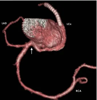
The 16-sliced multi-detector computed tomography sho- wed the LAD, LCx and RCA coronary arteries branching off separately from the right coronary sinus(Fig. 2). The patient has remained asymptomatic and there was no evidence of instent restenosis on the 6-month follow-up coronary angiography.
Discussion
Failure to identify the anomalous origin of coronary arteries can lead to inadequate diagnosis and prolonged procedures, which can result in serious complications,
Received:February 27, 2006 Revision Received:May 2, 2006 Accepted:July 6, 2006
Correspondence:Hyun-O Cho, MD,Department of Cardiology, Handong University, Sunlin Hospital, #69-7, Daesin-dong, Buk-gu, Pohang 791- 704, Korea
Tel: 82-54-245-5481, Fax: 82-54-245-5547 E-mail: [email protected]
818
·Korean Circulation J 2006;36:817-819and especially during acute myocardial infarction.
5)7)It is a huge mistake to assume that a vessel is occluded when, in fact, it has not been visualized due to an ano- malous origin.
5)When the origin of vessels has not been identified by initial angiography, the interventionist has to consider the anomalous origin of a coronary artery and then try to identify the anomalous origin of such a vessel by immediate left ventriculography or aortogra- phy.
6)There are four subtypes of anomalous origin of the left main coronary artery from the right coronary sinus (Fig. 3A).
7)The present case does not belong to any of these subtypes(Fig. 3B).
It has been reported that anomalous coronary arteries are prone to atherosclerosis.
8)9)The coronary blood flow would be disturbed in anomalous coronary arteries ori-
ginating from the opposite side coronary sinus, which is located between the pulmonary trunk and the ascen- ding aorta.
6)10-12)About half of the patients with ano- malous LCA arising from the right coronary sinus die before the age of 20 years, and usually during or shortly after vigorous exertion.
13-15)However, our patient did not have any cardiac symptoms until the age 48 years, even though there was a significant atherosclerotic chan- ge in the LCx coronary artery. It appears that the cause
RCA
LAD LCx
Fig. 2. Multi-detector computed tomography coronary angiography showing all 3 vessels arising from the right coronary sinus (arrow).
LAD: left anterior descending artery, LCx: left circumflex artery, RCA: right coronary artery.
RVOT LAD
RCA
LCx LAD LCx
RCA
A B
Fig. 1. Anterioposterior caudal views. A: pre-intervention angiography shows a large right coronary artery (RCA) and a left coronary artery (LCA) branching off separately from the right coronary sinus, and there was a diffuse, critical lesion in the distal left circumflex (LCx) coronary artery (arrow). B: post-intervention angiography shows a good appearance with TIMI 3 flow. The arrowhead indicates the stent de- ployed in the LCx coronary artery. LAD: left anterior descending artery, LCx: left circumflex artery, TIMI: thrombolysis in myocardial infarction.
Fig. 3. A: schema of four subtypes of anomalous origin of left main coronary artery from right coronary sinus.16) B: schema of the present case.
LMCA: left main coronary artery, LCCA: left circumflex coronary artery, LADCA: left anterior descending coronary artery, RCA: right coronary artery, LAD: left anterior descending, RVOT: right ventricular outflow tract, P: posterior sinus, R: right sinus, L: left sinus, A: anterior sinus.
Aorta
Pulmonary trunk RCA
P LCCA
LAD A
L L R
P RCA
LADCA RCA
RCA
RCA
LADCA
LADCA LADCA
LCCA LMCA
LCCA LMCA
LMCA
LCCA LCCA
Aorta
Pulmonary trunk A A
A L L
L L L
L
L
P R R R
R
P P P P
P
P
RVOT Ventricular
septum
B
A
Hyun-O Cho, et al:Anomalous Coronary Artery and AMI·819
of AMI in this patient might be related to coronary ar- tery risk factors such as hypertension and smoking rather than to the anomalous coronary artery itself.
Angiographic recognition of unsuspected coronary anomalies is considered important for making an app- ropriate diagnosis and managing acute myocardial in- farction. We report here on a case of successful primary percutaneous coronary intervention for acute myocar- dial infarction combined with an anomalous origin of the LAD and LCx coronary arteries that arose separately from the right coronary sinus. This coronary artery ano- maly did not belong to any of the four classical subtypes of left main coronary artery arising from the right co- ronary sinus.
REFERENCES
1) Kardos A, Babai L, Rudas L, et al. Epidemiology of congenital coronary anomalies: a coronary arteriography study on a central European population. Cathet Cardiovasc Diagn 1997;42:270-5.
2) Yamanaka O, Hobbs RE. Coronary artery anomalies in 126,595 patients undergoing coronary arteriography. Cathet Cardiovasc Diagn 1990;21:28-40.
3) Moreno R, Ramirez CB, Garcia J, Macaya C. Primary stenting of an anomalous left main trunk originating from the right coro- nary artery during acute myocardial infarction. J Invasive Cardiol 2004;16:159-61.
4) Yang DG, Cha KS, Kim BG, et al. Anomalous origin of the left main coronary artery from the right sinus of valsalva. Korean Circ J 2000;30;1165-9.
5) Serota H, Barth CW 3rd, Seuc CA, et al. Rapid identification of the course of anomalous coronary arteries in adults: the “dot and eye” method. Am J Cardiol 1990;65:891-8.
6) Cheitlin MD, de Carstro CM, McAllister HA. Sudden death as comlication of anomalous left coronary origin from the anterior sinus of valsalva: a not-so-minor congenital anomaly. Circulation 1974;50:780-7.
7) Ishikawa T, Brandt PW. Anomalous origin of the left main coro- nary artery from the right anterior aortic sinus: angiographic identification of anomalous course. Am J Cardiol 1985;55:770-6.
8) Harikrishnan S, Sonney PJ, Jaganmohan T, et al. Congenital co- ronary anomalies of origin and distribution in adults: a coronary arteriographic study. Indian Heart J 2002;54:271-5.
9) Wilkins CE, Betancourt B, Mathur VS, et al. Coronary artery anomalies. Texas Heart J 1988;15:166-73.
10) Choi YM. Congenital anomalies of the coronary arteries detected in adulthood. Korean Circ J 1997;27:287-95.
11) Mustafa I, Gula G, Radley-Smith R, Durrer S, Yacoub M. Ano- malous origin of the left coronary artery from the anterior aortic sinus: a potential cause of sudden death. J Thorac Cardiovasc Surg 1981;82:297-300.
12) Benge W, Martins JB, Funk DC. Morbidity associated with ano- malous origin of the right coronary artery from the left sinus of valsalva. Am Heart J 1980;99:96-100.
13) Angelini P, Velasco JA, Flamm S. Coronary anomalies: incidence, pathophysiology, and clinical relevance. Circulation 2002;105:
2449-54.
14) Angelini P. Coronary artery anomalies: current clinical issues.
Texas Heart Inst J 2002;29:271-8.
15) Virmani R, Burke AP, Farb A. The pathology of sudden cardiac death in athletes. In: Williams RA, editor. The Athlete and Heart Disease: diagnosis, evaluation and management. Philadelphia:
Lipincott Williams & Wilkins; 1999. p.249-72.
16) Roberts WC, Shirani J. The four subtypes of anomalous origin of the left main coronary artery from the right aortic sinus(or from the right coronary artery). Am J Cardiol 1992;70:119-21.