https://doi.org/10.5423/RPD.2021.27.1.38
Research in Plant Disease pISSN 1598-2262, eISSN 2233-9191 www.online-rpd.org
The Korean Society of Plant Pathology
This is an open access article distributed under the terms of the Creative Commons Attribution Non-Commercial License (http://creativecommons.org/licenses/ by-nc/4.0/), which permits unrestricted non-commercial use, distribution, and reproduction in any medium, provided the original work is properly cited.
Research Article Open Access
Development of Diagnostic Technology of Xylella fastidiosa Using
Loop-Mediated Isothermal Amplification and PCR Methods
Suyoung Kim, Yujin Park, and Gidon Kim*
Plant Quarantine Technology Center, Animal and Plant Quarantine Agency, Gimcheon 39660, Korea
Xylella fastidiosa is the most damaging pathogen in many parts of the world. To increase diagnostic capability of X. fastidiosa in the field, the loop-mediated isothermal amplification (LAMP) and polymerase chain reaction (PCR) assay were developed to mqsA gene of citrate-synthase (XF 1535) X. fastidiosa and evaluated for speci-ficity and sensitivity. Both assays were more robust than current published tests for detection of X. fastidiosa when screened against 16 isolates representing the four major subgroups of the bacterium from a range of host species. No cross reaction with DNA from healthy hosts or other species of bacteria has been observed. The LAMP and PCR assays could detect 10-4 pmol and 100 copies of the gene, respectively. Hydroxynaphthol blue was evaluated as an endpoint detection method for LAMP. There was a significant color shift that sig-naled the existence of the bacterium when at least 100 copies of the target template were present.
Keywords: Diagnostic, Loop-mediated isothermal amplification, PCR, Xylella fastidiosa
Introduction
Xylella fastidiosa (Wells et al., 1987) is a pathogen of bacte-rial plants that cause many economically significant diseases, including grapevine disease of Pierce, citrus veinal chlorosis, almond leaf scorch, phony peach, and leaf scorch on a range of ornamental plants and shade trees (Hopkins and Purcell, 2002). X. fastidiosa is a globally regulated and banned patho-gen. Leafhoppers of the subfamily Cicadellinae (Hemiptera: Cicadellidae) and spittle bugs or frog hoppers of the family Cercopidae (Hemiptera) are the most common known vec-tors (Purcell, 1997). The distribution of X. fastidiosa is usually restricted to the Americas (Purcell, 1997), with two excep-tions, in Vitis vinifera in Kosovo (Berisha et al., 1998) and pear in Taiwan (Leu, 1993). X. fastidiosa is known to be vulnerable to low temperatures, which has confined its movement to
regions with temperate climates and, in particular, to cold winters (Purcell, 1997). Many colder parts of the world, how-ever, have one or more vector species, such as the spittlebug (Philaenus spumarius), so there is a possibility for X. fastidiosa to spread to such areas if strains are cold-tolerant, such as al-mond leaf scorch, become established (Purcell, 1997). From a quarantine viewpoint, the main aspect of any exclusion technique is rapid identification and diagnosis. Present X. fastidiosa diagnostic tests include bacterial cell culture, tradi-tional polymerase chain reaction (PCR) (Huang, 2009; Huang et al., 2006; Minsavage et al., 1994; Pooler and Hartung, 1995; Rodrigues et al., 2003) and real-time PCR (Francis et al., 2006; Schaad et al., 2002). While many of these methods have been widely used in the laboratory, most of these methods are not readily transferable to the field. In addition, the PCR assay was developed over 15 years ago. When the X. fastidi-osa DNA sequence was little usable, this assay is widely used for quarantine screening and, thus, checking that it reliably detects all isolates of the bacterium is especially essential. Alternative methods of detection have been considered in
*Corresponding author Tel: +82-54-912-0675 Fax: +82-54-912-0688 E-mail: [email protected] ORCID https://orcid.org/0000-0002-0560-4714 Received November 10, 2020 Revised March 24, 2021 Accepted March 26, 2021
and evaluation of a LAMP assay for X. fastidiosa is presented here in order to improve diagnostic capacity by allowing sur-veillance activities, improving response times during incur-sions, and enabling testing at the border for imported goods. During the development of the LAMP assay, the potential for developing PCR was based on the identification of the same
from commercial sources (Korea Agricultural Culture Collec-tion, Suwon, Korea).
PCR primer design and PCR condition. PCR primers are
the method to diagnose a large number of samples quickly, and fast and accurate diagnostics can be made without
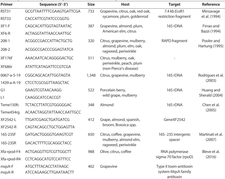
Table 1. Sequence of PCR primers for Xylella fastidiosa used in this study
Primer Sequence (5’-3’) Size Host Target Reference
RST31 GCGTTAATTTTCGAAGTGATTCGA 733 Grapevine, citrus, oak, red oak, sycamore, plum, goldenrod
7.4 kb EcoR1 restriction fragment
Minsavage et al. (1994)
RST33 CACCATTCGTATCCCGGTG
XF1-F CAGCACATTGGTAGTAATAC 387 Grapevine, almond, plum,
American elm, citrus
16S rDNA Firrao and
Bazzi (1994)
XF6-R ACTAGGTATTAACCAATTGC
208-1 ACGGCCGACCATTACTGCTG 320 Citrus, grapevine, mulberry,
almond, plum, elm, oak, ragweed, periwinkle
RAPD fragment Pooler and Hartung (1995)
208-2 ACGGCCGACCCGGAGTATCA
XF176f AAACAATCACAGGGGACTGC 511 Citrus, mulberry, oak,
periwinkle, peach, plum (non-Pierce’s disease)
-
XF686r ATATTCATAGATTCCGTCGA
0067-a-S-19 CGGCAGCACATTGGTAGTA 1,348 Citrus, grapevine, mulberry 16S rDNA Rodrigues et al.
(2003) 1439-a-A-19 CTCCTCGCGGTTAAGCTAC
G1 GAAGTCGTAACAAGG 522 Porcelain berry,
wild grape, mulberry
16S rDNA Huang and
Sherald (2004)
L1 CAAGGCATCCACCGT
Teme150fc TCTACCTTATCGTGGGGGAC 348 Almond 16S rDNA Chen et al.
(2005) Teme454rg ACAACTAGGTATTAACCAATTGCC
XF2542-L TTGATCGAGCTGATGATCG 412 Grape, almond, spanish,
broom, Brassica spp.
GeneXF2542
XF2542-R CAGTACAGCCTGCTGGAGTTA
16S-23SF GATGACTGGGGTGAAGTCGT 650 Citrus, coffee, grapevine, mulberry, almond elm, ragweed, periwinkle 16S-23S intergenic spacer Martinati et al. (2007) 16S-23SR GACACTTTTCGCAGGCTACC
Xfa-rpod-F4 ACTGAGGTTGTCGTTGGCTT 988 Olive, citrus, coffee RNA polymerase
sigma-70 factor (rpoD)
Bleve et al. (2016) Xfa-rpod-R4 CCTCAGGCATGTCCATTTCC
mqsA-F ATGCTTTACACCTATAAGC 402 Grapevine Type II toxin-antitoxin
system MqsA family antitoxin
mqsA-R ATCCAGAAGCTTGAATAACTT
complicated equipment. After exploring different gene sites linked to the diagnosis of X. fastidiosa, primers were therefore prepared after the optimal target gene was selected (Table 1). The mqsA gene sequence of the standard strain X. fastidi-osa was used to prepare PCR primers (Fig. 1). For PCR, the primer is 20 pM and AccuPower PCR premix (1 U Taq DNA polymerase, 250 μm dNTP, 10 mM Tris-HCI, 40 mM KCI, 1.5 mM MgCI2, stabilizer and tracking dye: Bioneer, Daejeon,
Korea), template genomic DNA, a reaction solution with a total volume of 20 μl was prepared (Francis et al., 2006). Initial denature (2 min at 94°C), denature (1 min at 94°C), annealing (1 min at 55°C), extension (1 min 30 sec at 72°C) After react-ing a total of 35 cycles, last extension (5 min at 72°C), reaction was carried out (Fukuta et al., 2003). Thereafter, the product was subjected to electrophoresis at 50 V for 40 min using 1.5% agarose gel added with SYBR Green I and 1× TAE buf-fer, stained with ethidium bromide, and observed under UV illumination.
LAMP primer design. In order to design the LAMP
PCR primer, the primer was designed by referring to the 10 kinds of 16S rDNA nucleotide sequences registered in Gen-Bank. LAMP DNA oligonucleotide primer of X. fastidiosa is the BLAST program (Berisha et al., 1998). The following sequence was designed by selecting a primer for LAMP of X. fastidiosa using Primer Explorer version 3 (https:// www.primerexplorer.jp/lamp3.0.0/index.html), a LAMP primer designing software (Fukuta et al. 2003). In the primer set, the following two inner primers (forward inner primer [FIP], backward inner primer [BIP]) that can recognize six specific regions of the target sequence composed of four primers are the complementary nucleotide sequence of the X. fastidi-osa sequence and loop. It was produced by combining the partial TTTT spacer of the sequence forming the reverse tran-scription LAMP (Fukuta et al., 2003). The following two outer primers (F3: forward outer primer, B3: backward outer prim-er) were designed to be located outside the inner primer, respectively. A total of 6 primers were produced by ordering
Fig. 1. mqsA gene region for the diagnosis of Xylella fastidiosa. Target gene selection; mqsA directly binds to the toxin-antitoxin system
op-eron and has a function to regulate opop-eron self-expression. Blue text is mqsA-F primer; green text is mqsA-R primer.
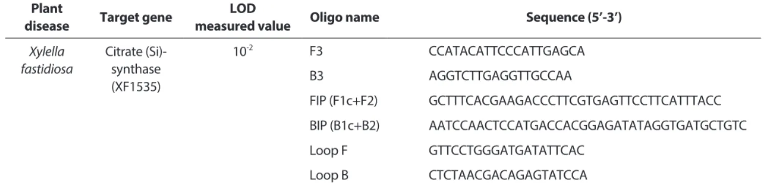
Table 2. Sequence of LAMP primers for Xylella fastidiosa used in this study Plant
disease Target gene measured value LOD Oligo name Sequence (5’-3’)
Xylella fastidiosa Citrate (Si)-synthase (XF1535) 10-2 F3 CCATACATTCCCATTGAGCA B3 AGGTCTTGAGGTTGCCAA FIP (F1c+F2) GCTTTCACGAAGACCCTTCGTGAGTTCCTTCATTTACC BIP (B1c+B2) AATCCAACTCCATGACCACGGAGATATAGGTGATGCTGTC Loop F GTTCCTGGGATGATATTCAC Loop B CTCTAACGACAGAGTATCCA
from Bioneer (Table 2).
Optimization of the XF 1535 gene LAMP assay.
Twen-ty pM F3, B3 primer and 40 pM FIP, BIP primer, reagent 1× reaction mix (20 mM Tris-HCI [pH 8.8], 10 mM (NH4)2SO4,
10 mM KCI, 2 mM MgSO4, 0.4% Triton X-100, 6 mM MgSO4
Green I (Life Technologies, Chicago, IL, USA). This is orange in the absence of an amplicon, and green in the presence of the LAMP amplification product.
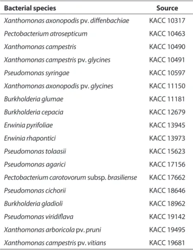
Sensitivity and specificity. Sensitivity and specificity
were tested with Xanthomonas axonopodis pv. diffenbachiae, Pectobacterium atrosepticum, X. campestris, X. campestris pv. glycines, Pseudomonas syringae, Xaxonopodis pv. glycines, Burkholderia glumae, Burkholderia cepacia, Erwinia pyrifoliae, E. rhapontic, Pseudomonas tolaasii, Pseudomonas agarici, Pectobacterium carotovorum subsp. brasiliense, Pseudomo-nas cichorii, Burkholderia gladioli, PseudomoPseudomo-nas viridiflava, X. arboricola pv. prun, and X. campestris pv. vitians (Table 3). DNA (2.0×10⁶ copies/ml) was diluted 10 times each, and the
sensitivity and specificity of PCR and LAMP were tested at a concentration of 2.0×10⁶ to 2.0×10² copies/ml.
Results
Specificity and sensitivity of created mqsA primer set. PCR was used to assess the specificity of the newly developed primer set. As a result of electrophoresis of the
Burkholderia glumae KACC 11181
Burkholderia cepacia KACC 12679
Erwinia pyrifoliae KACC 13945
Erwinia rhapontici KACC 13973
Pseudomonas tolaasii KACC 15623
Pseudomonas agarici KACC 17156
Pectobacterium carotovorum subsp. brasiliense KACC 17662
Pseudomonas cichorii KACC 18646
Burkholderia gladioli KACC 18962
Pseudomonas viridiflava KACC 19142
Xanthomonas arboricola pv. pruni KACC 19495
Xanthomonas campestris pv. vitians KACC 19681
Fig. 2. Specificity of created mqsA primer set. Verification of the ability to detect Xylella fastidiosa for one type of primer (mqsA-F/mqsA-R)
PCR product, the amplified result was obtained only in X. fastidiosa, and there was no non-specific reaction with other strains (Fig. 2). The mqsA site contained in toxin-antitoxin is a physiological regulatory site mainly involved in pathogenic-ity and motilpathogenic-ity, and it is considered to be highly useful as a species-specific primer for X. fastidiosa. Their detection ability was confirmed according to the PCR conditions (Fig. 3). The detection capability of Pierce’s disease bacteria X. fastidiosa was tested on 16 positive control and 16 negative control strains in total.
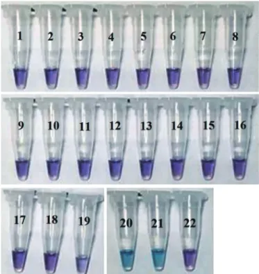
Confirmation of specificity of LAMP method and PCR method. As a result of testing the specificity of LAMP PCR using standard and similar strains, only X. fastidiosa showed a specific reaction (Fig. 4). In the case of LAMP PCR, it is neces-sary to observe the time because it may cause a false-positive reaction after the standard reaction time.
Confirmation of sensitivity of LAMP method and PCR method. After establishing the optimal conditions, the
sensitivity of the LAMP detection method is determined by diluting the Streptococcus uberis culture solution to a concen-tration of 1.0×10 to 1.0×10⁸ cfu/ml, and using the Genomic
DNA Extraction Kit (Bioneer) After extraction, the detection limit values of the LAMP detection method and the PCR method were compared. In the case of PCR, it can be de-tected only up to a concentration of 1.0×10⁴ cfu/ml, while as a result of measuring the detection limit of LAMP, it was con-firmed that it could detect up to a concentration of 1.0×10⁸ cfu/ml. It was confirmed that the LAMP method has a higher detection limit than the general PCR method.
In order to measure the sensitivity of the developed LAMP, the extracted DNA was diluted 10 times in steps, and then developed LAMP and PCR were performed on the genes extracted from each dilution. It was detected that the devel-oped LAMP was 10 times more sensitive than the existing PCR (Fig. 5).
Discussion
LAMP PCR is a technology that uses basic laboratory equip-ment to diagnose a variety of research products, such as in-fectious diseases, in the field (Chen et al., 2008). This time the
Fig. 3. Sensitivity of created mqsA primer set. Verification of
detec-tion ability by 10 times, diluted concentradetec-tion of positive control.
Fig. 4. Loop-mediated isothermal amplification visualized using
hydroxynapthal blue dye showing the sky-blue color change (tubes 20, 21) observed with Xylella fastidiosa-positive samples. Negative samples in which no amplification occurred remain violet (tubes 1 to 19, 22). The list of strains to be used is as follows; 1, Xanthomonas axonopodis pv. diffenbachiae; 2, Pectobacterium atrosepticum; 3, Xanthomonas campestris; 4, Xanthomonas campestris pv. glycines; 5, Pseudomonas syringae; 6, Xanthomonas axonopodis pv. glycines; 7, Burkholderia glumae; 8, Burkholderia cepacia; 9, Erwinia pyrifoliae; 10, Erwinia rhapontici; 11, Pseudomonas tolaasii; 12, Pseudomonas agarici; 13, Pectobacterium carotovorum subsp. brasiliense; 14, Pseu-domonas cichorii; 15, Burkholderia gladioli; 16, PseuPseu-domonas viridi-flava; 17, Xanthomonas campestris pv. vitians; 18, Xanthomonas arbo-ricola pv. pruni; 19, negative control; 20, ATCC 700964D-5 (Xyrella fasitidiosa); 21, ATCC 35881D (Xyrella fasitidiosa); 22, negative control.
LAMP PCR method was developed for use in epidemic and quarantine research. The analysis shows that there was no difference from the conventional PCR method, because of its sensitivity and specificities (Fig. 3). Consequently, the find-ings of this study are considered sufficient for X. fastidiosa di-agnoses with various hosts and symptoms. It is important to observe the time in the case of LAMP PCR, since it can cause a false positive reaction after the standard reaction time. For future more reliable and effective experiments, detailed analysis of the reaction conditions and reaction reagents is necessary.
In addition, microscopy, selective medium, and PCR tech-niques are primarily used in the diagnosis of plant diseases. Recently, by shortening the diagnosis time and using simple diagnosis methods away from competent and complicated diagnosis methods, there is a trend to create diagnostic kits. The LAMP PCR approach is not derived and amplified by the conjugation and expansion at isothermal temperatures and has the advantage, by using mainly 4-6 primers, of increas-ing the PCR specificity in the target species. Since DNA am-plification is possible if the temperature is kept isothermal, only an isothermal maintenance system such as a water bath and heat block can be detected without an expensive PCR device. The detection result can be tested with the naked eye when a fluorescent dye is used, so it can be used im-mediately in the field, and the number of applications of the diagnostic method LAMP PCR is increasing in the field.
X. fastidiosa is a high-risk pathogen that causes disease in a wide variety of plants. Test methods have been developed in European and Mediterranean Plant Protection Organiza-tion and others to diagnose these pathogens (Notomi et al., 2000). However, in recent years, as new genetic knowledge on pathogens has been discovered, it is important to check existing test methods and to diagnose new methods.
There-fore, a new test method was developed to complement the previously developed diagnostic method. The newly developed test method detects the mqsA gene of X. fastidi-osa. The findings showed high specificity and sensitivity as a consequence of checking the specificity and sensitivity of 16 related strains (Figs. 2, 3). It is important to establish a quanti-tative test method (real-time PCR) for more accurate testing in the future.
Conflicts of Interest
No potential conflict of interest relevant to this article was reported.
References
Berisha, B., Chen, Y. D., Zhang, G. Y., Xu, B. Y. and Chen, T. A. 1998. Isolation of Peirce’s disease bacteria from grapevines in Europe. Eur. J. Plant Pathol. 104: 427-433.
Chen, J., Civerolo, E., Tubajika, K., Livingston, S. and Higbee, B. 2008. Hypervariations of a protease-encoding gene, PD0218 (pspB), in Xylella fastidiosa strains causing almond leaf scorch and Pierce’s disease in California. Appl. Environ. Microbiol. 74: 3652-3657.
Chen, J., Groves, R., Civerolo, E. L., Viveros, A., Freeman, A. and Zheng, Y. 2005. Two Xylella fastidiosa genotypes associated with almond leaf scorch disease on the same location in California. Phytopathology 95: 708-714.
Firrao G. and Bazzi C. 1994. Specific identification of Xylella fastidi-osa using the polymerase chain reaction. Phytopathol. Mediterr. 33: 90-92.
Francis, M., Lin, H., Rosa, J. C.-L, Doddapaneni, H. and Civerolo, E. L. 2006. Genome-based PCR primers for specific and sensitive detection and quantification of Xylella fastidiosa. Eur. J. Plant Pathol. 115: 203.
Fukuta, S., Iida, T., Mizukami, Y., Ishida, A., Ueda, J., Kanbe, M. et al. 2003. Detection of Japanese yam mosaic virus by RT-LAMP.
Arch. Virol. 148: 1713-1720.
Goto, M., Honda, E., Ogura, A., Nomoto, A. and Hanaki, K.-I. 2009. Colorimetric detection of loop-mediated isothermal amplifica-tion reacamplifica-tion by using hydroxy naphthol blue. Biotechniques 46: 167-172.
Hopkins, D. L. and Purcell, A. H. 2002. Xylella fastidiosa: cause of Pierce’s disease of grapevine and other emergent diseases. Plant Dis. 86: 1056-1066.
Huang, Q. 2009. Specific detection and identification of Xylella fas-tidiosa strains causing oleander leaf scorch using polymerase chain reaction. Curr. Microbiol. 58: 393-398.
Huang, Q., Bentz, J. and Sherald, J. L. 2006. Fast, easy and efficient DNA extraction and one-step polymerase chain reaction for the detection of Xylella fastidiosa in potential insect vectors. J. Plant Pathol. 88: 77-81.
Huang, Q. and Sherald, J. L. 2004. Isolation and phylogenetic analy-sis of Xylella fastidiosa from its invasive alternative host, porce-lain berry. Curr. Microbiol. 48: 73-76.
Leu, L. S. 1993. Isolation, cultivation, and pathogenicity of Xylella fastidiosa, the causal bacterium of pear leaf scorch disease in Taiwan. Plant Dis. 77: 642-646.
Minsavage, G. V., Thompson, C. M., Hopkins, D. L., Leite, R. M. V. B. C. and Stall, R. E. 1994. Development of a polymerase chain reac-tion protocol for detecreac-tion of Xylella fastidiosa in plant tissue. Phytopathology 84: 456-461.
Notomi, T., Okayama, H., Masubuchi, H., Yonekawa, T., Watanabe, K., Amino, N. et al. 2000. Loop-mediated isothermal amplification of DNA. Nucleic Acids Res. 28: e63.
Pooler, M. R. and Hartung, J. S. 1995. Genetic relationships among strains of Xylella fastidiosa from RAPD-PCR data. Curr. Microbiol. 31: 134-137.
Purcell, A. H. 1997. Xylella fastidiosa, a regional problem or global threat? J. Plant Pathol. 79: 99-105.
Rodrigues, J. L. M., Silva-Stenico, M. E., Gomes, J. E., Lopes, J. R. S. and Tsai, S. M. 2003. Detection and diversity assessment of Xy-lella fastidiosa in field-collected plant and insect samples by us-ing 16S rRNA and gyrB sequences. Appl. Environ. Microbiol. 69: 4249-4255.
Schaad, N. W., Opgenorth, D. and Gaush, P. 2002. Real-time poly-merase chain reaction for one-hour on-site diagnosis of Pierce’s disease of grape in early season asymptomatic vines. Phytopa-thology 92: 721-728.
Tomlinson, J. and Boonham, N. 2008. Potential of LAMP for detec-tion of plant pathogens. CAB Rev. Perspect. Agric. Vet. Sci. Nutr. Nat. Resour. 3: 66.
Wells, J. M., Raju, B. C., Hung, H.-Y., Weisburg, W. G., Mandelco-Paul, L. and Brenner, D. J. 1987. Xylella fastidiosa gen. nov., sp. nov: gram-negative, xylem-limited, fastidious plant bacteria related to Xanthomonas spp. Int. J. Syst. Bacteriol. 37: 136-143.