Ethanol Extract from Asparagus Cochinchinensis Attenuates Glutamate-Induced Oxidative Toxicity in HT22 Hippocampal Cells
Malk Eun Pak1 and Byung Tae Choi1,2*
1Department of Korean Medical Science, School of Korean Medicine, Pusan National University, Yangsan 50612, Korea
2Division of Meridian and Structural Medicine, School of Korean Medicine, Pusan National University, Yangsan 50612, Korea Received July 5, 2016 /Revised August 23, 2016 /Accepted August 23, 2016
We investigated the neuroprotective effect of an ethanol extract from Asparagus cochinchinensis (AC) against glutamate-induced toxicity in the HT22 hippocampal cell, which is an ideal in vitro model for oxidative stress. The neuroprotective effects of AC in HT22 cells were evaluated by analyzing cell via- bility, lactate dehydrogenase (LDH), flow cytometry for cell death types, reactive oxygen species (ROS), mitochondria membrane potential (MMP), and Western blot assays. In the cell death analysis, AC treatment resulted in significantly attenuated glutamate-induced loss of cell viability with a de- crease in LDH release. AC treatment also reduced glutamate-induced apoptotic cell death. In the ROS and MMP analysis, AC treatment inhibited the elevation of intracellular ROS induced by glutamate exposure and the disruption of MMP. In oxidative stress-related proteins analysis, AC treatment in- hibited the expression of poly ADP ribose polymerase and heme oxygenase-1 by glutamate. These re- sults indicate that AC exerts a significant neuroprotective effect against glutamate-induced hippo- campal damage by decreasing ROS production and stabilizing MMP. Thus, AC potentially provides a new strategy for the treatment of oxidative stress-related diseases.
Key words : Asparagus cochinchinensis (AC), hippocampal cell, mitochondria membrane potential, oxidative stress, reactive oxygen species (ROS)
*Corresponding author
*Tel : +82-51-510-8475, Fax : +82-51-510-8437
*E-mail : [email protected]
This is an Open-Access article distributed under the terms of the Creative Commons Attribution Non-Commercial License (http://creativecommons.org/licenses/by-nc/3.0) which permits unrestricted non-commercial use, distribution, and reproduction in any medium, provided the original work is properly cited.
Journal of Life Science 2016 Vol. 26. No. 12. 1458~1465 DOI : http://dx.doi.org/10.5352/JLS.2016.26.12.1458
서 론
Glutamate는 척추동물의 신경계에서 가장 널리 분포하는 대표적인 흥분성 신경전달물질로서[20], 이의 과분비는 신경 독성 즉 excitotoxicity를 유발하며 허혈성 뇌졸중, 간질, 알츠 하이머병, 파킨스병 등 다양한 뇌질환에 관여한다[4, 8]. 신경 세포에서 glutamate에 의한 독성은 두 가지 주된 경로와 관련 되어 있다. 잘 알려진 경로는 glutamate 수용체에 의한 독성으 로 특히 N-methyl-D-aspartate (NMDA) 수용체의 과활성을 통해 칼슘이 과도하게 세포내로 유입되어, protein kinase C를 활성화시키며 free radical형성을 함으로 세포죽음에 이르는 다양한 효소를 발현 시키는 것이다[4, 5].
또 다른 경로는 glutamate 수용체를 거치지 않는 경로로 glutamate/cystine antiporter에 의하여 cysteine의 유입이 억 제됨으로서 세포내에서 cystine의 선구체인 glutathione의 함 량이 감소된다. 이로 인한 산화적 스트레스(oxidative stress)
로서 reactive oxygen species (ROS) 생성물을 증가시켜 이와 관련된 연속적인 반응들로 인해 세포죽음이 일어난다[9, 20, 26]. HT22 해마세포는 기능적인 NMDA와 같은 ionotropic glutamate 수용체의 결여로 인하여 수용체를 통한 독성이 배 제되어 있어 glutamate에 의한 산화적 스트레스를 통해 세포 죽음에 이른다[7]. 따라서 HT22세포는 glutamate에 산화적 스 트레스를 연구하고 이와 관련된 신경퇴행성 질환에 대한 물질 탐색의 이상적인 모델이다[9, 19, 20].
천문동(Asparagus cochinchinensis)은 중국, 일본, 한국 등에 서 자라는 다육생의 식물로서 그 뿌리는 한의학에서 폐와 비 장과 관련된 질병을 치료하고 면역기능을 향상시키는 치료약 으로 사용되어 왔다. 천문동 추출물에 대한 연구로는 노화 실 험모델에서 강한 radical scavenging 능력으로 항산화효과를 가지며[15], TNF-α 분비를 억제시켜 중추신경계에서 항염증 효과가 있다고 발표되었다[13, 14]. 최근에는 천문동의 메탄올 추출물이 세포와 동물 실험을 통해 신경보호효과가 있다고 알려져 있다[12].
천문동 추출물의 신경보호 가능성에 대한 연구와 더불어 [22, 28], 본 연구팀은 동의보감의 textmining기법을 통한 선험 연구에서 천문동을 인지기능 향상에 관여하는 한약재로서 선 별하였으며 기본적인 기능검색에서 해마신경세포 보호의 가 능성을 탐지하였다. 따라서 본 연구는 천문동 추출물이 산화 적 스트레스에 대한 해마신경세포 보호기능을 나타냄으로서 이와 관련된 질환에 적용가능한지를 알아보기 위하여 HT22
해마세포를 이용하여 세포죽음, ROS형성 및 mitochondria membrane potential (MMP) 및 Western blot 분석을 통해 신 경보호효과를 연구하였다.
재료 및 방법
시약 및 재료
L-glutamate, 3-(4,5-dimethylthiazol-2-yl)-2,5-diphenylte- trazolium bromide (MTT), β-actin 항체는 Sigma-Aldrich (St.
Louis, MO, USA)에서 구입하였다. Dulbecco’s modified Eagle’s medium (DMEM), fetal bovine serum (FBS)은 Wel- gene Inc. (Gyeongsan, Korea)에서 구입하였고 나머지 세포주 배양을 위한 시약들은 Gibco/Invitrogen (Carlsbad, CA, USA) 에서 구입하였다. Caspase-3, poly ADP ribose polymerase (PARP)에 대한 항체들은 Cell Signaling Technology (Dan- vers, MA, USA)에서 구입하였다. Heme oxygenase-1 (HO-1) 항체는 Enzo Life Sciences (Farmingdale, NY, USA)에서 구입 하였으며 optic atrophy 1 (Opa-1) 항체는 BD Bioscience (San Diego, CA, USA)에서 구입하였다. 이차항체들은 Enzo Life Sciences (Farmingdale, NY, USA)에서 구입하였다. FITC Annexin Ⅴ apoptosis dectection kit는 BD Bioscience에서 구 입하였고, Lactate dehydreogenase (LDH) cytotoxicity assay kit는 Promega (Madison, WI, USA)에서 구입하였다. ROS de- tection reagent, 5-(and-6)-carboxy-2’,7’-dichloro- dihydro- fluorescein diacetate (carboxy-H2DCFDA), Tetramethyrho- damineethylester (TMRE)은 Invitrogen (Carlsbad, CA, USA) 에서 구입하였다.
천문동의 에탄올추출
천문동(Asparagus cochinchinensis)의 말린 덩이줄기는 화림 제약(Pusan, Korea)에서 구입하였다. 에탄올 추출물은 마른 천문동 100 g을 70% 에탄올 300 ml에 3시간 간격으로 유리막 대로 저어서 하루 동안 실온에 침전하였다. 그 후 0.8-μm Whatman 여과지(No.2)로 두번 여과하고 40℃ water bath에 서 rotary evaporator을 사용하여 농축하였고 마지막으로 동 결건조 하였다. 최종 추출물 25.06 g을 수득하였고 –20도에 보관하여 증류수에 녹여 시료(AC)로 사용하였다.
세포배양 및 추출물처리
HT22 세포는 DMEM medium에 10% FBS, 1% penicillin/
streptomycin을 첨가하여 37℃ 5% CO2가 유지되는 incubator 에서 배양하였다. 세포는 실험적인 처리 전 24시간 동안 배양 하였으며 그 후, 여러 농도의 천문동 추출물을 24시간 동안 전처리하였다. 이후 5 mM glutamate를 천문동 추출물과 함께 24시간 동안 처리하였다.
MTT분석
HT22세포에 대한 생존율은 MTT분석을 이용하여 측정하 였다. 마지막 추출물처리가 끝난 후, 배지를 제거하고 DMEM 에 희석한 0.5 mg/ml MTT용액 100 μl를 각 well에 첨가하였 다. 37℃에 4시간 동안 어두운 곳에서 배양시키고 MTT용액을 조심스럽게 제거하고 DMSO를 사용하여 blue formazan dye 을 녹였다. 그리고 SpectraMAX 190 spectrometer (Molecular Devices, Sunnyvale, CA, USA)를 사용하여 570 nm에서 흡광 도를 측정하였다. 결과는 대조군에 대한 퍼센트로 표시하였다.
LDH분석
세포손상으로 인해 분비되는 LDH는 LDH kit (CytoTox96® Non-Radioactive Cytotoxicity assay, Promega, Madison, WI, USA)를 사용하여 측정하였다. 세포들을 1x lysis용액에 45-60 분 동안 두어 최대한으로 lysis를 하였다. 그 배양액을 96 well plate로 옮겨 각 sample에 LDH substrate용액을 첨가하였다.
실온에서 30분 동안 어두운 곳에서 반응한 후 반응정지액을 넣어 반응이 끝나면 plate를 Spectra MAX 190 spectrometer (Molecular Devices)을 이용하여 490 nm에서 흡광도를 측정 하였다. 실험값은 대조군에 대해 LDH분비량의 퍼센트로 표시 하였다.
Flow cytometry분석
세포들을 12,000 rpm으로 2분 동안 원심분리하여 모으고 1×10⁴ cells/ml 농도로 binding buffer로 재부유하였다. 세포 죽음의 유형을 분석하기 위해 용액 100 μl를 flow cytometry tube에 옮겨서 Annexin Ⅴ-FITC와 propidium iodide (PI) 용 액을 넣어 어두운 상태에서 15분간 실온상태로 배양하였다.
그 다음, 400 μl의 binding buffer을 더 넣어 각 sample들을 flow cytometer (FACS CantoTMⅡ; Becton-Dickinsom, San Jose, CA, USA)을 사용하여 분석하였다.
ROS분석
ROS는 carboxy-H2DCFDA로 염색하여 flow cytometer로 측정하였다. 처리 후, 세포들을 모으고 20 μM carboxy- H2DCFDA로 희석한 PBS 1 ml로 섞었다. 그런 다음, 37℃인 어두운 곳에서 1시간 동안 배양하였다. PBS로 씻은 후, 세포 내 fluorescence는 flow cytometer를 사용하여 측정하였으며 각 sample당 1,000개의 cell들을 얻어 분석하였다.
MMP분석
Mitochondrial membrane potential (MMP, Ψm)은 TMRE로 염색하여 flow cytometer를 사용하여 측정하였다. 처리 후, 세 포들을 모으고 PBS로 1번 씻어주었다. MMP측정하기 위해 TMRE를 100 nM의 농도로 희석한 PBS 600 μl를 섞은 뒤 30분 동안 37℃조건의 어두운 장소에서 배양하였다. 그런 다음, 세
A B
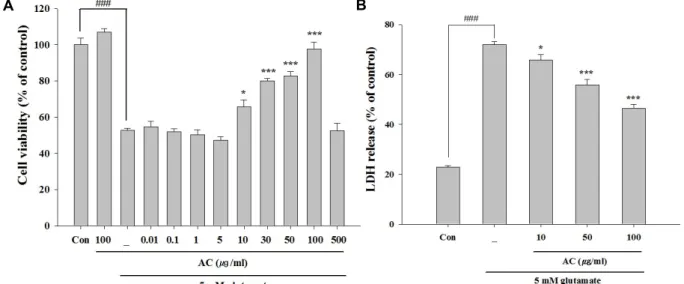
Fig. 1. Protective effects of the ethanol extract from Asparagus cochinchinesis (AC) against glutamate-induced cytotoxicity in HT22 cells. Cell viability and cytotoxicity were determined by MTT (A) and LDH assay (B). HT22 cell were pretreated with various concentrations for 24 hr, followed by treatment with glutamate (5 mM) for 24 hr. ###p<0.001 vs. control group, *p<0.05 and
***p<0.001 vs. glutamate-treated group. Data are presented as the mean±SEM from three independent experiments.
포들을 PBS로 2번 씻고 flow cytometer를 사용하여 각 sample 들을 분석하였으며 sample에서 10,000개의 cell을 얻어 분석하 였다.
Western blot 분석
세포들은 lysis buffer [200 mM Tris (pH8.0), 150 mM NaCl, 2 mM EDTA, 1 mM NaF, 1% NP40, 1 mM PMSF, 1 mM Na3VO4, proteaseinhibitorcocktail]로 균질화하였다. 동일한 양의 단백질을 10-12%의 sodium dodecyl sulfate-polyacryla- mide gel electrophoresis (SDS-PAGE)을 이용해 분리하고, ni- trocellulose membrane (Whatman GmbH, Dassel, Germany) 으로 transfer시켰다. Membrane은 PBST로 만든 5% skim milk를 사용하여 1시간 동안 block시킨 다음, 적합한 항체들로 overnight시켰다. 이 후, horseradish peroxidase로 이루어진 이차항체를 1시간 동안 배양시켰다. 모든 특정단백질 밴드는 enhanced chemiluminescence system (Pierce Biotech, Rock- ford, IL, USA)를 사용하여 볼 수 있게 하였고 Image Quant LAS-4000 imaging system (GE healthcare Life Science, Uppsala, Sweden)을 사용하여 사진을 찍었다.
통계 처리
실험 자료 분석은 SigmaPlot statistical program version 11.2 프로그램(Systat Software, San Jose, CA, USA)을 사용하 였다. 각 실험 데이터는 백분율과 mean ± SEM으로 나타내었 고 모든 측정값은 One-way ANOVA를 실시하여 F값을 구하 고 Turkey’s test를 이용하여 유의성을 검증하였다. 통계적 유 의성 판정을 위한 유의 수준은 p<0.05로 하였다.
결 과
천문동 추출물의 세포생존율에 미치는 영향
MTT분석을 통해 천문동 추출물의 전처리가 glutamate에 의한 세포독성에 대한 영향을 알아보았다. Fig. 1A에서 보는 바와 같이 glutamate처리군은 대조군에 비해 세포생존율이 47% 감소하였다. 그러나 천문동 추출물을 처리하였을 때 10 μg/ml에서 100 μg/ml에 이르는 농도에서 glutamate에 의한 독성을 유의성 있게 감소시켰다. 이러한 세포생존율을 LDH분 석을 통해 재확인 한 결과, Fig. 1B에서 보는 바와 같이 gluta- mate처리군에서 71.9%로 증가하였으나 10, 50 및 100 μg/ml 농도의 천문동추출물 처리는 LDH양을 각각 65.8%, 55.8% 및 46.4%로 유의성있게 감소시켰다. 이상의 결과는 천문동 추출 물이 HT22세포에서 glutamate에 의한 독성에 대해 보호효과 를 가짐을 확인할 수 있었다.
천문동 추출물의 세포죽음유형에 미치는 영향
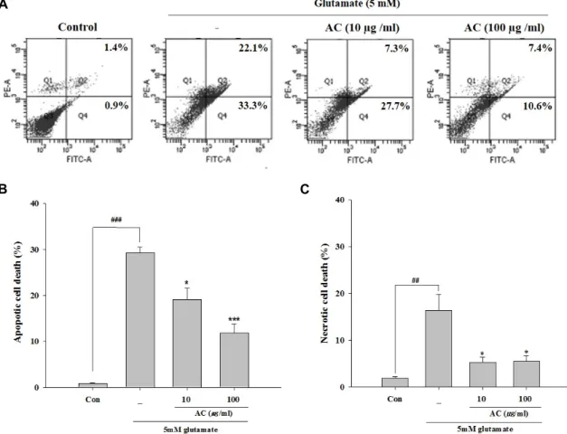
세포죽음의 유형을 분석하기 위해 Annexin Ⅴ/PI staining 을 사용하여 flow cytometer분석을 실시하였다. 세포생존율에 서 유의성을 보이는 10 μg/ml 및 100 μg/ml의 농도를 사용하 였다. Apoptotic cell death와 necrotic cell death는 대조군의 0.87%와 1.83%에 비해 glutamate처리군에서 29.3%와 16.4%
로 유의성 있게 증가하였으며 apoptotic cell death가 더욱 현 저하였다. 그러나 천문동 추출물의 처리는 Fig. 2에서 보는 바 와 같이 두 가지 농도 모두에서 apoptotic cell death가 19.1%
와 11.8%로 유의성 있게 감소하였다. 또한, necrotic cell death 도 천문동 추출물의 두 가지 농도 모두에서 5.27%와 5.53%로 유의성 있게 감소하였다. 이상의 결과는 천문동 추출물이 glu-
A
B C
Fig. 2. Effect of the ethanol extract from Asparagus cochinchinesis (AC) on the types of glutamate-induced cell death in HT22 cells.
Cells were pretreated with two concerntrations (10 and 100 μg/ml) of AC for 24 hr, followed by exposure to 5 mM glutamate for 24 hr. Cells labeled with FITC-Annexin Ⅴ and PI, and then measured by flow cytometry. Representative flow cytometry analysis scatter-grams for Annexin Ⅴ and PI staining (A). Quantitative analysis of the histograms expressed as the percentage of apoptotic or necrotic cell death in total cells (B and C). #p<0.05 and ###p<0.001 vs. control group, *p<0.05 and ***p<0.001 vs. glutamate-treated group. Data are presented as the mean±SEM from three independent experiments.
tamate에 의한 apoptotic cell death와 necrotic cell death를 억제함으로서 신경세포 보호효과를 나타냄을 확인 할 수 있었 다.
천문동 추출물의 ROS형성에 미치는 영향
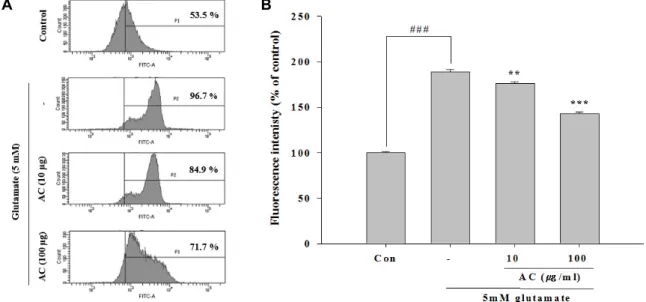
천문동 추출물이 glutamate에 의한 ROS생성에 미치는 영 향을 detection reagents를 사용하여 측정하였다. Fig. 3에서 보는 바와 같이 HT22세포에 대한 glutamate처리는 대조군과 비교하여 189.16%로 현저한 ROS생성을 형성하였다. 그러나 천문동 추출물의 처리는 두 가지 농도 모두에서 이러한 ROS 형성을 176.08% 및 142.86%로현저히 감소시켰다. 이상의 결과 는 천문동 추출물은 glutamate에 의한 ROS형성을 저해함으로 서 신경세포 보호작용을 나타냄을 확인할 수 있었다.
천문동 추출물의 MMP에 미치는 영향
높은 ROS농도는 미토콘드리아에서 MMP에 영향을 미쳐 proton기울기와 membrane depolarization의 소실을 형성한
다[23]. 따라서 미토콘드리아 membrane potential을 측정하기 위해 TMRE staining 분석을 사용한 결과 Fig. 4에서 보는 바와 같이 glutamate처리군에서 1.42%로 현저한 TMRE fluo- rescence의 감소를 볼 수 있었다. 그러나 천문동 추출물을 처 리하였을 때 특히 100 μg/ml 농도에서 82.74%로 증가하므로 대조군과 유사한 membrane potential을 나타내었다. 이상의 결과로 보아 천문동 추출물은 glutamate에 의한 미토콘드리아 MMP를 회복시킴으로서 미토콘드리아의 손상를 저해하여 신 경세포 보호작용을 나타냄을 확인할 수 있었다.
천문동 추출물의 관련 단백질발현에 미치는 영향
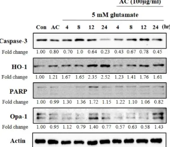
천문동 추출물의 관련 단백질발현에 미치는 영향을 알아보 기 위해, glutamate를 처리한 후 4, 8, 12, 24시간별로 세포죽음 의 caspase-3 및 PARP, 산화적 스트레스의 HO-1, 그리고 미토 콘드리아융합과 세포죽음의 Opa-1 등 관련 단백질의 발현을 연구하였다. Fig. 5에서 보는 바와 같이 glutamate처리 후 24시 간에 total caspase-3의 발현이 23%로 감소하나 천문동 추출물
A B
Fig. 3. Effect of ethanol extract from Asparagus cochinchinesis (AC) on ROS production in glutamate-treated HT22 cells. Cells were pretreated with two concerntrations (10 and 100 μg/ml) of AC for 24 hr, followed by exposed to 5 mM glutmate for 24 hr. Cells were stained with carboxy-H2DCFDA and production of ROS was measured using a flow cytometer (A). Quantitative analysis of the histograms expressed as the percentage of ROS production in HT22 cells (B). ###p<0.001 vs. control group,
**p<0.01 and ***p<0.001 vs. glutamate-treated group. Data are presented as the mean±SEM from three independent experiments.
A B
Fig. 4. Effect of ethanol extract from Asparagus cochinchinesis (AC) on MMP in glutamate-treated HT22 cells. Cells were pretreated with two concerntrations (10 and 100 μg/ml) of AC for 24 hr, followed by exposure to 5 mM glutmate for 24 hr. The MMP was examined using TMRE staining by flow cytometry (A). Quantitative analysis of the histograms expressed as the percentage of mitochondrial inner membrane potential in HT22 cells (B). ###p<0.001 vs. control group, ***p<0.001 vs. gluta- mate-treated group. Data are presented as the mean±SEM from three independent experiments.
에 의해 45%로 회복되며, 12 및 24시간에 천문동 추출물에 의해 PARP발현의 감소를 볼 수 있었으며 HO-1도 유사한 양 상을 보였다. Opa-1은 glutamate처리에 의해 감소하고 12시간
후 140%로 일시적 증가를 보였으나, 천문동 추출물은 24시간 째에 143%로 증가시키는 것을 확인하였다. 이상의 결과로 보 아 천문동 추출물은 관련 단백질 발현을 조절함으로서 신경세
(hr)
Fig. 5. Effect of ethanol extract from Asparagus cochinchinesis (AC) on oxidative toxicity-related proteins in gluta- mate-treated HT22 cells. Cells were pretreated with 100 μg/ml of AC for 24 hr, followed by exposed to 5 mM glutamate. After cell treatment, each groups were main- tained in the original medium for 4, 8, 12 and 24 hr.
Equal amounts of proteins in each group were subjected to western blot using the indicated antibodies. Equal protein loading was confirmed by actin expression.
포 보호작용을 나타냄을 알 수 있었다.
고 찰
천문동은 한의학에서 숨이 차고 기침하는 것을 치료하고 마음을 진정시키고 배뇨를 잘하게 하는 치료제로써 사용되어 져 왔다. 최근 천문동은 항염증효과 및 면역반응을 활성화 시 킬 뿐 아니라[28] 노화모델에서 강한 radical scavenging능력 으로 항노화효과가 알려져 있다[15]. 특히, 신경계에서는 as- trocyte의 TNF-α분비를 억제시키는 항염증효과[14], micro- glia세포에서 항염증효과[13], 그리고 신경세포와 허혈성 뇌졸 중모델에서 보호효과를 가진다는 것이 확인되고 있다[12]. 본 연구팀에서도 동의보감에 대한 textmining기법을 통한 인지 기능 향상에 관여하는 한약재를 도출하였으며, 기본적인 검색 결과 천문동 추출물이 해마세포에 대한 신경보호효과 가능성 이 있음을 제시하였다[22].
중추신경계에서 신경세포 바깥으로의 과도한 glutamate분 비는 전형적인 excitotoxicity를 형성하는데 초기에는 necrotic cell death를 일으키고 이후 apoptotic cell death를 촉진한다 [2]. HT22세포에서도 glutamate에 의한 oxidative pathway를 통해 전형적인 oxidative toxicity를 일으키며[8, 27], 뇌졸중, 파킨슨병, 알츠하이머병을 포함하는 신경퇴행성 과정에 중요 한 특징 중 하나이다[16, 18]. 본 연구의 MTT 및 LDH분석결
과, 천문동 추출물은 glutamate에 의한 신경독성으로부터 보 호하는 효과가 있음을 확인할 수 있으며 oxidative toxicity으 로부터 보호 기능을 알 수 있었다. 나아가 세포죽음에 대한 유형 분석을 위한 Annexin Ⅴ와 PI 염색에 의한 flow cy- tometer분석 결과, 천문동 추출물은 apoptotic cell death를 현 저히 감소시킴을 확인 할 수 있었다.
미토콘드리아는 세포죽음을 이끄는 과정에서 중요한 역할 을 하며 glutamate로 유도된 oxidative toxicity에서 중요한 표 적이 된다[11]. Glutamate로 인한 세포질내 칼슘유입이 증가 하면 미토콘드리아가 손상을 입게 되는데, 먼저 미토콘드리아 의 전자전달계로부터 ROS가 생성되고 이로 인해 세포 죽음이 일으킨다[9, 25]. 또한 glutamate에 의한 산화적 스트레스로 인해 미토콘드리아의 MMP가 감소하거나 파괴되어 세포죽음 에 이른다. 즉 미토콘드리아의 permeability transition pore (MPTP)가 산화적 스트레스로 인해 일시적으로 열리게 되어 삼투압의 팽창, 기질 대산산물이 방출되면서 MMP의 붕괴가 일어난다[10, 11]. 본 연구의 ROS 및 MMP분석 결과를 살펴보 면, 천문동 추출물은 glutamate에 의한 ROS생성을 현저히 감 소시킬 뿐 아니라 미토콘드리아의 MMP를 조절함으로서 신 경세포를 보호하는 것을 확인 할 수 있었다.
Caspase-3는 세포질에 비활성상태로 존재하다가 절단되어 활성상태가 되면 세포죽음에 관여하며[3, 6], glutamate에 의 한 세포죽음은 caspase-3활성에 의한 미토콘드리아의 cy- tochorome C의 방출과 연관이 있다[30]. 또한 glutamate에 의 해 칼슘의 과다 유입은 강력한 산화제 peroxinitrie를 생성하고 이는 PARP의 활성화를 통한 세포죽음에 관여한다[21]. HO-1 는 스트레스 단백질로 산화적 스트레스관련 질병모델에서 유 발된다. 지속적인 고산소증(hyperoxia)과 같은 자극은 ROS형 성을 통한 산화적 스트레스에 의한 HO-1의 발현을 증가한다 [24]. 안쪽 미토콘드리아 막에 위치하고 있어 미토콘드리아의 융합과 크리스테 구조를 조절하면서 APT합성과 세포죽음에 관여하는 Opa-1은 활성을 잃게 되면 크리스테의 결합이 열리 게 되고 이에 cytochrome c 방출을 일으킨다. 특히 산화적 스 트레스에 Opa-1의 발현을 감소한다[1, 17, 29].
본 실험의 산화적 세포죽음과 관련된 대표적인 단백질의 발현을 살펴 본 결과, 천문동 추출물의 처리는 전체 caspase-3 의 발현 증가와 더불어 PARP발현의 감소를 보였다. Caspase- 3의 cleavged된 활성은 관찰할 수 없었으나, 전체 caspase-3의 양 및 PARP를 보아 간접적인 활성저하를 유추할 수 있었다.
또한 glutamate에 의해 증가된 HO-1은 천문동 추출물에 의해 감소하였으며, 이와 반대로 Opa-1은 glutamate처리 후반부에 증가하였다. 이로 보아 천문동 추출물은 산화적 스트레스와 관련된 단백질의 발현 또한 조절함으로서 신경세포 죽음에 대한 보호효과를 가진다는 것을 확인 할 수 있었다.
이상의 결과로 보아, 천문동 추출물은 해마세포에서 gluta- mate에 의한 oxidative toxicity에 대해 ROS형성저해 및 MMP
회복에 의해 세포죽음을 완화시킴으로서 신경세포 보호작용 을 나타내는 것으로 생각된다. 이러한 천문동 추출물의 해마 신경세포 보호작용은 glutamate에 의한 산화적 독성에 의해 나타내는 뇌졸중, 파킨스병, 알츠하이머병 등과 같은 퇴행성 신경질환 및 인지장애에 대한 치료와 예방에 적용 가능한 약 재로 생각된다.
감사의 글
이 논문은 부산대학교 기본연구지원사업(2년)에 의하여 연 구되었음.
References
1. Anand, R., Wai, T., Baker, M. J., Kladt, N., Schauss, A. C., Rugarli, E. and Langer, T. 2014. The i-AAA protease YME1L and OMA1 cleave OPA1 to balance mitochondrial fusion and fission. J. Cell. Biol. 204, 919-929.
2. Ankarcrona, M., Dypbukt, J. M., Bonfoco, E., Zhivotovsky, B., Orrenius, S., Lipton, S. A. and Nicotera, P. 1995.
Glutamate-induced neuronal death: a succession of necrosis or apoptosis depending on mitochondrial function. Neuron 15, 961-973.
3. Budihardjo, I., Oliver, H., Lutter, M., Luo, X. and Wang, X. 1999. Biochemical pathways of caspase activation during apoptosis. Annu. Rev. Cell. Dev. Biol. 15, 269-290.
4. Choi, D. W. 1988. Glutamate neurotoxicity and diseases of the nervous system. Neuron 1, 623-634.
5. Choi, D. W. 1992. Excitotoxic cell death. J. Neurobiol. 23, 1261-1276.
6. Colussi, P. A., Harvey, N. L., Shearwin-Whyatt, L. M. and Kumar, S. 1998. Conversion of procaspase-3 to an autoacti- vating caspase by fusion to the caspase-2 prodomain. J. Biol.
Chem. 273, 26566-26570.
7. Davis, J. B. and Maher, P. 1994. Protein kinase C activation inhibits glutamate-induced cytotoxicity in a neuronal cell line. Brain Res. 652, 169-173.
8. Fukui, M., Song, J. H., Choi, J., Choi, H. J. and Zhu, B. T.
2009. Mechanism of glutamate-induced neurotoxicity in HT22 mouse hippocampal cells. Eur. J. Pharmacol. 617, 1-11.
9. Fukui, M. and Zhu, B. T. 2010. Mitochondrial superoxide dismutase SOD2, but not cytosolic SOD1, plays a critical role in protection against glutamate-induced oxidative stress and cell death in HT22 neuronal cells. Free Radic. Biol. Med.
48, 821-830.
10. Halestrap, A. P., Doran, E., Gillespie, J. P. and O'Toole, A.
2000. Mitochondria and cell death. Biochem. Soc. Trans. 28, 170-177.
11. Henchcliffe, C. and Beal, M. F. 2008. Mitochondrial biology and oxidative stress in Parkinson disease pathogenesis. Nat.
Clin. Pract. Neurol. 4, 600-609.
12. Jalsrai, A., Numakawa, T., Kunugi, H., Dieterich, D. C. and Becker, A. 2016. The neuroprotective effects and possible
mechanism of action of a methanol extract from Asparagus cochinchinensis: In vitro and in vivo studies. Neuroscience 322, 452-463.
13. Jian, R., Zeng, K. W., Li, J., Li, N., Jiang, Y. and Tu, P. 2013.
Anti-neuroinflammatory constituents from Asparagus co- chinchinensis. Fitoterapia 84, 80-84.
14. Kim, H., Lee, E., Lim, T., Jung, J. and Lyu, Y. 1998. Inhibitory effect of Asparagus cochinchinensis on tumor necrosis fac- tor-alpha secretion from astrocytes. Int. J. Immunopharmacol.
20, 153-162.
15. Lei, L., Ou, L. and Yu, X. 2016. The antioxidant effect of Asparagus cochinchinensis (Lour.) Merr. shoot in d-gal- actose induced mice aging model and in vitro. J. Chin. Med.
Assoc. 79, 205-211.
16. Lin, M. T. and Beal, M. F. 2006. Mitochondrial dysfunction and oxidative stress in neurodegenerative diseases. Nature 443, 787-795.
17. Liu, H., Mao, P., Wang, J., Wang, T. and Xie, C. H. 2015.
Allicin protects PC12 cells against 6-OHDA-induced oxida- tive stress and mitochondrial dysfunction via regulating mi- tochondrial dynamics. Cell. Physiol. Biochem. 36, 966-979.
18. Lo, E. H., Moskowitz, M. A. and Jacobs, T. P. 2005. Exciting, radical, suicidal: how brain cells die after stroke. Stroke 36, 189-192.
19. Maher, P. and Davis, J. B. 1996. The role of monoamine me- tabolism in oxidative glutamate toxicity. J. Neurosci. 16, 6394-6401.
20. Murphy, T. H., Miyamoto, M., Sastre, A., Schnaar, R. L. and Coyle, J. T. 1989. Glutamate toxicity in a neuronal cell line involves inhibition of cystine transport leading to oxidative stress. Neuron 2, 1547-1558.
21. Nikolova, S., Lee, Y. S., Lee, Y. S. and Kim, J. A. 2005. Rac1- NADPH oxidase-regulated generation of reactive oxygen species mediates glutamate-induced apoptosis in SH-SY5Y human neuroblastoma cells. Free Radic. Res. 39, 1295-1304.
22. Pak, M. E., Kim, Y. R., Kim, H. N., Ahn, S. M., Shin, H.
K., Baek, J. U. and Choi, B. T. 2016. Studies on medicinal herbs for cognitive enhancement based on the text mining of Dongeuibogam and preliminary evaluation of its effects.
J. Ethnopharmacol. 179, 383-390.
23. Pallast, S., Arai, K., Wang, X., Lo, E. H. and van Leyen, K. 2009. 12/15-Lipoxygenase targets neuronal mitochondria under oxidative stress. J. Neurochem. 111, 882-889.
24. Pietrofesa, R. A., Velalopoulou, A., Lehman, S. L., Arguiri, E., Solomides, P., Koch, C. J., Mishra, O. P., Koumenis, C., Goodwin, T. J. and Christofidou-Solomidou, M. 2016. Novel double-hit model of radiation and hyperoxia-induced oxida- tive cell damage relevant to space travel. Int. J. Mol. Sci.
17, 953-975.
25. Starkov, A. A., Chinopoulos, C. and Fiskum, G. 2004.
Mitochondrial calcium and oxidative stress as mediators of ischemic brain injury. Cell Calcium 36, 257-264.
26. Tan, S., Schubert, D. and Maher, P. 2001. Oxytosis: A novel form of programmed cell death. Curr. Top. Med. Chem. 1, 497-506.
27. Tan, S., Wood, M. and Maher, P. 1998. Oxidative stress in-
초록:HT22 해마세포의 oxidative toxicity에 대한 천문동 유래 에탄올추출물의 보호 효과
박맑은1․최병태1,2*
(1부산대학교 한의학전문대학원 한의과학과, 2부산대학교 한의학전문대학원 경락구조의학부)
본 연구는 oxidative stress에 의한 세포죽음 분석의 이상적인 모델로 사용되는 HT22세포를 이용하여 천문동 에탄올추출물의 glutamate에 의한 oxidative toxicity에 대한 신경보호 효과를 살펴보았다. 이를 위해 cell viability, lactate dehydrogenase (LDH), 그리고 세포죽음형태, reactive oxygen species (ROS), mitochondria membrane potential (MMP) 등에 대한 flow cytometry 및 Western blot분석을 이용하였다. 천문동 추출물의 처리는 cell via- bility 및 LDH분석에서 glutamate에 의한 cell toxicity를 저하시키며, 특히 apoptotic cell death를 현저히 감소시켰 다. ROS 및 MMP분석 결과, 천문동 추출물은 ROS의 형성을 저하시키며 glutamate에 의해 저하된 MMP를 현저 히 회복시켜 주었다. 이와 관련된 단백질 발현을 보면, 천문동 추출물은 PARP 및 HO-1의 발현을 억제하였다.
이상의 결과는 천문동 추출물이 HT22해마세포에서 ROS형성저해 및 MMP회복에 의해 세포죽음을 완화시켜 보 호작용을 하는 것으로 사료되며 oxidative toxicity관련 질환에 적용 가능할 것으로 보여 진다.
duces a form of programmed cell death with characteristics of both apoptosis and necrosis in neuronal cells. J. Neuro- chem. 71, 95-105.
28. Xiong, D., Yu, L. X., Yan, X., Guo, C. and Xiong, Y. 2011.
Effects of root and stem extracts of Asparagus cochinchinensis on biochemical indicators related to aging in the brain and liver of mice. Am. J. Chin. Med. 39, 719-726.
29. Yan, M. H., Wang, X. and Zhu, X. 2013. Mitochondrial de-
fects and oxidative stress in Alzheimer disease and Parkin- son disease. Free Radic. Biol. Med. 62, 90-101.
30. Zhang, Y. and Bhavnani, B. R. 2005. Glutamate-induced apoptosis in primary cortical neurons is inhibited by equine estrogens via down-regulation of caspase-3 and prevention of mitochondrial cytochrome c release. BMC Neurosci. 6, 13- 35.