저작자표시-비영리-변경금지 2.0 대한민국 이용자는 아래의 조건을 따르는 경우에 한하여 자유롭게 l 이 저작물을 복제, 배포, 전송, 전시, 공연 및 방송할 수 있습니다. 다음과 같은 조건을 따라야 합니다: l 귀하는, 이 저작물의 재이용이나 배포의 경우, 이 저작물에 적용된 이용허락조건 을 명확하게 나타내어야 합니다. l 저작권자로부터 별도의 허가를 받으면 이러한 조건들은 적용되지 않습니다. 저작권법에 따른 이용자의 권리는 위의 내용에 의하여 영향을 받지 않습니다. 이것은 이용허락규약(Legal Code)을 이해하기 쉽게 요약한 것입니다. Disclaimer 저작자표시. 귀하는 원저작자를 표시하여야 합니다. 비영리. 귀하는 이 저작물을 영리 목적으로 이용할 수 없습니다. 변경금지. 귀하는 이 저작물을 개작, 변형 또는 가공할 수 없습니다.
Master's Thesis
Neuroprotective effects of Carpinus
tschonoskii leaves extract against
glutamate-induced oxidative stress in
HT22 hippocampal cells
Ji-Woon Hwang
Department of Biotechnology GRADUATE SCHOOL JEJU NATIONAL UNIVERSITY
HT22 해마 세포의 글루타메이트
유발 산화 스트레스에 대한
개서어나무
(Carpinus tschonoskii)잎
추출물의 신경 보호 효과
.
지도교수 김 재 훈
황 지 운
이 논문을 이학 석사학위 논문으로 제출함
2020 년 07 월황지운의 이학 석사학위 논문을 인준함
심사위원장 운노타쯔야 위 원 김 창 숙 위 원 김 재 훈 제주대학교 대학원 2020 년 07 월Neuroprotective effects of Carpinus
tschonoskii leaves extract against
glutamate-induced oxidative stress in HT22
hippocampal cells.
Ji-Woon Hwang
(Supervised by Professor Jae Hoon Kim)
A thesis submitted in partial fulfillment of the requirement For the degree of Master of Science
July, 2020
This thesis has been examined and approved.
Chairperson of the supervising committee
Professor Tatsya Unno, Ph.D., College of Applied Life Sciences, Jeju National University Professor chang Sook Kim, Ph.D., College of Applied Life Sciences, Jeju National University Professor Jae Hoon Kim, Ph.D., College of Applied Life Sciences, Jeju National University
Department of Biotechnology GRADUATE SCHOOL JEJU NATIONAL UNIVERSITY
i
CONTENTS
CONTENTS... i
LIST OF FIGURES...ii
ABSTRACT...1
1. INTRODUCTION...2
2. MATERIALS AND METHODS...4
2.1. Materials ………..……….…….………...……....4
2.2. Cell culture ……….……….…………...…...…...4
2.3. Cell viability assay …………...…...4
2.4. Cell ROS analysis……….….….……...…...…...4
2.5. Cell apoptosis analysis ………...5
2.6. Western blot assay ……….…….5
2.7. GC-MS/MS analysis ………...5
3. RESULTS ……….………... ……..7
3.1. CTE inhibits glutamate-induced cytotoxicity………..7
3.2. CTE inhibits glutamate-induced ROS production………..….…9
3.3. CTE inhibits glutamate-induce apoptotic cell death...11
3.4. Measurement of phosphorylation in MAPK pathways...13
3.5. Measurement of protein expression in pro-apoptotic pathways...15
3.6. Analysis of GC-MS/MS from CTE...17
4. DISCUSSION...19
5. REFERENCES...21
ii
LIST OF FIGURES
Figure 1. Protective effects CTE against glutamate-induced cytotoxicity in HT22 cells.
Figure 2. Effect of CTE on glutamate-induced ROS production in HT22 cells. Figure 3. Effect of CTE on types of glutamate-induced apoptotic cell death in HT22
cells.
Figure 4. Effect of CTE on glutamate-induced oxidative toxicity relative proteins in HT22 cells.
Figure 5. Effect of CTE on glutamate-induced pro-apoptotic relative proteins in HT22 cells.
1
ABSTRACT
The present study investigated the neuroprotective effect of the intracellular protection mechanism using C.tschonoskii leaves extract (CTE) against glutamate-induced oxidative damage in HT22 cells. We found that pretreatment of HT22 cells with CTE significantly inhibited the cell death induced by glutamate in a dose dependent manner. CTE reduced ROS production and apoptosis induced by glutamate. In addition, CTE inhibited phosphorylation of p38 and Erk, transcription factors important for neuronal survival, and reduced the expression of Bax and AIF, apoptosis-related proteins. The result is that CTE in HT22 cells can prevent glutamate-induced apoptosis through ROS production, phosphorylation inhibition of oxidative stress related proteins, and reduction of expression. Therefore, it suggests that it can be applied to the development of therapeutic agents for diseases of the brain and nervous system.
2
1. INTRODUCTION
It is expected that the number of patients with degenerative cerebral nervous system disease will increase rapidly due to the prolonged aging of humans, and it lead development of treatment urgently.
Dementia is not a disease caused by a specific diagnosis, but by various causes such as a defect of memory, cognitive dysfunction and other loss of a function indication. There are Alzheimer’s dementia, Vascular dementia, dementia with lewy bodies and frontotemporal lobar dementia.
In the Korea, Alzheimer’s disease(AD) is estimated 50-60% of whole dementia patients and vascular dementia is estimated 15-20% of them, so the study of prevention and treatment of AD is a very important issue.
Although there is no clear cause for the major causes of AD, various causative hypotheses are being argued[29]. several studies, especially neuropathological studies, have emphasized the neuron oxidation of AD[30-31].
Injury of neuron by oxidative damage results in oxidative damage of major component such as lipid, protein and nucleic acid and eventually causes fatal cell death or necrosis.
Carpinus is composed of 40 species and is distributed in temperate zone of the
northern hemisphere. Only 5 species of C. tschnoskii, C. lausiflora, C. cordata, C.
turcjaninowi, and C. Coreana grow in the Korea. Recent research has shown that Carpinus leaves contain flavonoid compounds such as Tannin, Myricetin, Kaempferol,
Apigenin and Luteroline[13], and have strong antioxidant and anti-inflammatory effects[14].
Glutamate plays an important role in learning and memory in neurons and is a major excitability neurotransmitter associated with acute and chronic neurodegenerative diseases as well as Alzheimer’s and Parkinson’s disease[1][15].
Physiologically, as brain glutamate concentration increase, intercellular ROS increases and neurotoxicity occurs. Recently, studies have been reported that cytotoxicity by glutamate causes mitochondria functional disorder and destroys mitochondrial dysfunction[3].
3
Abnormal Ca2+ flow in the cell activate Calpaine to activate apoptosis protein Bax[4-5].
In addition, mitochondrial functional disorder by oxidative stress translocated the pro-apoptosis factor (AIF) to the nucleus, eventually activating the apoptosis pathway[6].
Reactive oxygen species (ROS) are associated with tissue complication and factor that causes neurological diseases such as inflammation, aging, cancer, arterisclerosis, hypertension, diabetes, ischemic stroke, Alzheimer’s disease and parkinson’s disease[7-8].
Over produced ROS cause apoptosis by activating MARK pathway[9]. Recent studies have shown that nerve damage from various cytokines, inflammation-inducing agents, heat shocks and etc phosphorylate the p38[10] and ERK is sensitive to oxidative stress in neuronal cell death. Therefore, according to important role of these MARK pathways, materials by suppressing phosphorylation of MARK pathways may be a candidate treatment for protecting neuronal cells.
This study was conducted to neuroprotective effect mechanism of C.tschnoskii extract by glutamate-induced oxidative stress in HT22 mouse hippocampal cell which has been widely used as a successful in vitro model.
4
2. MATERIALS AND METHODS
2.1. Materials.
EtOH extract (80 %) and several solvent fractions from C. tschonoskii
were purchased from Jeju Biodiversity Research Institue of Jeju Technopark (JBRI), which has professional facilities and provides extracts from plants growing on Jeju Island for research purposes in biotechnology and related industrial fields.
2.2. Cell culture.
Mouse HT22 hippocampal cells were gifted by Jae-ran Lee from Korean Research Institute of Bioscience and Biotechnology (KRIBB).The HT22 cells were maintained in DMEM supplemented with 10 % fetal bovine serum (Gibco-BRL, Gaithersburg, MD, USA), 100 μg/ml streptomycin (Invitrogen), 100 U/mL penicillin at 37℃. 2.3. Cell viability assay.
We measured cell viability using the WST-1
(2-(4-iodophenyl)-3-(4-nitrophenyl)-5-(2,4-disulfophenyl)-2H-tetrazolium) solution (Boechringer Mannheim, Mannheim , Germany). Cells were seeded 2.5×10⁴in
24-well plates. After cells were incubated for 24 h, we were treated with CTE for 24 h at 37℃. Each well was added to a final concentration of 10% WST-1 solution after 15 min incubation at room temperature. The absorbance was measured at 450 nm using the microplate reader.
2.4. Cell ROS analysis.
Intracellular ROS levels were determined using the fluorescent probe 2’,7’-dichlorofluorescin diacetate (DCFH-DA) (Rosenkranz et al., 1992). This molecule is cleaved intra-cellularly by esterases to form non-fluorescent
2’,7’-dichlorofluorescin (DCFH), which is transformed to the fluorescence compound 2’,7’-dichlorofluorescein (DCF) upon oxidation by ROS. HT22 cells were seeded 5.0×10⁴in 6-well plates for 12 h. The cells were pretreated with 20 μg/ml of CTE and 4mM glutamate for treatment for 12 h. After collecting the cells, we were treated 10
5
μM DCFH-DA for 15 min at 37o C under 5% CO2 in the dark. The apoptotic cells were detected by flow cytometry (LSRFortessa, BD)
2.5. Cell apoptosis analysis.
For apoptosis analysis, HT22 cells were seeded in 6-well plates. After 24 h, they were treated with CTE for 24 h. After collecting the cells, we incubated the cells with Annexin V-FITC and PI (FITC Annexin V apoptosis detection kit, BD pharmigen). The apoptotic cells were detected by flow cytometry (LSRFortessa, BD).
2.6. Western blot assay
Cells were lysed in M-PER lysis buffer (Thermo science, Bonn, Germany)
containing protease inhibitor cocktail (Roche), 2 mM sodium vanadate, 30mM sodium pyrophosphate, and 100mM sodium fluoride. After total protein quantification,
proteins were separated by 10 % SDS-PAGE (sodium dodecyl sulfate-polyacrylamide gel electrophoresis) and transferred to nitrocellulose membranes (Amersham
Bioscience, Little Chalfont, Buckinghamshire, UK). Membranes were blocked with 5 % skim milk in TBST. Primary antibodies, such as phospho-Erk(44/42) (Thr202/204), phosphor-p38 (Thr180/Tyr182), Bax, AIF and GAPDH (Cell signaling technology, USA), were diluted 1:1000 in TBST and incubated overnight at 4 °C. Secondary antibodies (Merck Millipore, Germany) were diluted in TBST 1:4000 and incubated for 1h. The protein bands were detected by the ECL kit (Biosesang).
2.7. GC-MS MS analysis.
The sample was eluted in ethanol. The analysis was performed using a gas chromatograph (GC 2010, Shimadzu Corporation). The GC was equipped with an AOC-20i auto-injector and a triple quadruple tandem mass spectrometer (TQ8040, Shimadzu Corporation). One µl volume of the sample was injected in splitless mode on Rtx-4MS (30 m x 0.25 mm, 0.25-µmfilm thickness) column (restek). The carrier gas and the collision gas were helium and argon, respectively. Oven temperature started from 80 ℃ (3 min hold time) to 310 ℃ (3 min hold time). The mass spectrometer was operated in multiple reaction monitoring (MRM) mode. The data acquisition was started at 20 min.
6 2.8. Statistical analysis.
Data were presented as average with standard deviation (SD). The differences between multiple groups were analyzed using turkey’s post hoc method. P < 0.05 was regarded statistically significant.
7
3. RESULTS
3.1. Measured of cell viability by WST-1 Assay.
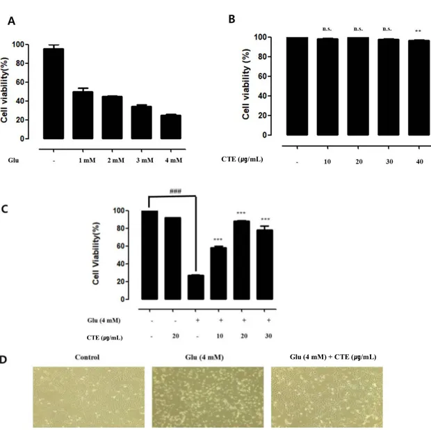
It was measured cell viability from CTE by Glutamate-induced cytotoxicity in HT22 cells. Through the analysis, it can be figured out the effect to cytotoxicity by glutamate treatment group and CTE pre-treated. HT22 cells were treated by Various concentration of glutamate and CTE. After 24hour, cell viability were measured by WST-1 assay. Cell viability was reduced to 75% in 4mM glutamate treatment group (Fig1. A and B). When various concentrated CTE treated in HT22 Cell, there isn’t any significant difference. HT22 cells were pre-treated with 10, 20, 30 μg /mL of CTE and 4 mM glutamate for 24 hr. Glutamate treatment group reduced cell viability to 72 %. Cell viability was increased to 92 % In twenty μg/mL of CTE and 4mM glutamate pretreated.
8
Figure 1. Protective effects of CTE against glutamate-induced cytotoxicity in HT22 cells.
Cytotoxicity was assessed after 24 hr incubation period with various concentrations (1, 2, 3 and 4 mM) of glutamate (A). Mouse HT22 hippocampal cells was treated with various concentrations (0, 10, 20, 30 and 40 μg/ml) of CTE for 24hr (B).Cells were pretreated with glutamate (4 mM) for 24 hr, followed by treatment with various concentrations for 24 hr. (C). Glutamate-induced toxicity in HT22 cells as assessed by phase contrast microscopy (100x) (D). Cell viability was determined by WST-1 assay. Graphs showed mean ±SEM, **P< 0.01 vs. control group.(A and B). ###P< 0.001 vs. control group, ***P<0.001 vs. glutamate-treated group.(C and D)
CTE (㎍/mL)
CTE (㎍/mL)
9
3.2. CTE inhibits glutamate-induced ROS production.
It was measured ROS effect of CTE by glutamate-induced oxidative stress in HT22 cells.
Fig 2. A and B show that production result of intercellular ROS by glutamate-induced. Through the analysis, it can be figured out the effect of glutamate-induced increases of ROS by glutamate treatment group. Intercellular ROS were measured and increased 6 times to compared control group. Twenty μg/mL CTE and 4 mM glutamate pretreated reduced intercellular ROS as 5 times compare 4 mM glutamate treated group in HT22 cells. NAC were used positive control group. This result of the analysis, twenty μg/mL of CTE effects neuroprotective by suppressing production of intercellular ROS.
10
Figure 2. Effect of CTE on glutamate-induced ROS production in HT22 cells.
Cells were pretreated with twenty μg/ml of CT for 12 hr, followed by exposed to 4mM glutamate for 12 hr. Cells were stained with carboxy-H2DCFDA and production of ROS was measured using a flow cytometer (A). Quantitative analysis of the histograms expressed as the realative fold chanage of ROS production in HT22 cells (B). Graphs showed mean ±SEM, ###P<0.001 vs. control group, ***P<0.001 vs. glutamate-treated group. CTE (20㎍/mL) Glu (4 mM) + CTE (20 ㎍/mL) Glu (4 mM) CTE (㎍/mL) NAC (1 mM)
11
3.3. CTE inhibits glutamate-induce apoptotic cell death.
.
Apoptosis reactivity from CTE by glutamate-induced production of apoptosis in HT22 cells was measured.
Fig 3. A and B show that production of apoptosis by glutamate-induced. Through the analysis, it can be figured out the effect to glutamate induced increases Apoptosis.
4mM glutamate treated group increased apoptosis to about 31 % compared control group.
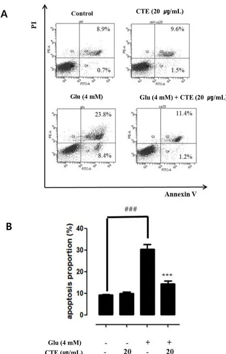
Twenty μg/ml of CTE and 4 mM glutamate pre-treated was reduced Apoptosis to about 12 % compare 4mM glutamate treated group in HT22 cells. Twenty ug/ml of CTE effects neuroprotective by suppressing Apoptosis.
12
Figure 3. Effect of CTE on types of glutamate-induced apoptotic cell death in HT22 cells.
Cells was pretreated with twenty μg/ml of CTE for 24 hr, followed by exposed to 4mM glutamate for 24 hr. Cells labeled with FITC-Annexin V and PI, and then measured by flow cytometer. Representative flow cytometry analysis scatter-grams for Annexin V and PI staining (A). Quantitative analysis of the histograms expressed as percentage of apoptotic or necrotic cell death in total cells (B). Graphs showed mean ±SEM, ###P<0.001 vs. control group, ***P<0.001 vs. glutamate-treated group.
A
B
Glu (4 mM) + CTE (20 ㎍/mL) CTE (20 ㎍/mL) Glu (4 mM) CTE (㎍/mL)13
3.4. Measurement of phosphorylation in MAPK pathways.
Protein phosphorylation from CTE by MAPK pathways protein of glutamate-induced oxidative stress in HT22 cells was measured.
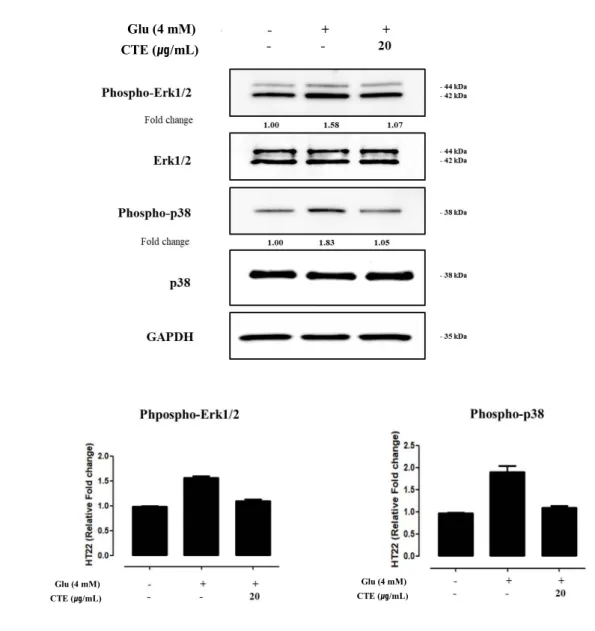
Fig 4. A and B shows phosphorylation of protein by glutamate-induced. 4mM glutamate-induced increased phosphorylation of protein which is included p38 and ERK in MAPK pathways.
Twenty μg/ml of CTE and 4mM glutamate’s pre-treated was reduced phosphorylation of protein compare 4mM glutamate treated group in HT22 cells.
14
Figure 4. Effect of CTE on glutamate-induced oxidative toxicity relative proteins in HT22 cells.
Cells were pretreated with twenty μg/ml of CTE for 12 hr, followed by exposing to 4 mM glutamate. After cell treatment, each groups were maintained in the original medium 12 hr. Equal amounts of proteins in each group were subjected to western blot using the indicated antibodies. Equal protein loading was confirmed by actin expression.
A
B
Glu (4 mM) CTE (㎍/mL) Glu (4 mM) CTE (㎍/mL) Glu (4 mM) CTE (㎍/mL)15
3.5. Measurement of protein expression in pro-apoptotic pathways
Protein expression from CTE by pro-apoptotic pathways protein of glutamate-induced oxidative stress in HT22 cells was measured.
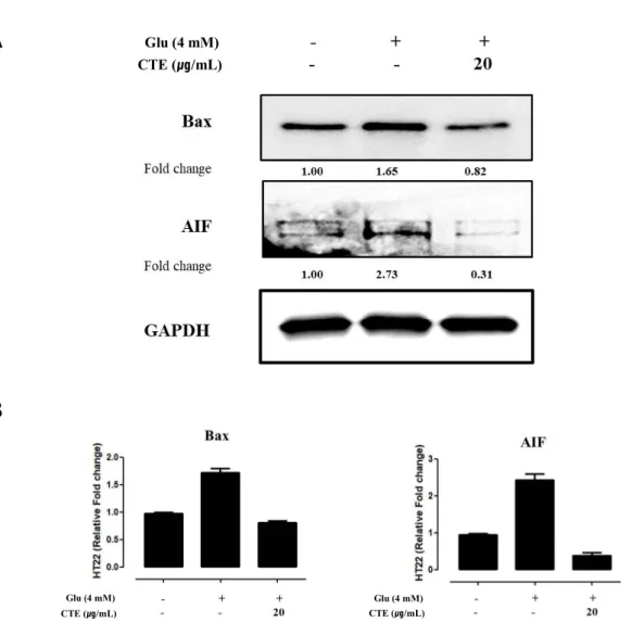
Fig 5. A and B shows protein expression by glutamate-induced. 4 mM glutamate-induced increased protein expression which is included pro-apoptotic protein (Bax) and pro-apoptotic factor (AIF). twenty ug/ml of CTE and 4mM glutamate’s pre-treated was reduced protein expression compare 4 mM glutamate treated group in HT22 cells.
Twenty μg/ml of CTE effects neuroprotective by reducing Bax and AIF of protein expression in apoptotic pathways.
16
Figure 5. Effect of CTE on glutamate-induced pro-apoptotic relative proteins in HT22 cells.
Cells were pretreated with twenty μg/ml of CTE for 12 hr, followed by exposing to 4 mM glutamate. After cell treatment, each groups were maintained in the original medium 12 hr. Equal amounts of proteins in each group were subjected to western blot using the indicated antibodies. Equal protein loading was confirmed by actin expression.
A
B
Glu (4 mM) CTE (㎍/mL) Glu (4 mM) CTE (㎍/mL) Glu (4 mM) CTE (㎍/mL)17
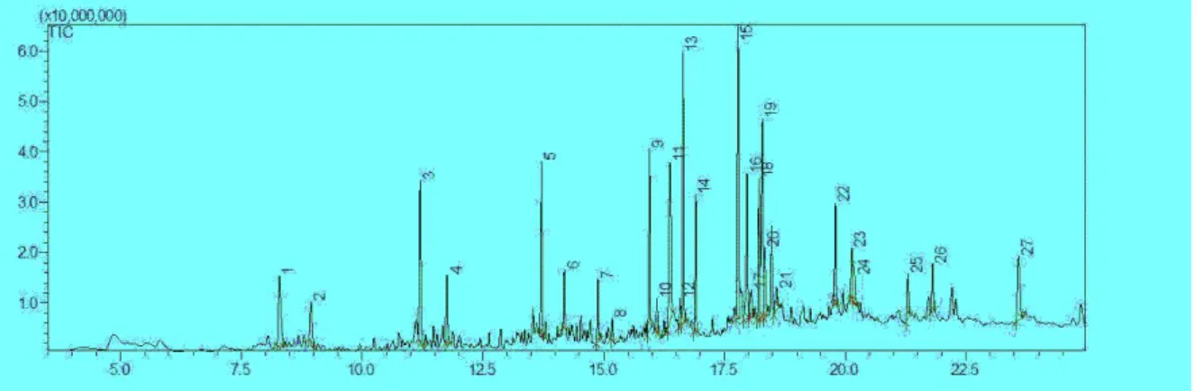
3.6. Analysis of GC-MS/MS from CTE.
Fig 6 was analysis result of GC-MS/MS from CTE. 13,15 and 19’s peak area among various peak is 8.4%, 9.5% and 9.0%. Table 1 shows compound name and activity about 3 peaks of Fig 6.
No.13 peak is anti-oxidant, no.15 peak is anti-cancer and no.19 is anti-inflammatory of activity.
18 Figure 6. Chromatogram of CTE by GC-MS/MS.
19
3. DISCUSSION
When a people get older, the blood vessels of the brain cells and myocardial cells become narrow and clogged, resulting in a fatal heart attack or cerebral infarction.
It also brings many changes to the blood vessels that make up the vascular system. Accordingly, fatal cerebrovascular occur such as dementia and stroke [21].
One of the main causes of Alzheimer's is oxidative stress and Glutamate is known to cause oxidative stress in cells in the central nervous system[15].
CTE has strong antioxidant and anti-inflammatory effects[14], It is known to effect a neuroprotective effect in the Parkinson model[11].
This study was conducted the cell viability and neuroprotective effect mechanism by using HT22 mouse hippocampal cell by Glutamate which caused oxidative damage to measure the protective and antioxidant effects of CTE effect.
As a result of the WST-1 analysis in this study, glutamate cause cytotoxicity in HT22 cell, (Fig. 1A), and CTE doesn’t cause cytotoxic in HT22 cell (Fig. 1B).
CTE and glutamate’s pre-treated shows to increase cell viability (Fig 1. C and D). From the above results, CTE has neuroprotective effect by glutamate-induced cytotoxicity in HT22 cells.
Mitochondria plays an important role in the process of apoptosis and are important targets for oxidative toxicity by glutamate[16].
Mitochondria is damaged when the influx of calcium by glutamate in the cytoplasm increases[4], First, ROS is generated from the mitochondrial electron transport system, which is caused apoptosis[17].
From the ROS analysis of this study, it is shown that CTE significantly reduces ROS by glutamate (Fig 2. A and B).
Over glutamate secretion in the central nervous system outside the neurons forms typical excitotoxicity, it produces early apoptosis in the early stage and promotes late apoptosis in the late stage[18]. As a result of the flow cytometer analysis by Annexin V and PI staining for the type analysis of apoptosis, it was shown that CTE was significantly reduced early and late apoptotic cell death (Fig. 3 A and B).
20
Apoptosis is essential for normal brain development, but it is also important for a variety of degenerative neurological disorders that result from the death of certain parts of the neuron.
The mitogen-activated protein kinase (MAPKs) involved in signaling pathways are serine/threonine kinases involved in regulating various cellular responses, such as cell proliferation, differentiation, and apoptosis[19]
Among the MAPKs, extracellular signal-regulated kinase (ERK), c-Jun N-terminal kinase (JNK/SAPK) and p38 kinase play an important role[20].
CTE suppress phosphorylation of ERK and P38 in signal mechanism study of CTE by glutamate-induced oxidative stress(Fig 4 A and B).
ERK phosphorylation increases apoptosis by the response to the stimulus such as hypoxia, growth factor deficiency and H202[11].
The abnormal Ca2+ flow inside the cell by oxidative stress activates calpaine to
activate the apoptosis proteine (Bax)[4-5].
Increased pro-apoptotic proteins(bax) expression forms pores in the mitochondrial membrane, causing cytochrome c and pro-apoptosis factor(AIF)[24-25].
Promotes the release of AIF is a flavoprotein located inside the mitochondria that plays an important role in ROS metabolism and has oxidoreductase activity. AIF is released from the mitochondria during apoptosis and migrates to the nucleus to induce DNA fragmentation, causing caspase-independent cell death[22-23]
CTE reduced the protein expression of Bax and AIF by glutamate-induced oxidative stress.(Fig 4. C and D)
GC-MS/ MS analysis confirmed that there were substances with antioxidant, anti-inflammatory and anti-cancer effects[33].
In view of the above results, CTE have neuroprotective effect through inhibition of ROS by glutamate-induced oxidative stress in HT22 cells, suppression of
phosphorylation of the MAPK pathway of ERK, P38, and reduction of pro apoptotic protein expression of Bax and AIF.
Therefore, in the future, the neuroprotective effects of CTE may be useful for the treatment and prevention of degenerative neurological diseases and cognitive disorders such as Parkinson's disease, and Alzheimer's disease, which are caused by
21
6. REFERENCE
1. Reynolds, I. J., & Hastings, T. G. (1995). Glutamate induces the production of reactive oxygen species in cultured forebrain neurons following NMDA receptor activation. Journal of Neuroscience, 15(5 I), 3318–3327. https://doi.org/10.1523/jneurosci.15-05-03318.1995
2. Dong, X. X., Wang, Y., & Qin, Z. H. (2009). Molecular mechanisms of excitotoxicity and their relevance to pathogenesis of neurodegenerative diseases. Acta Pharmacologica Sinica, 30(4), 379–387. https://doi.org/10.1038/aps.2009.24
3. Riedel, G., Platt, B., & Micheau, J. (2003). Glutamate receptor function in learning and memory. Behavioural Brain Research, 140(1–2), 1–47. https://doi.org/10.1016/S0166-4328(02)00272-3
4. Polster, B. M., Basańez, G., Etxebarria, A., Hardwick, J. M., & Nicholls, D. G. (2005). Calpain I induces cleavage and release of
apoptosis-inducing factor from isolated mitochondria. Journal of Biological Chemistry, 280(8), 6447–6454. https://doi.org/10.1074/jbc.M413269200
5. Choi, D. W. (1985). Glutamate neurotoxicity in cortical cell culture is calcium dependent. Neuroscience Letters, 58(3), 293–297.
https://doi.org/10.1016/0304-3940(85)90069-2
6. Cabon, L., Galán-Malo, P., Bouharrour, A., Delavallée, L., Brunelle-Navas, M. N., Lorenzo, H. K., Gross, A., & Susin, S. A. (2012). BID regulates AIF-mediated caspase-independent necroptosis by promoting BAX activation. Cell Death and Differentiation, 19(2), 245–256.
https://doi.org/10.1038/cdd.2011.91
7. Ray, P. D., Huang, B. W., & Tsuji, Y. (2012). Reactive oxygen species (ROS) homeostasis and redox regulation in cellular signaling. Cellular Signalling, 24(5), 981–990. https://doi.org/10.1016/j.cellsig.2012.01.008
8. Tan, S., Wood, M., & Maher, P. (2002). Oxidative Stress Induces a Form of Programmed Cell Death with Characteristics of Both Apoptosis and Necrosis in Neuronal Cells. Journal of Neurochemistry, 71(1), 95–105. https://doi.org/10.1046/j.1471-4159.1998.71010095.x
22
9. Pearson, G., Robinson, F., Gibson, T. B., Xu, B. E., Karandikar, M., Berman, K., & Cobb, M. H. (2001). Mitogen-activated protein (MAP) kinase pathways: Regulation and physiological functions. Endocrine Reviews, 22(2), 153–183. https://doi.org/10.1210/er.22.2.153
10. Murphy, T. H., Miyamoto, M., Sastre, A., Schnaar, R. L., & Coyle, J. T. (1989). Glutamate toxicity in a neuronal cell line involves inhibition of cystine transport leading to oxidative stress. Neuron, 2(6), 1547–1558.
https://doi.org/10.1016/0896-6273(89)90043-3
11. Kim, M. K., Kim, S. C., Kang, J. Il, Boo, H. J., Hyun, J. W., Koh, Y. S., Park, D. B., Yoo, E. S., Kang, J. H., & Kang, H. K. (2010). Neuroprotective effects of carpinus tschonoskii max on 6-hydroxydopamine-induced death of pc12 cells. Biomolecules and Therapeutics, 18(4), 454–462.
https://doi.org/10.4062/biomolther.2010.18.4.454
12. Lee, P.W., Kim, H.S., and eom, Y.G, Wood anatomy of Genus Carpinus grow in Korea. Seoul Natl. Univ. J. Agric. Sci. 14, 41-48 (1989)
13. Chang, C. S. and Jeon, J. I. (2004). Foliar flavonoids of Carpinus, sect. Distegocarpus in eastern Asia. Biochem. Syst. Ecol. 32, 35-44.
14. Yin, J., Ahn, H. S., Ha, S. Y., Hwang, I. H., Yoon, K. D., Chin, Y. W., & Lee, M. W. (2019). Anti-skin ageing effects of phenolic compounds from
Carpinus tschonoskii. Natural Product Research, 33(22), 3317–3320. https://doi.org/10.1080/14786419.2018.1497026
15. Hynd, M. R., Scott, H. L., & Dodd, P. R. (2004). Glutamate-mediated excitotoxicity and neurodegeneration in Alzheimer’s disease. Neurochemistry International, 45(5), 583–595. https://doi.org/10.1016/j.neuint.2004.03.007
16. Henchcliffe, C. and Beal, M. F. 2008. Mitochondrial biology and oxidative stress in Parkinson disease pathogenesis. Nat. Clin. Pract. Neurol. 4, 600-609.
17. Starkov, A. A., Chinopoulos, C. and Fiskum, G. 2004. Mitochondrial calcium and oxidative stress as mediators of ischemic brain injury. Cell Calcium 36, 257-264
18. Ankarcrona, M., Dypbukt, J. M., Bonfoco, E., Zhivotovsky, B., Orrenius, S., Lipton, S. A. and Nicotera, P. 1995. Glutamate-induced neuronal
23
death: a succession of necrosis or apoptosis depending on mitochondrial function. Neuron 15, 961-973.
19. Pearson, G., Robinson, F., Beers Gibson, T., Xu, B.E., Karandikar, M., Berman, K. and Cobb, M.H. Mitogenactivated protein (MAP) kinase pathways. regulation and physiological functions. Endocr. Rev. 22: 153-183, 2001.
20. Xia, Z., Dickens, M., Raingeaud, J., Davis, R.J. and Greenberg, M.E. Opposing effects of ERK and JNK-p38 MAP kinases on apoptosis. Science 270: 1326-1331, 1995
21. Zhu, X., Smith, M.A., Honda, K., Aliev, G., Moreira, P.I., Nunomura, A., Casadesus, G., Harris, P.L., Siedlak, S.L., Perry. G. Vascular oxidative stress in Alzheimer disease. J Neurol Sci., Jun 15, 257(1-2):240-246. Epub 2007 Mar 6, 2007.
22. Joza N, Susin SA, Daugas E, Stanford WL, Cho SK, Li CY, Sasaki T, Elia AJ, Cheng HY, Ravagnan L, Ferri KF, Zamzami N, Wakeham A, Hakem R, Yoshida H, Kong YY, Mak TW, Zuniga-Pflucker JC, Kroemer G, Penninger JM. Essential role of the mitochondrial apoptosis-inducing factor in programmed cell death. Nature 401, 549-554, 2001.
23. Arnoult D, Gaume B, Karbowski M, Sharpe JC, Cecconi F, Youle RJ. Mitochondrial release of AIF and EndoG requires caspase activation downstream of Bax/Bak-mediated permeabilization. EMBO J 22, 4385-4399, 2003.
24. Danial NN, Korsmeyer SJ. Cell death: critical control points. Cell 116, 205-219, 2004.
25. Adams JM, Cory S. The Bcl-2 protein family: arbiters of cell survival. Science 281, 1322-1326, 1998
26. Shimizu S, Narita M, Tsujimoto Y. Bcl-2 family proteins regulate the release of aoptogenic cytochrome c by the mitochondrial channel VDAC. Nature 399, 483-487, 1999.
27. Jooghan Park. Causes and Management of Dementia. Korean J Psychophamacol. 1992; 3(1):33-42
24
29. Cummings JL, Berson DF. Dementia. A Clinical Approach. 2nd ed. Boston. Butterworth-Heinemann. 1992
30. Coyle JT, Puttfarcken P. Oxidative stress. glutamete. and neurodegenerative disorder. Science. 1993;262:689-69
31. Markesbery WR. Oxidative stress hypothesis in Alzheimer's disease. Free Radic Biol Med. 1997;23:134-147
32. Ertas, A., Yilmaz, M. A., & Firat, M. (2015). Chemical profile by LC-MS/MS, GC/MS and antioxidant activities of the essential oils and crude extracts of two Euphorbia species. Natural Product Research, 29(6), 529–534. https://doi.org/10.1080/14786419.2014.954113
25