Glutamate가 유도하는 세포독성으로부터 신경세포를 보호하는 상백피 추출물의 효과
김현정․김지현․손은순․이정민․박해룡*
경남대학교 식품생명학과
Received April 15, 2009 /Accepted June 18, 2009
Neuroprotective Effect of Extracts from Root Bark of Morus alba on Glutamate-induced Cytotoxicity in Neuronal Cells. Hyun-Jung Kim, Ji-Hyun Kim, Eun-Soon Son, Jeung-Min Lee and Hae-Ryong Park*. Department of Food Science and Biotechnology, Kyungnam University, Masan 631-710, Korea - This study evaluated the neuroprotective effect of extracts from the root bark of Morus alba (MA) against glutamate-induced cytotoxicity in neuronal cells. Glutamate-induced cytotoxicity was shown by MTT reduction assay. The neuroprotective effects of methanol, ethanol, and acetone extracts from MA against glutamate-induced cytotoxicity were measured. Among the three extracts, the methanolic ex- tracts showed the highest protective effect, as determined by the results of an morphological assay, a lactate dehydrogenase release assay. Furthermore, the methanol extracts were fractionated sequen- tially with hexane, diethyl ether, ethyl acetate, and water layer according to degree of polarity. The hexane fractions exhibited a neuroprotective effect against glutamate-stressed N18-RE-105 cells.
Therefore, these results suggest that extracts of MA could be a new potential candidate as a protective substance against glutamate-induced cytotoxicity.
Key words : Neuroprotective effect, Morus alba, glutamate, cytotoxicity, N18-RE-105 cell
*Corresponding author
*Tel:+82-55-249-2689, Fax:+82-55-249-2995
*E-mail : [email protected]
서 론
Glutamate는 척추동물의 중추신경계에서 가장 중요한 흥 분성 신경전달물질(excitatory neurotransmitter) [1,3]로 뇌 조 직 중에서 기억력과 학습력에 관여한다고 알려진 소뇌편도, 해마 등에 집중적으로 분포되어 있는 세 종류의 수용체, 즉 quisqualate, N-methyl-D-aspartate (NMDA) 및 kainite 수용 체에 작용하여 다양한 생리작용의 발현에 관여한다[19]. 뇌의 원활한 기능 유지에 꼭 필요하나 일단 뇌가 손상을 받으면 glutamate는 신경세포 내에서 밖으로 그 유리가 증가되나 안 으로의 유입은 감소되어 결과적으로 세포 밖의 glutamate 농 도를 급속히 증가하게 된다. 세포 내의 농도가 10
-3M 이내와 세포 밖의 농도가 10
-6에서 10
-5M일 때가 정상 수치이나 50 M까지 상승하게 되면[23] 독성을 유발함에 따라 glutamate는 cystine의 유출을 증진시키거나 glutmate-cysteine의 anti- porter을 억제함으로써 경쟁적으로 cystine의 흡수를 막음으 로써[4] glutathione (GSH)는 점점 감소하게 되고 ROS는 점점 과잉으로 축적되어 신경세포의 괴사를 초래하는 산화적 스트 레스를 유발하게 된다[8,9,24]. 또한 항산화 시스템의 기능이 저하되면서 체내의 산화계와 항산화계의 불균형으로 일어나 기도 한다[3,10,17].
현대사회의 눈부신 발달과 더불어 생체조직의 노화를 비롯 한 퇴행성 신경질환은 사회적 문제로까지 크게 대두되고 있으
며, 산화적 스트레스가 알츠하이머 증후군[2,14], 파킨슨 증후 군, 헌팅턴 증후군과 같은 중추신경계의 퇴행성 뇌 질환의 중 요한 요인으로 밝혀졌다[12,18,22]. 퇴행성 신경 질환을 치료하 기 위해서 항산화물 처리, 세포 이식, 외과적 수술 등 다양한 치료법이 제시되고 있지만 대부분이 위험요소와 부작용을 나 타내고 있으며 뇌 조직의 경우 일단 손상이 되면 기능 회복이 어렵기 때문에 퇴행성 신경질환에 있어서 신경세포를 보호할 수 있는 치료제의 개발이 요구되고 있다[25].
본 연구팀에서는 국내․외에서 자생하는 약용식물을 대상 으로 신경세포 보호효과를 가지는 물질을 탐색한 결과 상백피 추출물로부터 신경세포 보호 활성을 확인할 수 있었다. 상백 피는 동속식물 또는 뽕나무 과에 속한 낙엽관목인 뽕나무의 뿌리껍질로부터 채취하여 코르크층을 제거하고 햇볕에 말린 것을 일컫는다. 예로부터 상백피는 진해제, 항염증제 및 이뇨 제 등으로 널리 사용되어 온 생약중의 하나이다[13,21,26]. 폐 의 열을 내리고 폐기를 내려주어 폐열로 인한 담이 많은 기침, 천식 등의 병증을 다스리며 이뇨작용이 있어 급성신염 초기나 허약성 부종, 유행성 간염 등에도 활용하고 혈압강화 작용도 있다[11]. 또한 상백피 추출물은 강력한 항산화 작용을 갖고 있는 것으로도 알려져 식품 및 화장품용으로 대량 이용되고 있다. 하지만 상백피에 관한 약리학적인 연구는 아직 초보적 인 단계에 불과하며, 특히 신경세포 보호효과와 관련된 연구 는 수행된 바가 없다.
따라서 본 연구에서는 천연자원으로부터 신경세포 보호효
과를 갖는 물질을 탐색하기 위한 목적으로 상백피 추출물을
이용하여 glutamate에 의해 유도되는 세포독성으로부터 신경
세포를 보호하는 효과를 밝히고자 하였다.
재료 및 방법
시약 및 실험 재료
본 실험에서 사용된 상백피는 경남 마산시 (주)금강제약으 로부터 제공받아 추출하여 실험에 사용하였다. 신경세포 보호 효과 실험에 사용된 시약으로 L-glutamic acid (monosodium salt hydrate), 3-[4,5-dimethylthiazol-2-yl]-2,5-diphenyl tetra- zolium bromide (MTT), ascorbic acid, dimethyl sulfoxide (DMSO)는 Sigma chemical Co. (St. Louis, MO, USA) 제품 을 구입하였으며, LDH (lactate dehydrogenase) release as- say kit는 WaKo Pure Chemical Industries, Ltd. (Osaka, Japan)로부터 구입하였다. 세포주 배양을 위해 필요한 Dulbecco's modified Eagle's medium (DMEM), fetal bovine serum (FBS), horse serum (HS) 및 HAT supplement 등은 Gibco- BRL (Grand Island, NT, USA)에서 구입하였으며, 그 외 연구에 사용된 용매 및 시약은 모두 일급 이상의 등급을 사용하였다.
시료의 추출 및 분획물 제조
상백피 5 g에 methanol, ethanol, acetone 용매를 각각 100 ml씩 가하여 상온에서 3일 동안 정치시켜 추출한 후, 여과지 (Advantec, Tokyo, Japan)로 여과 하였다. 여과된 추출여액은 회전감압농축기(EYELA N-1000, Tokyo, Japan)를 이용하여 40
oC에서 감압 농축하여 각 용매 추출물을 얻었다. 이 중 methanol 추출물은 용매의 극성에 따라 순차적으로 분획하였 다. 즉, hexane과 물을 같은 비율로 하여 분획여두에서 hexane 층을 분획하고, 동일한 방법으로 남은 수용액 층에 diethyl ether, ethyl acetate, water 층으로 분획하여 각각의 용매 분획 물을 얻어 감압 농축 하였다[15]. 각 추출물 및 분획물은 10 mg/ml로 DMSO에 현탁하여 적당한 농도로 희석하여 세포에 처리하였다.
세포주 및 배양
본 실험에 사용된 세포주는 hybridoma N18-RE-105 cell로 써 한국생명공학연구원에서 분양 받아 사용하였다. 사용된 배 지는 DMEM medium에 10% FBS, 5% HS 및 1× HAT supple- ment를 첨가하여 사용하였고, 95%의 습도가 유지되는 37
oC, 5% CO
2incubator (MCO-18AIC, Sanyo, Osaka, Japan)에서 배양하였다.
MTT reduction assay
상백피 추출물의 신경세포 보호효과를 측정하기 위하여 MTT reduction assay를 실시하였다. 세포주를 5×10
4cells/ml 로 맞추고 96-well plate에 각각 100 μl씩 첨가하여 24시간 동
안 37
oC, 5% CO
2incubator에서 배양한 후, DMSO에 현탁한 상백피 추출물을 농도별로 처리하였다. 그리고 30분 동안 배 양한 후 20 mM glutamate를 처리하여 24시간 배양하고 각 well에 PBS 완충액에 녹인 MTT (5 μg/ml) 용액을 10 μl씩 첨가하여 1시간 동안 다시 배양하였다. 반응 후 well 바닥에 형성된 formazan이 흩어지지 않게 상등액을 제거하고 DMSO 100 μl 첨가하여 녹이고 ELISA reader (Model 680, BioRad, USA)를 이용하여 540 nm에서 흡광도를 측정하였다. 대조구 세포수를 100%로 하였을 때 상대적인 세포성장 억제율을 구 하였다.
LDH release assay
상백피 추출물의 세포독성을 측정하기 위하여 LDH release assay를 실시하였다. N18-RE-105 세포주를 5×10
4cells/ml로 맞춘 후, 100 μl씩 96-well plate에 분주하여 CO
2incubator에 서 24시간 배양한 뒤 상백피 추출물을 세포주에 처리하였다.
30분 후 20 mM glutamate를 처리하여 24시간 배양한 다음, 배양액을 새로운 96-well plate에 50 μl 분주하고, 이 배양액에 LDH reagent를 50 μl씩 첨가하여 상온에서 정치시킨 후, 20분 간 반응하였다. 반응이 완료되면 stop solution인 1 N HCl을 100 μl씩 첨가하여 반응을 중지시킨 후, ELISA reader를 이용 하여 540 nm에서 흡광도를 측정하였다. 살아남은 세포의 LDH 측정을 위해 배양액을 제거한 후, 0.5% Triton X-100용액 을 50 μl 첨가하여 40 rpm으로 10분 동안 shaking시켜 세포벽 을 깨트린 다음, 같은 방법으로 LDH reagent 50 μl을 첨가하여 반응시키고, 반응이 끝나면 반응 정지액을 넣은 뒤, 540 nm에 서 흡광도를 측정하였다. LDH에 의한 세포독성의 백분율은 배양액과 살아있는 세포에서 유리된 총 LDH에 대한 배양액으 로부터 유리된 LDH의 값으로 계산하여 glutamate 실험군과 비교한 값을 나타내었다.
신경세포의 형태학적 변화 관찰
N18-RE-105 세포주의 형태학적인 관찰을 위해 6-well plate에 1×10
5cells/well로 24시간 동안 배양하였다. 상백피 추출물 농도별로 처리한 후 20 mM의 glutamate를 24시간 노출시켜 phase-contrast microscope (TS 100-F, Nikon, Tokyo, Japan)로 각 well의 세포형태를 관찰하고 100배로 사 진 촬영하였다.
통계처리
모든 자료의 처리는 SPSS-PC+ 통계 package를 사용하여
처리하였다. 각 항목에 따라 백분율과 평균치 ± 표준편차(SD)
를 구하고 각 추출물의 세포독성 정도를 비교하기 위해
one-way 분산분석(ANOVA)을 시행하여 F값을 구하고
Scheffe's test를 이용하여 대조군과 각 구간의 유의성 차이를
검증하였다. 통계적 유의성은 5% 수준에서 평가하였다.
결과 및 고찰
상백피 추출물의 신경세포 보호효과
뇌신경계 세포주 hybridoma N18-RE-105를 이용하여 glu- tamate로 유도된 세포독성으로부터 신경세포를 보호하는 물 질을 찾기 위하여 천연물을 대상으로 탐색을 시도한 결과 상 백피(Morus alba) 추출물로부터 강력한 신경세포 보호효과를 확인할 수 있었다. 먼저, 상백피를 methanol, ethanol, acetone 으로 추출하여 10, 50 μg/ml의 농도로 N18-RE-105 세포주에 처리하여 glutamate에 의해 유도된 스트레스 상태에서 신경세 포 보호효과를 확인하였다[6](Fig. 1). 먼저 ethanol 추출물 10 μg/ml에서는 50% 정도의 약한 활성을 보였지만 methanol과 acetone 추출물 10 μg/ml에서는 70% 이상의 높은 활성을 보 였다. 특히 methanol 추출물 50 μg/ml에서 80% 이상의 활성 을 보이면서 신경세포를 보호하고 있는 것을 확인할 수 있었 다. 또한 동일한 조건하에서 세포독성의 정도를 확인하기 위 하여 세포 배양액 중에 들어있는 LDH (lactate dehydrogen- ase)의 방출 양을 측정하였다[7](Table 1). Glutamate가 처리된 세포주에 methanol 추출물을 1, 5, 10, 50 μg/ml을 처리하여 glutamate 처리군과 비교한 결과, LDH 방출량이 53.9%, 45.8%, 40.3%, 37.2%로 감소하여 정상군 수준으로 회복되었다 는 것을 확인할 수 있었다. 이상의 결과로부터 상백피 meth- anol 추출물에는 신경세포를 보호하는 물질이 있다는 것을 확 인 할 수 있었으며 이는 상백피 내에 존재하는 flavonoid 유도 체인 morusin, mulberrin, mulberrochromene 또는 미지의 화 합물에 의한 항산화 효과로 추정할 수 있었다[16].
상백피 추출물의 세포형태학적 영향
신경세포가 외부환경으로부터 자극을 받게 되면 세포사의 발생으로 인한 형태학적인 변화가 나타나는데 본 실험에서는 glutamate로 유도된 세포독성 상태에서 상백피의 methanol 추출물을 가지고 N18-RE-105 세포주의 형태학적 변화에 미치 는 영향을 알아보기 위해 광학 현미경 하에서 관찰하였다 [5](Fig. 2).
그 결과, 정상군에 비해 glutamate 처리군은 스트레스로 인 해 신경돌기가 거의 소멸된 양상을 보이며 형태학적으로 신경 세포에 큰 변화가 유도된 것을 확인할 수 있었으나, 상백피 methanol 추출물 10 μg/ml, 50 μg/ml로 처리하였을 때 농도 의존적으로 신경세포의 생존을 확인할 수 있었다. 이상의 결 과로부터 상백피 methanol 추출물은 세포생존율을 확인한 Fig. 1과 Table 1의 결과와 마찬가지로 형태학적인 변화를 확 인하는 실험에서도 신경세포 보호효과가 일치하는 결과를 얻 을 수 있었다.
상백피 분획물의 신경세포 보호효과
상백피의 신경세포 보호효과를 가지는 물질의 특성을 알아
Fig. 1.
Morus alba
(MA) extracts protected N18-RE-105 cells from glutamate-induced cytotoxicity in N18-RE-105 cells. The cells pretreated for 30 min with various concentrations (10, 50 μg/ml) of extracts from MA (methanol, ethanol, and acetone extracts). The cells were then treated with 20 mM of glutamate for 24 hr. After MTT assay, the MTT reduction rate (mean±SD of triplicate determination) were calculated by setting each of control survival rate. ***significant vs.glutamate-treated control group (
p
<0.001).Table 1. Protective effect of methanolic extracts from MA (MAM) on the glutamate-stressed N18-RE-105 cells Sample Concentration
(μg/ml) LDH release (%) Control
Glutamate (20 mM) -
20 44.3±1.41
67.6±0.17 + Glutamate (20 mM)MAM
1 105 50
53.9±0.97 45.8±1.16 40.3±0.78 37.2±2.02 The cells were treated with various concentrations (1, 5, 10, 50 μg/ml) of MAM. Data were normalized to the activity of LDH release from vehicle-treated cells (100%) and expressed as percentage of the glutamate-treated control group (obtained separate plating).
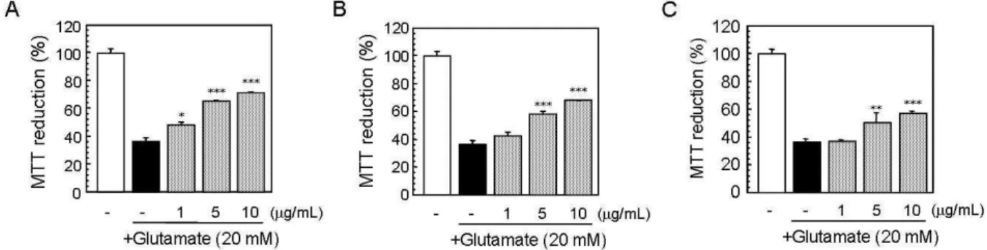
보기 위하여 추출물 중 가장 강력한 활성을 가지는 methanol 추출물을 이용하여 극성도에 따라 hexane, diethyl ether, eth- yl acetate 및 water 순으로 분획하여 그 분획물을 얻었다(Fig.
3). 각 분획물의 신경세포 보호효과에 대한 결과는 Fig. 4와 같다. N18-RE-105 세포주에 각 분획물을 1, 5, 10 μg/ml의 농 도로 처리하였을 때, 여러 용매 분획층 중에서 hexane 분획물 에서의 활성이 가장 높았다. 즉, hexane 분획물 5 μg/ml을 처리했을 때 65.5%의 생존율을 보였고 10 μg/ml에서는 71.5%
의 생존율을 보였으며, diethyl ether 분획물에서도 10 μg/ml
의 농도 처리 시 67.9%로 활성이 보였으나 hexane 분획물과
비교하였을 때 활성이 약하다는 것을 알 수 있었다. Ethyl ace-
tate 층에서는 아주 미약한 활성을 보였으며 water 층에서는
활성이 없었다. 이상의 결과로부터 상백피 추출물에서 신경세
Fig. 4. Protective effect of each fraction in glutamate-stressed N18-RE-105 cells. A; hexane, B; diethyl ether, C; ethyl acetate. The cells pretreated for 30 min with 1, 5 and 10 μg/ml of fractions from MAM. The cells were then treated with 20 mM of glutamate for 24 hr. After MTT assay, the MTT reduction rate (means±SD of triplicate determination) were calculated by setting each of control survival rate. Significant vs. glutamate-treated control group (*:
p
<0.05; **:p
<0.01; ***:p
<0.001).Fig. 2. Analysis of phase-contrast microscopic morphological changes by MAM in N18-RE-105 cells. The cells were exposed to various concentrations of MAM and mor- phological changes were monitored for 24 hr (a: control, b: 20 mM glutamate, c: 20 mM glutamate/10 μg/ml MAM, and d: 20 mM glutamate/50 μg/ml MAM).
Photographs were taken with a phase-contrast micro- scope at 100× magnification.
Fig. 3. Fractionation procedure of MAM. The extracts were frac- tionated in sequence with hexane, diethyl ether, and eth- yl acetate according to degree of polarity.
포 보호효과를 나타내는 물질은 대체로 비극성의 성질을 가짐 을 확인할 수 있었다.
본 연구를 통하여 상백피 추출물은 신경독성을 유도하고 흥분성 신경전달물질로 잘 알려져 있는 glutamate에 의한 산 화적 스트레스로부터 신경세포를 보호하는 효과를 확인 할 수 있었으며 뇌신경계 질환을 치료하거나 예방할 수 있는 새 로운 의약품의 개발이 명확하게 제시되지 않고 있는 시점에 퇴행성 신경질환의 새로운 치료제로의 가능성을 제시 하고자 하였다.
요 약
본 연구에서는 glutamate로 유도된 세포독성으로부터 신 경세포를 보호하는 상백피(Morus alba) 추출물의 활성을 확 인하기 위하여 N18-RE-105 세포주를 이용하여 MTT reduc- tion assay, LDH release assay 및 광학 현미경을 이용하여 형태학적인 변화를 관찰하였다. 그 결과, 상백피 methanol 추출물에서 농도 의존적으로 신경세포 보호효과가 나타났으 며, 50 μg/ml 농도에서는 80% 이상의 세포생존율을 확인할 수 있었다. 이 결과는 N18-RE-105 세포주의 LDH release as- say와 형태학적 변화에서도 일치하는 결과를 확인하였다. 가 장 높은 활성을 보인 상백피 methanol 추출물을 hexane, di- ethyl ether, ethyl acetate, water 층으로 분획하여 각 각 1, 5, 10 μg/ml 농도로 처리 시 hexane 층에서 48.0%, 65.6%, 71.5%로 가장 높은 신경세포 보호효과를 확인할 수 있었다.
따라서 상백피 추출물은 glutamate에 의한 세포독성으로부 터 신경세포 손상을 억제하며 신경세포를 보호하는 효과가 있다는 것을 알 수 있었다.
감사의 글
이 연구결과물은 2009학년도 경남대학교 학술연구장려금
지원에 의한 것임.
References
1. Chen, L. and L. Y. Hunag. 1991. Sustained potentiation of NMDA receptor-mediated glutamate responses through ac- tivation of protein kinase C by a mu opioid.
Neuron
7, 319-326.2. Cowburn, R., J. Hardy, P. Roverts, and R. Briggs. 1988.
Regional distribution of pre- and postsynaptic gluta- matergic function in Alzheimer’s disease.
Brain Res.
452, 403-407.3. Coyle, J. T. and P. Puttfarecken. 1993. Oxidative stress, glu- tamate, and neurodegenerative disorders.
Science
262, 689-695.4. Choi, B. W., E. M. Hur, J. H. Lee, D. J. Jun, and K. T. Kim.
2005. Protein kinase Cδ-mediated proteasomal degradation of MAP kinase phosphatase-1 contributes of glutamate-in- duced neuronal cell death.
J. Cell Sci.
119, 1329-1340.5. Dillon-Carter, O., C. Conejero, M. Poltorak, and W. J. Freed.
1998. N18-RE-105 cells: differentiation and activation of p53 in response to glutamate and adriamycin is blocked by SV40 large T antigen tsA58.
Cell Tissue Res.
291, 191-205.6. D'Orlando, C., M. R. Celio, and B. Schwaller. 2002.
Calretinin and calbindin D-28k, but not parvalbumin pro- tect against glutamate-induced delayed excitotoxicity in trasfected N18-RE 105 neuroblastoma-retina hybrid cells.
Brain Res.
945, 181-190.7. Duffy, S., A. So, and T. H. Murphy. 1998. Activation of en- dogenous antioxidant defenses in neuronal cells prevents free radical-madiated damage.
J. Neurochem.
71, 69-77.8. Fonnum, F. 1984. Glutamate: a neurotransmitter in mamma- lian brain.
J. Neurochem.
42, 1-11.9. Hara, J., D. Gerashchenko, J. P. Wisor, T. Sakurai, X. Xie, and T. S. Kilduff. 2009. Thyrotropin-releasing hormone in- creases behavioral arousal through modulation of hypo- cretin/orexin neurons.
J. neruosci.
29, 3705-3714.10. Harman, D. 1981. The aging process.
P. Natl. Acad. Sci. USA
78, 7124-7128.11. Hikino, H., T. Mizuno, Y. Oshima, and C. Konno. 1985.
Isolation and hypoglycemic activity of Moran A, a glyco- protein of
Mousalba
root barks.Planta Med.
51, 159-160.12. Jin, D. Q., C. S. Lim, J. K. Hwang, I. H. Ha, and J. S. Han.
2005. Anti-oxidant and anti-inflammatory activity of maceli- gnan in murine hippocampal cell line and primary culture of rat microglial cells.
Biochem. Biophy. Res. Co.
331, 1264-1269.13. Kang, T. H., H. R. Oh, S. M. Jung, J. H. Ryu, M. W. Park, Y. K. Park, and S. Y. Kim. 2006. Enhancement of neuro- protection of mulberry leaves (
Morus alba
L.) prepared by the anaerobic treatment against ischemic damage.Biol.
Rharm. Bull.
29, 270-274.14. Kim, M. J., S. J. Choi, H. K. Kim, C. J. Kim, B. Hong, and
Y. J. Kim. 2007. Activation effects of
allium tuberosum
Rottl.on choline acetyltrasnferase.
Biosci. Biotech. Bioch.
71, 226-230.15. Kim, J. Y., J. H. Hwang, M. R. Cha, B. D. Choi, S. U. Choi, H. P. Park, and Y. I. Hwnag. 2006. Growth-inhibitory effects of the
plocamium telfairiae
extracts on cancer cells.Jorunal of Life Science
4, 659-663.16. Kim, S. Y., H. S. Lee, K. S. Ryu, E. J. Lee, and Y. C. Kim.
1999. protective effets of extracts of Mori Cortex Radicis on corbon tetrachloride-induced hepatotoxicity.
Yakhak Hoeji
. 43, 391-396.17. Lee, M. R., C. S. Han, D. Y. Han, E. Park, S. C. Lee, and H. R. Park. 2007. Protective effect of neuronal cell on gluta- mate-induced oxidative stress from
viola mandshurica
extracts.J. Korean Soc. Appl. Biol. Chem.
51, 79-83.18. Lim, C. S., D. Q. Jin, J. Y. Sung, J. H Lee, H. G. Choi, I.
H. Ha, and J. S. Han. 2006. Antioxidant and anti-in- flammatory activities of the nethanolic extract of
neo- rhodomela aculeate
in hippocampal and microglial cells.Biol.
Pharm. Bull.
29, 1212-1216.19. Monaghan, D. T., R. J. Bridges, and C. W. Cotman. 1989.
The excitatory amino acid receptors: their classes, pharma- cology and distinct properties in the function of the central nervous system.
Annu. Rev. Pharmacol.
29, 365-402.20. Noh, H. S., Y. S. Hah, R. Nilufar, J. Han, J. H. Bong, S.
S. Kang, G. J. Cho, and W. S. Choi. 2006. Acetoacetate pro- tects neuronal cells from oxidative glutamate toxicity.
J.
Neurosic. Res.
83, 702-709.21. Nomura, T., T. Fukai, S. Yamada, and M. Katayanagi. 1976.
Phenolic constituents of the cultivated Mulberry tree (
Morus alba
L.
).Chem. Pharm. Bull.
24, 2898-2900.22. Olanwo, C. W. and W. G. Tatton. 1999. Etiology and patho- genesis of Parkinson’s disease.
Annu. Rev. Neurosic.
22, 123-144.23. Parfenova, H., S. Basuroy, S. Bhattacharya, D. Tcheranova, Y. Qu, R. F. Regan, and C. W. Leffler. 2005. Glutamate in- duces oxidative stress and apoptosis in cerebral vascular endothelial cells: contributions of HO-1 and HO-2 to cytoprotection.
Am. J. Physiol. Cell Physiol.
290, 1399-1401.24. Weiloch, T. 1985. Hypoglycemai-induced neuronal damage prevented by an N-methyl-D-aspartate antagonists.
Science
230, 681-683.25. Yoon, M. Y., J. Y. Kim, J. H. Hwang, M. R. Cha, M. R. Lee, K. J. Jo, and H. R. park. 2007. Protective effect of methanolic extracts from
Dendrobium nobile
Lindl. On H2O2-induced neurotoxicity in PC12 cells.J. Korean soc. Appl. Chem.
50, 63-67.26. Zhang, W., F. Han, J. He, and C. Duan. 2008.
HPLC-DAD-ESI-MS/MS analysis and antioxidant activities of nonanthocyanin phenolics in mulberry (