저작자표시-비영리-변경금지 2.0 대한민국 이용자는 아래의 조건을 따르는 경우에 한하여 자유롭게 l 이 저작물을 복제, 배포, 전송, 전시, 공연 및 방송할 수 있습니다. 다음과 같은 조건을 따라야 합니다: l 귀하는, 이 저작물의 재이용이나 배포의 경우, 이 저작물에 적용된 이용허락조건 을 명확하게 나타내어야 합니다. l 저작권자로부터 별도의 허가를 받으면 이러한 조건들은 적용되지 않습니다. 저작권법에 따른 이용자의 권리는 위의 내용에 의하여 영향을 받지 않습니다. 이것은 이용허락규약(Legal Code)을 이해하기 쉽게 요약한 것입니다. Disclaimer 저작자표시. 귀하는 원저작자를 표시하여야 합니다. 비영리. 귀하는 이 저작물을 영리 목적으로 이용할 수 없습니다. 변경금지. 귀하는 이 저작물을 개작, 변형 또는 가공할 수 없습니다.
Master's Thesis
The neuroprotective effects of Archidendron
jiringa(Jack)I.C.Nielsen extract inhibit glutamate-induced
apoptosis in hippocampal HT22 cells.
Cheol LEE
Department of Biotechnology
GRADUATE SCHOOL
JEJU NATIONAL UNIVERSITY
The neuroprotective effects of Archidendron
jiringa(Jack)I.C.Nielsen extract inhibit glutamate-induced
apoptosis in hippocampal HT22 cells.
지도교수 김 재 훈
이 철
이 논문을 이학 석사학위 논문으로 제출함
2020 년 7 월이 철의 이학 석사학위 논문을 인준함
심사위원장 운노타쯔야
위 원
김 창 숙
위 원 김 재 훈
제주대학교 대학원
2020 년 7 월The neuroprotective effects of Archidendron
jiringa(Jack)I.C.Nielsen extract inhibit glutamate-induced
apoptosis in hippocampal HT22 cells.
Cheol LEE
(Supervised by Professor Jae Hoon Kim)
A thesis submitted in partial fulfillment of the requirement For the degree of Master of Science
July, 2020
This thesis has been examined and approved.
Chairperson of the supervising committee
Professor Tatsya Unno, Ph.D., College of Applied Life Sciences, Jeju National University
Professor Chang Sook Kim, Ph.D., College of Applied Life Sciences, Jeju National University
Professor Jae Hoon Kim, Ph.D., College of Applied Life Sciences, Jeju National University
Department of Biotechnology
GRADUATE SCHOOL
JEJU NATIONAL UNIVERSITY
i
The neuroprotective effects of Archidendron
jiringa(Jack)I.C.Nielsen extract inhibit glutamate-induced
apoptosis in hippocampal HT22 cells.
CONTENTS
CONTENTS... i
LIST OF FIGURES...ii
ABSTRACT...1
1. INTRODUCTION...2
2. MATERIALS AND METHODS...5
2.1. DPPH radical scavenging assay………...5
2.2. Cell culture and reagents…………..……….………….……....……….5
2.3. Preparation of sample…...…...………5
2.4. Cell viability assay………....………….………...5
2.5. Flow cytometry analysis...……...…...6
2.6. Measurement of intracellular ROS………...…………..……...6
2.7. Western blot assay ……….……….….……...…...…...6
2.8. Statistical analysis...……….………....…………..………...…...….7
3. RESULTS ……….………...………...8
3.1. Subtropical natural product screening for radical scavenging………8
3.2. A. jiringa extract protects glutamate-induced HT22 neuronal cell death...10
3.3. A. jiringa extract reverses glutamate-induced apoptotic HT22 cell death….…...12
3.4. A. jiringa extract inhibits glutamate-induced ROS production……...13
3.5. A. jiringa extract regulates signal pathway related MAPK and ER stress………...15
4. DISCUSSION...17
5. REFERENCES...20
ii
LIST OF FIGURES
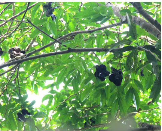
Figure 1.The picture of Archidendron jiringa.
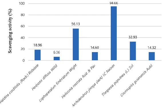
Figure 2. Subtropical natural product screening for radical scavenging activity
Figure 3. Pretreatment with A.jiringa extract protected HT22 cells against glutamate-induced cell death.
Figure 4. Glutamate-induced apoptotic cell death was inhibited by A. jiringa extract.
Figure 5. A. jiringa extract reduced glutamate-induced ROS production.
Figure 6. Pretreatment of A. jiringa extract inhibits the phosphorylation activities of ERK,p38, JNK activation.
Figure 7. A. jiringa extract regulates the Calpain 1, Caspase-12, Bax, Bcl-2, AIF
LIST OF TABLES
1
ABSTRACT
Archidendron Jiringa is a leguminous tree growing mainly in Southeast Asia. A
recent study found that A. jiringa has the effect of anticancer, anti-inflammatory, anti-diabetic and insect repellents. However, the neuroprotective effect of the A.
jiringa extract on glutamate induced cell apoptosis was not studied. Glutamate
activates the formation of reactive oxygen species(ROS) and the pathways of the mitogen-activated protein kinase (MAPK). MAPK is the oxidative stress pathway, and simultaneously regulates Calpain 1, Caspase 12, Bax, Bcl-2 and AIF which are the Endoplasmic Reticulum(ER) stress pathway. In this study, the effects of A.
jiringa extract on glutamate induced oxidative stress and ER stress in HT22 mouse
hippocampal cells were investigated.
It has been found that pretreatment of A. jiringa extract reduces ROS production and recovers HT22 cells from glutamate induced cell apoptosis. In addition, our study confirmed that A. jiringa extract inhibited MAP kinase signaling pathway, activated Calpain 1, Caspase 12, Bax, AIF and inhibited Bcl-2. Our results suggest that the A.
jiringa extract prevents damage from glutamate induced oxidative stress and ER
stress. It provides experimental evidence that it can be used as a potential
phytochemical for the treatment and prevention of neurodegenerative diseases due to the neuroprotective effects of A. jiringa.
2
1. INTRODUCTION
Recently, the temperature has been rising every year due to global warming. If the current condition is maintained, the average temperature will rise by more than 5℃ and most regions of South Korea, except for mountainous areas, will change to a subtropical climate. The average temperature rise in South Korea is 2.4 times steeper than the global average. As a result, the existing crop plantations are moving
northward, and new crops are being introduced in the southern regions of the country[1, 2]. In preparation for this situation, there is a need to collect and develop subtropical genetic resources that are valuable as future income crops. We are studying overseas plant resources that were difficult to study due to technical or capital reasons, and was conducted to establish a sustainable bio-economic foundation by securing resources to cope with the changing climate.
The Archidendron Jirninga is a Southeast Asian leguminous tree traditionally used to treat toothache, chest pain and skin conditions, as well as to clean up blood and induce urination [3, 4].
A study on A. jiringa extract reported that increasing SOD, an enzyme important for protecting gastrointestinal mucous membranes, has an antimicrobial effect, an
antioxidant effect by a strong DPPH radical scavenging ability, and an anti-cancer, an insect repellent and an anti-diabetic effect[3, 5, 6]. Despite many positive effects, the A. jiringa extract has not yet been studied for potential cell protection of the hippocampal nerve cells. This led to the possibility of A. jiringa as a candidate for brain disease treatment.
Glutamate is a major neurotransmitter for nerve stimulation and synapse
formation[7, 8], and Glutamate-induced toxicity is a major cause of serious brain diseases such as ischemic stroke, epilepsy, Parkinson's disease, Alzheimer's disease, etc.[9, 10]. Glutamate is tightly regulated in the central nervous system, but when the extracellular glutamate level increases rapidly, reactive oxygen species (ROS) are generated, which increases the formation of intracellular Ca2+ levels necessary for
3
oxidative glutamate toxicity, leading to neurodegenerative disorders and neuron cell death[11].
In neurons, glutamate toxicity occurs in two main ways. One is glutamate receptor-mediated excitatory toxicity, which causes N-methyl-D-aspartate (NMDA) receptors to become overactive, where Ca2+ is excessively introduced into cells to form free radicals, thereby expressing various enzymes leading to cell death[12]. Also, another pathway does not pass through the glutamate receptor, and the content of glutathione which is the precursor of the cysteine in the cell is reduced by suppressing the inflow of the cysteine by the glutamate/cystine antiporter. Consequently, the ROS product is increased by the oxidative stress, and a vicious cycle causing redox imbalance and leading to ER stress is repeated to form a self-amplified loop to further activate the cell death[13]. As a result, in the process of killing neuron cells, an increase in ROS concentration in cells stimulates oxidation stress and ER stress, and plays an
important role in developing neurological diseases[12].
In the HT22 cell, toxicity due to glutamate is eliminated due to lack of an ionotropic glutamate receptor functionally identical to that of the NMDA receptor. So, it is a model that can study the cell death caused by oxidative stress by suppressing the cellular uptake of cysteine caused by excessive accumulation of glutamate. In order to determine whether the A. jiringa extract is applicable to diseases
associated with it by showing a neuroprotective effect function on the hippocampal nerve cells, the neuroprotective effect was verified by using HT22 cells through cell survival, cell apoptosis analysis, intracellular ROS formation and western blot analysis.
4
5
2. MATERIALS AND METHODS
2.1 DPPH radical scavenging assay
The DPPH radical scavenging activity was measured by the method of Shimada et al., 1992. Sample extract was dissolved in 80% MeOH to a final concentration of 50 𝜇g/ml. 0.2 mM DPPH in MeOH was added to 96-well plates. The absorbance was measured at 525 nm after 30 min. MeOH was used as control. The
scavenging activity of DPPH radicals by the sample was calculated according to the following equation: DPPH radical scavenging activity (%) = (1-absorbance of sample/absorbance of control) × 100.
2.2 Cell culture and reagents
We obtained a HT22 cells line (mouse hippocampal neuronal cells) from Jae-ran Lee of Korea Research Institute of Bioscience and Biotechnology (KRIBB). The cells were cultured in Dulbecco’s Modified Eagle Medium (DMEM) supplemented with 10% fetal bovine serum, penicillin-streptomycin (Gibco-BRL, Gaithersburg, MD, USA) and 37℃ in 5% CO2 humidified atmospheres. Each cell suspension was
subcultured by treatment with trypsin-EDTA(Gibco-BRL) every 2 days in order to maintain exponential growth.
2.3 Preparation of sample
The dry extract of Archidednron jiringa was provided by International Biological
Material Research Center(KRIB, Korea). The sample was dissolved in 80% EtOH and used for the experiment.
2.4 Cell viability assay
HT22 cells were seeded into the 24-well plates at a density 5×10⁴ cells/well, and pre-treated with A. jiringa extract from 0 to 20 ug/ml for 2 h, and then 4 mM of L-Glutamate(Sigma-Aldrich, MO, USA) were added. After incubation for 24 h, Cell viability was measured by using the
WST-1(2-(4-iodophenyl)-3-(4-nitrophenyl)-5-6
(2,4-disulfophenyl)-2H-tetrazolium) assay (EZ-Cytox, DoGen, Korea). Each well was added to a final concentration of 10% WST-1 solution after 30 min incubation at room temperature. The absorbance was measured at 450 nm by using the microplate reader.
2.5 Flow cytometry analysis
For apoptosis analysis, HT22 cells were seeded in 6-well plates. After 24 h, they were pre-treated with A. jiringa extract at doses of 5 and 15 ug/ml for 2 h, and then added 4 mM L-Glutamate for 12 h. After collecting the cells, we incubated the cells with Annexin V-FITC and PI (FITC Annexin V apoptosis detection kit, BD
pharmigen). The apoptotic cells were detected by flow cytometry (LSRFortessa, BD).
2.6 Measurement of intracellular ROS
To evaluate the levels of intracellular ROS, HT22 cells were pre-incubated with A.
jiringa extract at doses 5 to 15 ug/ml for 2 h, and added 4 mM L-Glutamate. After 12
h, Fluorescent probe dye was diluted with basic medium and warmed up in a water bath at 37℃. Cells were stained with 10 mM of 2’-7’-dichlorodihydrofluorescein diacetate (DCFH-DA, Sigma-Aldrich, St. Louis, MO, USA) for 15 min in darkness at 37℃, and then harvested and washed with PBS three times. The green
fluorescence, which reflects the intracellular ROS level, was measured by flow cytometry (LSRFortessa, BD).
2.7 Western blot
Cells were lysed in M-PER lysis buffer (Thermo science, Bonn, Germany) containing protease inhibitor cocktail(Sigma-Aldrich, MO, USA), 2 mM sodium vanadate, 30 mM sodium pyrophosphate, and 100 mM sodium fluoride. After total protein quantification, proteins were separated by 10 - 12% SDS-PAGE (sodium dodecyl sulfate polyacrylamide gel electrophoresis) and transferred to nitrocellulose membranes (Amersham Biosience, Little Chalfont, Buckinghamshire, UK).
Membranes were blocked with 5% skim milk in Tris-buffered saline with 0.05% Tween 20 (TBST). Primary antibodies, such as, Calpain 1, Bax, , Bcl-2, CHOP,
7
Caspase12, GAPDH (Cell signaling technology, USA), phosphory-p38, p38, phosphory-ERK, ERK, phosphory-JNK, JNK were diluted 1:1000 in TBST and incubated overnight at 4°C. Secondary antibodies (Merck Millipore, Germany) were diluted in TBST 1:4000 and incubated for 1h. The immune complexes were detected by the ECL detection kit (Biosesang, Korea) according to the manufacturer’s
protocols.
2.8 Statistical analysis
Error bars represent ± SD. The statistical analysis was conducted using GraphPad Prism 5 to identify significant differences based on the one-way analysis of variance (ANOVA) followed by post hoc multiple comparisons (Turkey’s test). The value of p< 0.05 was considered to indicate significant differences.
8
3. RESULTS
3.1 Subtropical natural product screening for radical scavenging
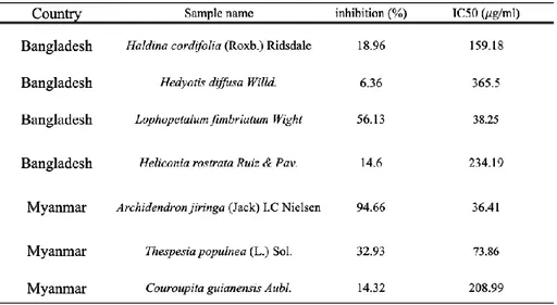
We screened various subtropical natural products based on DPPH radical scavenging ability. In Fig. 2, Archidendron jiringa extract showed the highest scavenging activity (94.6%). Next, lophopetalum fimbriatum extract showed the second highest scavenging activity (56.1%). In other extracts, the scavenging ability was less than 40%. Also, in Table.1, the value of IC50 of Archidendron jiringa is the lowest (36.41 𝜇g/ml). Therefore, we selected Archidendron jiringa and used it for experiments.
9
10
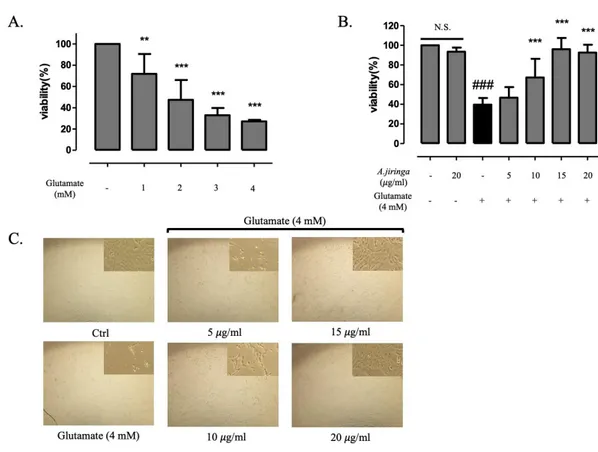
3.2 A. jiringa extract protects glutamate-induced HT22 neuronal cell death
we examined the neuroprotective effect of A. jiringa extract against glutamate-induced apoptosis in hippocampal HT22 cells by WST-1 assay.
Before evaluating the neuroprotective effect of the A. jiringa extract on the HT22 cells, the cell viability was measured after treatment with glutamate for each concentration for 24 h in order to establish an appropriate concentration of the glutamate on the HT22 cells. A survival rate of the cell exposed to glutamate is remarkably reduced by 60% at 4 mM (Fig.2A).
Next, WST-1 assay was performed to evaluate the cell viability of the A. jiringa extract for glutamate induced HT22 cells. After pre-treatment of A. jiringa extract by concentration, glutamate was treated and then cell viability was measured. We confirmed that the A. jiringa extract recovered 46.7%, 67.3%, 96% and 92.5% at 5, 10, 15, 20 𝜇g/ml , respectively, and showed a values similar to the the control group at 15 𝜇g/ml. Additionally, at concentration 20 𝜇g/ml, it was confirmed that it did not adversely affect cell viability (Fig.2B).
A morphological change of the HT22 cells are measured by a microscope for the glutamate induced cell death inhibition effect, and compared with the result of Figure 2B, it is determined that the cell viability and the morphological results are consistent (Fig.2C). These results suggest that treatment with A. jiringa extract can protect HT22 cells in glutamate-induced toxicity.
11
Figure 3.Pretreatment with A. jiringa extract protected HT22 cells against
glutamate-induced cell death. (A) WST-1 assay was performed after treatment with glutamate ( 0 – 4 mM). (B) WST-1 assay was performed after treatment with A.
jiringa extract ( 0 – 20 𝜇g/ml) for 2 h, followed by glutamate (4 mM) treatment on HT22 cells. (C) The cells of pretreatment with A. jiringa extract morphology was detected by microscopy. Each bar represents the mean ± SD (n=3) from three independent experiments per group. ##p<0.01 and ###p<0.001 vs. Ctrl. *p < 0.05, **p < 0.01, ***p < 0.001 vs. glutamate treated cells.
12
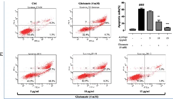
3.3 A. jiringa extract reverses glutamate-induced apoptotic HT22 cell death.
To analyze the type of cell apoptosis, Annexin V / PI staining was measured with a flow cytometer. Concentrations of 5, 10 and 15 ug/ml, which showed significance in cell viability, were used. Apoptotic cell death increased significantly to 61.6% in the glutamate induced group compared to 4% in the control group. However, the
treatment of A. jiringa extract decreased to 54.7% and 24.9%, 7.9% at 5,10, 15 𝜇g/ml, respectively. The above results confirmed that the A. jiringa extract exhibits neuroprotective effects by inhibiting apoptotic cell death by glutamate.
Figure 4. Glutamate-induced apoptotic cell death was inhibited by A. jiringa extract.
A. jiringa reduced apoptosis of glutamate-induced HT22 cells, as determined by flow
cytometry. Each bar represents the mean ± SD (n=3) from three independent experiments per group. ##p<0.01 and ###p<0.001 vs. Ctrl. *p < 0.05, **p < 0.01, ***p < 0.001 vs. glutamate treated cells.
13
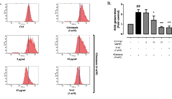
3.4 A. jiringa extract inhibits glutamate-induced ROS production.
Changes in ROS in the early stages of glutamate induced cell death are important indicators. It is well established that glutamate toxicity is mediated by ROS
production and cell death induced by oxidative stress. And excessive production of ROS can be an important mediator that damaged cell structures [14]. A recent study also found that ROS is involved in ER stress [15, 16, 17].
The effect of glutamate on the ROS was measured using flow cytometry. Already, another study reported there was a high increase in the ROS at the time of 12 h exposure, and we conducted experiments using this as a reference [9].
An N-acetylcysteine (NAC) is treated as a positive control in order to compare the effect of A. jiringa extract on the accumulation of ROS in glutamate induced HT22 cells. As shown in Fig.3A and 3B, the amount of ROS production in HT22 cells increased by about 2.2 times when glutamate was treated alone. In contrast, the A.
jiringa extract is reduced from 5, 10, 15 𝜇g/ml to 2.1 times, 1.4 times, 0.7 times, respectively, compared to the glutamate treated group and showed similar effects to the A. jiringa extract (15 𝜇g/ml) during NAC (1mM) treatment. The results suggest that the A. jiringa extract effectively protects HT22 cells from cell apoptosis caused by ROS generated by glutamate.
14
Figure 5. A. jiringa extract reduced glutamate-induced ROS production. HT22 cells
were pretreated with different concentrations of A. jiringa extract(5, 10, 15 𝜇g/ml) for 2 h, and then followed by glutamate (4 mM). after 24 h, cells were treated with 10 𝜇M DCF-DA for 15 min, and DCF fluorescence, as an indicator of the amount of ROS, was measured by flow cytometry. Each bar represents the mean ± SD (n=3) from three independent experiments per group. ##p<0.01 and ###p<0.001 vs. Ctrl. *p < 0.05, **p < 0.01, ***p < 0.001 vs. glutamate treated cells.
15
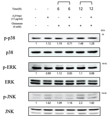
3.5 A. jiringa extract regulates signal pathway related MAPK and ER stress.
Another study suggests that MAP kinase activation and oxidative stress contribute to pathology of neurodegenerative diseases[8, 15, 18]. Accordingly, the effect of A.
jiringa extract on the activation of ERK, p38, JNK by glutamate was investigated.
As a result, ERK, p38 and JNK phosphorylation levels of A. jiringa extract treated group were reduced (Fig.4). The above results indicate that the A. jiringa extract shows a neuroprotective activity by regulating oxidative stress-related proteins.
Figure 6. Pretreatment of A. jiringa extract inhibits the phosphorylation activities of
ERK, p38, JNK activation. Cells were pre-treated for 2 h with 15 𝜇g/ml A. jiringa extract, and then exposed for 12 h to 4 mM of glutamate.
16
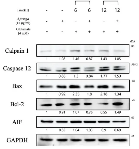
In other studies, the claim that ROS produced by oxidative stress amplifies ER stress is involved in cell death [15, 16]. We investigated the expression of the ER stress-related proteins Calpain 1, Caspase 12, Bax, Bcl-2 and AIF.
Figure 7. A. jiringa extract regulates the Calpain 1, Caspase-12, Bax, Bcl-2, AIF
Calpain 1 activated when Ca2+ homeostasis collapses and Caspase 12 activated only by ER stress have been confirmed to decrease the protein expression in the A. jiringa extract treatment group compared to the control group.
In addition, the activation of Calpain activates Bax and suppresses Bcl-2, thereby releasing AIF from the mitochondria[19, 20]. The A. jiringa extract treated group has decreased in Bax and AIF compared to the control group, and Bcl-2 increased the protein band at 12 h as compared with the control group(Fig.5). These results suggest that A. jiringa extract suppresses cell apoptosis pathway caused by ER stress.
17
4. DISCUSSION
Archidendron jiringa has traditionally been used to purify blood and to treat
dysentery. Recently, A. jiringa showed an increase in SOD, an important enzyme for gastrointestinal mucosa protection, and decreased the development of gastric lesions. Other studies have reported that phenol components, flavonoids, terpenoids and alkaloids are present and have strong DPPH radical scavenging activity. In addition, there are reports that it has anti-diabetic, anti-cancer, and antibacterial effects[3-6]. Based on this, we conducted a study to examine the potential of neuroprotection related to cell death induced by glutamate.
Several studies have shown that the initiation and progression of neurodegenerative diseases are associated with oxidative and ER stress[16, 17]. The mechanisms of glutamate induced oxidative toxicity disrupts glutamate/cysteine antiporter, depleting the cellular antioxidant GSH. After the depletion of GSH, the weakened antioxidant capacity causes a series of biological processes such as ROS accumulation and Ca2+ influx[10]. Ultimately, the accumulation of ROS induces oxidative stress and ER stress, leading to cell death[8, 16]. Although the mechanism between oxidative stress and ER stress has not been specifically identified, there have been reports that ROS generated by oxidative stress accelerates ER stress, which also leads to an increase in ROS cycle, and evidence has been shown that there are many connections between the two pathways[16, 17, 22]
We have successfully confirmed the protection effect against glutamate induced apoptosis, as previously suggested, by improving cell viability, reducing apoptosis rates, inhibiting ROS overproduction, and regulating protein expression using HT22 neuronal cells.
Through the WST-1 assay, it was confirmed that the A. jiringa extract had an effect of protecting from toxicity by glutamate. Additionally, the flow cytometer analysis by Annexin V and PI staining for cell apoptosis analysis showed that the A. jiringa extract significantly reduced cell apoptosis.
18
Mitochondria are damaged when the influx of Ca2+into the cytoplasm increases due
to glutamate. Ca2+affects both ER stress and oxidative stress, and ROS is produced
and released from the mitochondria to the cytoplasm, resulting in cell death [23]. Our results confirmed that the A. jiringa extract significantly reduces the production of ROS by glutamate.
Excessive ROS production activates the MAP kinase pathway, induces
phosphorylation of ERK, p38, JNK to participates in the cell apoptosis pathway. MAP kinase protein is known as a representative oxidative stress-related protein[24]. In previous studies, it has been reported that sustained activation of ERK causes oxidative stress induced cell death[25]. In particular, a study found that ERK is sensitive to oxidation stress in nerve cell death [8, 9].
The JNK and p38 pathways have a variety of mechanisms that triggered by stress and are involved in cell differentiation and apoptosis [26].
Consistent with these studies, it was found that A. jiringa extract inhibits phosphorylation of ERK, JNK, p38, blocking the cell apoptosis pathway.
MAPKs have also been reported to be associated with ER stress[16, 17]. A recent study suggests that activation of the JNK pathway occurs before inducing ER stress[27].
Endoplasmic reticulum (ER) is an organ that synthesizes secretory organs and membrane proteins, and ER stress is generated by the homeostatic variation of Ca2+ [28]. Ca2+ overload is the main mediator of the mitochondria-related glutamate
induced toxicity and activates Calpain 1. Many studies suggest that calpain plays an important role in apoptosis pathways[29, 30]. When the ER function is severely damaged, apoptosis is induced and caspase 12 is activated [31]. In addition, Ca2+
emitted from ER increase the ROS production of mitochondria, Bax is activated and Bcl-2 is suppressed, destroying the mitochondrial membrane. After then the
apoptosis factor, AIF, is activated[32].
In this study, it was confirmed that the A. jiringa extract treatment suppresses Calpain 1 and Caspase 12 from the result of protein expression associated with ER stress. This can be inferred that the unbalanced Ca2+ influx has been restored. In addition, it was confirmed that both Bax and AIF, which are cell apoptotic factors activated by Calpain 1, were decreased by A. jiringa extract.
19
In total, it is found that glutamate induced oxidative stress and ER stress by ROS production in HT22 hippocampal neuron cells, and A. jiringa extract reduces ROS accumulation and suppresses expression of proteins related to apoptosis pathways such as Calpain 1, Caspase 12, Bax, AIF and MAPKs to prevent cell death. This indicates that the A. jiringa extract has neuroprotective activity and suggests potential as a natural neuroprotective agent for the treatment of neurodegenerative diseases.
20
5. REFERENCE
1 Shim, K. M., Kim, G. Y., Jeong, H. C., Lee, J. T., Korean, T., & For, S. (2008). Adaptation and Assessment of the impacts of Global Warming on
Agricultural Environment in Korea.
2 Kim, C., Kim, Y., Han, S., Lee, S., & Ko, H. (2017). Current Situations and
Prospects on the Cultivation Program of Tropical and Subtropical Crops in Korea.
3 Bunawan, H., Dusik, L., Bunawan, S. N., & Amin, N. M. (2013). Botany, traditional uses, phytochemistry and Pharmacology of Archidendron jiringa: A review. Global Journal of Pharmacology, 7(4), 474–478.
https://doi.org/10.5829/idosi.gjp.2013.7.4.824
4 Shukri, R., Mohamed, S., Mustapha, N. M., & Hamid, A. A. (2011). Evaluating the toxic and beneficial effects of jering beans (Archidendron jiringa) in normal and diabetic rats. Journal of the Science of Food and Agriculture.
https://doi.org/10.1002/jsfa.4516
5 Charungchitrak, S., Petsom, A., Sangvanich, P., & Karnchanatat, A. (2011). Antifungal and antibacterial activities of lectin from the seeds of Archidendron jiringa Nielsen. Food Chemistry. https://doi.org/10.1016/j.foodchem.2010.11.114 6 Lubis, M. Y., Marpaung, L., Nasution, M. P., & Simanjuntak, P. (2018). Gallic acid from pods of Jiringa (Archidendron Jiringa [Jack] I.C. Nielsen) and its antioxidant. Asian Journal of Pharmaceutical and Clinical Research.
https://doi.org/10.22159/ajpcr.2018.v11s1.26582
7 Lee, J. S., Kim, W. Y., Jeon, Y. J., Lee, S. K., & Son, C. G. (2018).
Aquilariae Lignum extract attenuates glutamate-induced neuroexcitotoxicity in HT22 hippocampal cells. Biomedicine and Pharmacotherapy, 106(July), 1031–1038. https://doi.org/10.1016/j.biopha.2018.07.032
8 Li, Z., Chen, X., Lu, W., Zhang, S., Guan, X., Li, Z., & Wang, D. (2017). Anti-oxidative stress activity is essential for amanita caesarea mediated
neuroprotection on glutamate-induced apoptotic HT22 cells and an Alzheimer’s disease mouse model. International Journal of Molecular Sciences, 18(8). https://doi.org/10.3390/ijms18081623
9 Yang, E. J., Kim, G. S., Kim, J., & Song, K. S. (2013). Protective effects of onion-derived quercetin on glutamate-mediated hippocampal neuronal cell death.
Pharmacognosy Magazine, 9(36), 302–308.
21
10 Lin, S. P., Li, W., Winters, A., Liu, R., & Yang, S. H. (2018). Artemisinin prevents glutamate-induced neuronal cell death via Akt pathway activation.
Frontiers in Cellular Neuroscience, 12(April), 1–9.
https://doi.org/10.3389/fncel.2018.00108
11 Shirlee Tan, B. S. P., David Schubert, B. S. P., & Pamela Maher, B. S. P. (2005). Oxytosis: A Novel Form of Programmed Cell Death. Current Topics in
Medicinal Chemistry, 1(6), 497–506. https://doi.org/10.2174/1568026013394741
12 Rodrigues, T. B., & Ballesteros, P. (2007). Acetoacetate Protects Neuronal Cells From Oxidative Glutamate Toxicity (2007). Journal of Neuroscience Research,
3253(April), 3244–3253. https://doi.org/10.1002/jnr
13 Chaudhari, N., Talwar, P., Parimisetty, A., d’Hellencourt, C. L., & Ravanan, P. (2014). A molecular web: Endoplasmic reticulum stress, inflammation, and oxidative stress. Frontiers in Cellular Neuroscience, 8(JULY), 1–15.
https://doi.org/10.3389/fncel.2014.00213
14 Kim, H. N., Kim, Y. R., Jang, J. Y., Choi, Y. W., Baek, J. U., Hong, J. W., Choi, Y. H., Shin, H. K., & Choi, B. T. (2013). Neuroprotective effects of
Polygonum multiflorum extract against glutamate-induced oxidative toxicity in HT22 hippocampal cells. Journal of Ethnopharmacology, 150(1), 108–115. https://doi.org/10.1016/j.jep.2013.08.014
15 Atlante, A., Calissano, P., Bobba, A., Giannattasio, S., Marra, E., &
Passarella, S. (2001). Glutamate neurotoxicity, oxidative stress and mitochondria. In
FEBS Letters. https://doi.org/10.1016/S0014-5793(01)02437-1
16 Jin, M. L., Park, S. Y., Kim, Y. H., Oh, J. Il, Lee, S. J., & Park, G. (2014). The neuroprotective effects of cordycepin inhibit glutamate-induced oxidative and ER stress-associated apoptosis in hippocampal HT22 cells. NeuroToxicology, 41, 102–111. https://doi.org/10.1016/j.neuro.2014.01.005
17 Chong, W. C., Shastri, M. D., & Eri, R. (2017). Endoplasmic reticulum stress and oxidative stress: A vicious nexus implicated in bowel disease
pathophysiology. International Journal of Molecular Sciences, 18(4), 1–19. https://doi.org/10.3390/ijms18040771
18 Chhunchha, B., Fatma, N., Kubo, E., Rai, P., Singh, S. P., & Singh, D. P. (2013). Curcumin abates hypoxia-induced oxidative stress based-ER stress-mediated cell death in mouse hippocampal cells (HT22) by controlling Prdx6 and NF-κB regulation. American Journal of Physiology - Cell Physiology, 304(7).
https://doi.org/10.1152/ajpcell.00345.2012
19 Pae, H. O., Jeong, S. O., Zheng, M., Ha, H. Y., Lee, K. M., Kim, E. C., Kim, D. H., Hwang, S. Y., & Chung, H. T. (2009). Curcumin attenuates ethanol-induced toxicity in HT22 hippocampal cells by activating mitogen-activated protein kinase
22
phosphatase-1. Neuroscience Letters, 453(3), 186–189. https://doi.org/10.1016/j.neulet.2009.02.025
20 Adams, J. M. (2003). Ways of dying: Multiple pathways to apoptosis. Genes
and Development, 17(20), 2481–2495. https://doi.org/10.1101/gad.1126903
21 Zhang, Y. M., & Bhavnani, B. R. (2006). Glutamate-induced apoptosis in neuronal cells is mediated via caspase-dependent and independent mechanisms involving calpain and caspase-3 proteases as well as apoptosis inducing factor (AIF) and this process is inhibited by equine estrogens. BMC Neuroscience, 7, 1–22. https://doi.org/10.1186/1471-2202-7-49
22 Choi, A. Y., Choi, J. H., Lee, J. Y., Yoon, K. S., Choe, W., Ha, J., Yeo, E. J., & Kang, I. (2010). Apigenin protects HT22 murine hippocampal neuronal cells against endoplasmic reticulum stress-induced apoptosis. Neurochemistry
International, 57(2), 143–152. https://doi.org/10.1016/j.neuint.2010.05.006
23 Prentice, H., Modi, J. P., & Wu, J. Y. (2015). Mechanisms of Neuronal Protection against Excitotoxicity, Endoplasmic Reticulum Stress, and Mitochondrial Dysfunction in Stroke and Neurodegenerative Diseases. In Oxidative Medicine and
Cellular Longevity. https://doi.org/10.1155/2015/964518
24 PARK, S. Y., JUNG, W. J., KANG, J. S., KIM, C.-M., PARK, G., & CHOI, Y.-W. (2015). Neuroprotective effects of α-iso-cubebene against glutamate-induced damage in the HT22 hippocampal neuronal cell line. International Journal of
Molecular Medicine, 35(2), 525–532. https://doi.org/10.3892/ijmm.2014.2031
25 Stanciu, M., Wang, Y., Kentor, R., Burke, N., Watkins, S., Kress, G.,
Reynolds, I., Klann, E., Angiolieri, M. R., Johnson, J. W., & DeFranco, D. B. (2000). Persistent activation of ERK contributes to glutamate-induced oxidative toxicity in a neuronal cell line and primary cortical neuron cultures. Journal of Biological
Chemistry. https://doi.org/10.1074/jbc.275.16.12200
26 Suh, H. W., Kang, S., & Kwon, K. S. (2007). Curcumin attenuates
glutamate-induced HT22 cell death by suppressing MAP kinase signaling. Molecular
and Cellular Biochemistry. https://doi.org/10.1007/s11010-006-9365-6
27 Go, B. S., Ahn, S. M., Shim, I., & Choe, E. S. (2010). Activation of c-Jun N-terminal kinase is required for the regulation of endoplasmic reticulum stress
response in the rat dorsal striatum following repeated cocaine administration. Neuropharmacology. https://doi.org/10.1016/j.neuropharm.2010.04.009
28 Choi, J. H., Choi, A. Y., Yoon, H., Choe, W., Yoon, K. S., Ha, J., Yeo, E. J., & Kang, I. (2010). Baicalein protects HT22 murine hippocampal neuronal cells against endoplasmic reticulum stress-induced apoptosis through inhibition of reactive oxygen species production and CHOP induction. Experimental and Molecular
23
29 Vosler, P. S., Brennan, C. S., & Chen, J. (2008). Calpain-mediated signaling mechanisms in neuronal injury and neurodegeneration. In Molecular Neurobiology. https://doi.org/10.1007/s12035-008-8036-x
30 Momeni, H. R. (2011). Role of calpain in apoptosis. Cell Journal, 13(2), 65– 72.
31 Toshiyuki, N., Hong, Z., Nobuhiro, M., En, L., Jin, X., Bruce, A. Y., & Junying, Y. (2000). Caspase-12 mediates endoplasmic reticulum specific apoptosis and cytotoxicity by amyloid-b. Nature, 403(January), 98–103.
32 Fleury, C., Mignotte, B., & Vayssière, J. L. (2002). Mitochondrial reactive oxygen species in cell death signaling. Biochimie. https://doi.org/10.1016/S0300-9084(02)01369-X
24