45(2) : 161∼ 167 (2014)
161
영지 에탄올 추출물의 마우스 유래 뇌 해마세포 보호효과
이승철1#·임남경1#·정혜영2·최은화1·전수명1·정길생1*
1계명대학교 약학대학, 2계명대학교 자연과학대학
Neuroprotective Effects of Ethanol Extract of Ganoderma lucidum L.
on murine hippocampal cells
Seung Cheol Lee1#, Nam-Kyung Im1#, Hye Young Jeong2, Eun Hwa Choi1, Soo Myeong Jeon1 and Gil-Saeng Jeong1
*
1College of Pharmacy, Keimyung University; Dae-gu 704-701, Korea
2College of Natural Sciences, Keimyung University; Dae-gu 704-701, Korea
Abstract− Ganoderma lucidum L. (GL) is a traditional oriental medicine that has been widely used as anti-inflammatory, anti- tumor and anti-oxidant in Korea and other Asian countries. In this study, we investigated the ethanol extract of GL has neu- roprotective effects in murine hippocampal HT22 cells. GL ethanol extract has the potent neuroprotective effects on glutamate- intoxicated cells by inducing the expression of heme oxygenase (HO)-1 in HT22 cells. GL ethanol extract increased JNK phos- phorylation. Obviously, When we treated the GL extract with c-Jun N-terminal kinase (JNK) inhibitor (SP600125), HO-1 expression was reduced. Moreover, we found that GL treatment caused the nuclear accumulation of Nrf2. In conclusion, the ethanol extract of GL significantly protects glutamate-induced oxidative damage by induction of HO-1 via Nrf2, JNK pathway in mouse hippocampal HT22. These results suggest that GL ethanol extract would be a good source for taking active com- pounds and may be a potential pharmaceutical products for brain disorder induced by neuronal damage and oxidative stress.
Key words− Ganoderma lucidum L., Anti-oxidant, Neuroprotective, HT22, Ethanol extract, Heme oxygenase-1
영지(Ganoderma lucidum L.)는 잔나비걸상과에 속하며, 근연종의 자실체를 약재로 사용한다. 색에 따라 흑지(黑芝), 황지(黃芝), 백지(白芝), 홍지(紅芝)로 불린다. 삿갓은 반원 또는 콩팥 모양으로 목질화 되어 딱딱하고, 바깥면은 붉은 색, 검은색, 푸른색, 흰색, 황색, 자색 등 각각 다른 색을 띠 는 여러 가지 종류가 있다. 온대이상의 북반구 지방에 분포 하며 활엽수의 그루터기에 자생하고 냄새가 거의 없으며 맛 은 약간 쓰다.1,2) 예로부터 민간에서는 고지혈증, 협심증, 부 정맥, 간염, 소화성궤양, 당뇨에 효과가 있다고 알려져 왔으 며, 불면, 신경쇠약증, 소화불량, 노인성 기관지염에 의한 기 침 등의 만성질환에 사용되어 왔다. 영지의 약리효과에 관한 보고는 항종양,3,4) 세포보호작용,5,6) 면역계에 대한 작용,7,8) 항 바이러스작용,9,10) 심혈관계에 대한 작용,11) 항산화 작용,12) 등이 있으며, 영지에서 분리한 주요 성분으로는 polysaccharide
성분인 ganoderan B, C, GL-1,13) triterpene류의 ganoderic acid A~T, ganoderiol F, lucidenic acid A~P, methyl lucidenate F, lucidenolactone 등이 보고 되어있다.14,15)
활성산소종(Reactive oxygen spesies, ROS)은 생체 대사 과정 및 호흡과정에서 자연스럽게 생성되며 여러 세포 반 응들을 조절한다.16) 과도한 ROS의 축적은 산화적 스트레스 를 유발하여 헌팅턴 증후군, 파킨슨 증후군, 알츠하이머 증 후군과 같은 중추 신경계의 퇴행성 뇌질환의 발병에 중요 한 원인이 될 수 있다.17)또한, 글루타메이트의 과다축적은 뇌 신경세포에 과산화 반응을 일으켜 세포사멸을 통한 뇌 손상을 일으킨다.18) 본 실험에 사용한 HT22세포는 글루타 메이트 수용체가 존재하지 않기 때문에 글루타메이트 처리 시 신경전달물질로서 기능하지 않고 산화적 스트레스를 유 도한다. 이와 같은 특성 때문에 HT22 세포는 뇌 세포 보호 물질을 찾는 실험 모델로 유용하게 이용되고 있다.19)
항산화 시스템에서 중요한 구성요소 중 하나인 HO-1은 세포내의 heme을 분해하여 일산화탄소, 철, billiverdin을 생
#These authors contributed equally to this work.
*교신저자(E-mail):[email protected] (Tel):+82-53-580-6649
성한다. 대사생성물인 일산화탄소, 철, billiverdin과 HO-1은 그 자체로 세포손상 및 사멸의 억제, 항염증 및 항산화 작 용을 하는 것으로 알려져 있다.20) 최근에는 HO-1과 그 부 산물의 작용으로 뇌세포 보호를 비롯한 다양한 생리활성을 가진다는 연구 보고가 있으며, 특히 HO-1은 뇌세포 보호활 성의 marker protein으로 많이 활용하고 있다.21) 따라서, 본 연구에서는 글루타메이트 처리를 통해 산화적 손상을 입은 HT22 세포주의 사멸에 대하여 영지 에탄올 추출물의 농도 에 따른 뇌세포 보호 효과의 유의성을 살펴 보았으며, 그 작 용 기전을 탐색하였다.
재료 및 방법
실험 재료 − 본 실험에 사용된 영지(Ganoderma lucidum L.)은 2014년 3월 대구광역시 약령 시장에서 건조된 것을 구입하여 실험에 사용하였다. 정길생 교수가 형태학적 평가 를 통하여 동정하였고 표본시료(KMP-2014-03)는 계명대학 교 약학대학 생약학연구실에 보관하였다.
시약 및 기기 − 세포배양에 사용된 DMEM 배지와 trypsin- ethylenediaminetetraacetic acid (EDTA)는 Gibco Laboratories 사에서 구입하였으며, fetal bovine serum (FBS)는 Hyclone Laboratories사에서 구입하였다. 96well tissue culture plates 와 기타 tissue culture dishes는 Nunc사 제품을 이용하였다.
흡광도는 BioRad사의 Microplate Reader를 이용하여 측정 하였다. L-glutamate, Trolox와 3'-(4,5-dimethylthiazol-2-yl)- 2,5-diphenyltetrazolium bromide (MTT)는 Sigma 사에서 구 입하였다.
추출 − 건조된 영지 400 g을 100 g씩 물, 30% 에탄올, 70% 에탄올, 100% 에탄올로 각각의 조성의 용매 1 L를 넣 어 추출, 여과하고 rotary vacuum evaporator로 농축하였다.
각 추출물들을 감압 농축하여 용매를 휘발시켜 물(5.54 g), 30% 에탄올(4.82 g), 70% 에탄올(6.44 g), 100% 에탄올(4.04 g) 추출물을 얻었다. 각 시료들은 -70oC에서 보관하여 사용하 였다.
세포배양 − 마우스 해마 유래의 HT22 세포주는 묵인희 교수(서울대학교)로부터 분양 받아 사용하였으며, DMEM 배지에 10% FBS와 1% antibiotics를 첨가하여 37oC의 5%
CO2 incubator에서 1-2일 마다 계대배양 하면서 실험에 사 용하였다.
활성산소종(Reactive Oxygen Species: ROS)측정 − 세 포내에서 활성산소가 발생되면 2',7'-dichlorofluorescein diacetate (DCFDA)가 esterase 또는 산화적 가수분해에 DCFH로 탈아세틸화 되고 비형광성인 DCFH는 활성 산소 에 의해 산화되어 2',7'-dichlorofluorescein (DCF)로 전환된 다. 이는 강한 형광을 나타낸다. ROS 생성정도의 측정은 위 의 기술된 원리를 이용하였다. 배양된 세포를 PBS로 세척
한 후, 10 µM 2',7'-dichlorofluorescein diacetate (DCFDA, 35845)를 포함하는 Hank' blanced salt 용액에서 30분 동안 암실에서 반응시켰으며, 세포의 형광도(excitation wave length 490 nm; emission wave lenth; 525 nm)를 microplate reader (SpectramaxGemini XS, Molecular Devices, Sunnyvale, CA, U.S.A.)를 이용하여 측정하였다.
글루타메이트 유도 뇌세포 보호효과 측정 − 글루타메이 트로 유도한 뇌세포 독성에 대한 보호 효과는 정등의 방법20) 에 따라 실시하였다. HT22 세포를 96well microplates에 2
×105cells/well이 되도록 분주한 후, 37oC의 5% CO2 incubator에서 24시간 배양한 다음, 5 mM의 글루타메이트 와 각 추출물(50, 200 µg/ml)을 농도별로 처리하였다. 시료 처리 12시간 후 세포 생존율을 MTT 방법으로 측정하였으 며, Trolox 50 µM을 양성대조군으로 하여 비교하였다. 모든 실험은 대조군에 대한 세포 보호율을 mean±S.D로 표시하 였으며, 각각 3회 반복 실험치를 이용하여 계산하였다.
Western Blot Analysis − HT22 세포를 60 mm dish에 3
×105 cell/well 이 되도록 배양한 후 시료를 농도별 시간별로 처리한 후 상등액을 제거하고 PBS로 세척하였다.
RIPA buffer를 첨가하여 세포를 용해시키고 원심 분리하 여 얻은 단백질은 Bradford assay로 정량하여 12% SDS- PAGE를 이용하여 전기 영동한 후 PVDF membrane에 옮 긴 다음 5% skim milk로 30분간 blocking 한 후 1차 항체 (HO-1)를 1:1000으로 희석하여 넣고 12시간 동안 반응한 다 음, 다시 2차 항체를 반응시키고, ECL 용액을 1:1로 잘 섞 어서 membrane 위에 가하여 발광시키고 LAS4000을 이용 하여 확인하였다.
핵과 세포질 분획 − HT22 세포에 Protease inhibitor cocktail I과 1 mM phenylmethylsulfonyl fluoride (PMSF) 를 첨가한 PER-mammalian protein extraction buffer를 첨가 하여 균질화 하고 4oC에서 16,000×g로 원심분리 하였다. 5 분간 원심분리 후 상등액을 세포질 분획으로 사용하기 위 하여 -80oC에서 저장하였다. 나머지 침전물은 PBS로 세척한 후 RIPA buffer [150 mM NaCl, 1% NP-40, 0.5% sodium deoxycholate, 0.1% SDS, 50 mM Tris-HCl (pH 7.4), 50 mM glycerophosphate, 20 mM NaF, 20 mM ethylene glycol tetraacetic acid (EGTA), 1 mM dithiothreitol (DTT), 1 mM Na3VO4, protease inhibitor]를 첨가하고 4oC에서 15 분간 혼합한 후 4oC, 16,000×g에서 10분간 원심분리 하였 다. 이후의 과정에서는 10% SDS-PAGE를 이용하여 앞에서 설명한 western blotting 방법을 이용하였다.
통계처리 − 본 실험의 통계처리는 GraphPad Prism, version 3.03 (GraphPad Software Inc, San Diego, CA, USA)을 사용하였다. 각 실험군의 결과는 평균치와 표준오 차로 나타내었으며, 각 실험군 간의 결과는 ANOVA test를 사용하여 분석하고 유의적인 차이가 있는 항목에 대해서만
검정하였다. 실험군 간의 차이는 95% 수준(P<0.05)에서 유 의성 있는 것으로 하였다.
결과 및 고찰
본 연구에서는 영지 추출물의 뇌세포보호 효과와 그 기전 을 연구하였다. 세포 구성성분인 당, 지질, 단백질 및 DNA 등은 반응성이 높은 ROS에 의해 비가역적으로 파괴된다.22-24)
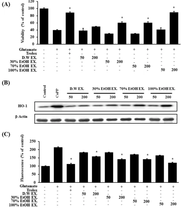
5 mM의 글루타메이트를 18시간 처리하였을 때 대조군에 비 해 세포생존율이 33.8±1.75%로 감소하는 세포 손상을 유도 하였으며, 영지 물 추출물, 30% 에탄올 추출물, 70% 에탄 올 추출물, 100% 에탄올 추출물과 글루타메이트를 함께 처 리 하였을 때 각 추출물에서 세포 보호 활성이 나타났으며, 100% 에탄올 추출물 처리군에서 세포 생존률이 가장 높게 측정되었다. 특히 200 µg/ml에서는 89±1.8%로 글루타메이 트 처리군과 비교 시 우수한 보호활성을 나타내었다(Fig. 1A).
Fig. 1. Effects of GL ethanol extract in a variety of concentration on glutamate-induced oxidative neurotoxicity and inhibition of reactive oxygen species generation and expression of HO-1. (A) Cells were treated with samples and then incubated for 18 h with glutamate (5 mM). (B) Expression of HO-1 was determined by western blot analysis, and representative blots of three independent experiments are shown. CoPP (20µM) was used for positive control. (C) Exposure of HT22 cells with 5 mM glutamate for 18 h increased reactive oxygen species production. Each bar represents the mean±S.D. of three independent experiments. *P<0.05.
Trolox (50µM) was used as the positive control.
또한 마우스 해마 유래의 HT22 세포에 영지 에탄올 추 출물을 18시간 처리한 후 뇌세포의 보호 역할을 담당하는 단백질인 HO-1 발현정도를 western blot analysis 방법으로 확인한 결과, 에탄올 함량이 높은 추출용매에서 얻어진 추 출물일수록 HO-1 발현이 증가하였다(Fig. 1B). ROS 소거 효과도 세포 보효효과와 같이 100% 에탄올 추출물 50, 200µg/ml에서 168±3.2%, 118±2.13%의 우수한 ROS 감소 효과를 나타내었다(Fig. 1C).
HO-1 발현정도가 에탄올 추출물의 농도에 의존적인가를 확인하기 위하여 25, 50, 100, 200 µg/ml 네 가지 농도로 18 시간 처리 하였으며, 양성 대조군으로는 cobalt protoporphyrin (CoPP) 20µM을 사용하였다. 그 결과, 추출물의 농도의 증 가에 따른 HO-1의 발현증가가 확인되었다(Fig. 2). 산화적 손상이 유도된 세포의 보호효과 역시 에탄올 추출물의 농 도에 따라 확연히 증가 되었으며(Fig. 3A), 농도 의존적으 로 ROS 감소효과를 나타내었다(Fig. 3B).
Nrf2는 transcription factor로 기능하며 이것의 핵 내 이동 은 HO-1발현에 관여하는 경로 중 중요한 인자로 알려져 있 으며, 외부 침입인 산화적 스트레스 방어기전의 주요한 역 할을 담당한다.25,26) 이를 바탕으로 영지 에탄올 추출물로 인
한 Nrf2 단백질의 핵 내 이동을 통해 HO-1 발현의 증가가 일어나는지 확인해보기 위해 HT22 세포에 200 µg/ml 농도 를 시간별로 처리하고 단백질 발현 정도를 확인해 본 결과, 시간이 증가함에 따라 세포질의 Nrf2 발현이 감소하는 양 상과는 대조적으로 핵에서의 Nrf2 발현은 증가하였다(Fig.
4). 이는 영지 에탄올 추출물이 Nrf2의 핵 내 이동을 촉진 시킴을 알 수 있는 결과이다. Mitogen activated protein kinase (MAPK) 경로 역시 HO-1발현에 주요한 역할을 하 는 것으로 알려져 있다. 일반적으로 MAPKs는 extracellular signal-regulated kinase (ERK), p38, c-Jun N-terminal kinase (JNK)의 세 가지 계열이 널리 알려져있으며, 세포 외부의 자극을 전달하여 세포내 반응을 일으키는 등의 다양한 역 할을 한다.27,28) 영지 에탄올 추출물의 작용으로 인한 HO-1 발현에 있어서 MAPK의 영향을 알아보기 위해 200 µg/ml
Fig. 2. Effect of GL 100% ethanol extract of HO-1 expression in HT22 cells. Cells were incubated for 18 h with ethanol extract. Expression of HO-1 was determined by western blot analysis, and representative blots of three independent experi- ments are shown. CoPP (20µM) was used for positive control.
Fig. 3. Effects of GL extract of 100% ethanol on glutamate- induced oxidative neurotoxicity and inhibition of reactive oxy- gen species generation. (A) Cells were treated with samples and then incubated for 18 h with glutamate (5 mM). (B) Expo- sure of HT22 cells with 5 mM glutamate for 18 h increased reactive oxygen species production. Each bar represents the mean±S.D. of three independent experiments. *P<0.05. Trolox (50µM) was used as the positive control.
의 영지 에탄올 추출물을 일정한 시간 간격으로 처리하였 다. 시간이 지남에 따라 영지 에탄올 추출물은 p38과 ERK 의 인산화에는 영향을 주지 못하였으나(Fig. 5B, 5C), JNK 의 인산화를 증가시키는 것을 확인할 수 있었다(Fig. 5A).
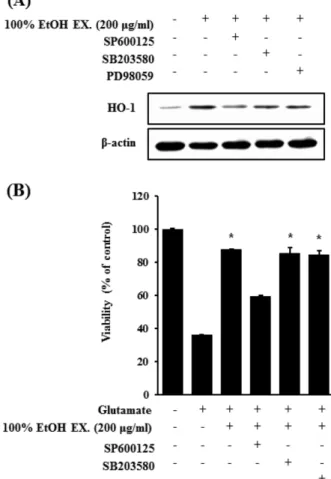
이러한 결과를 바탕으로 영지 에탄올 추출물 처리로 인한 HO-1의 발현 증가가 JNK의 인산화를 통하여 나타나는지 살펴보기 위하여 JNK, p38, ERK에 대한 각각의 억제제를 전 처리한 후 영지 에탄올 추출물을 처리하여 HO-1 발현과 세포 생존율을 확인하였다. 그 결과, p38의 억제제인 SB203580과 ERK의 억제제인 PD98059를 전 처리 하였을 Fig. 4. Effect of GL ethanol extract on Nrf2 nuclear translo-
cation in HT22 cells. Cells were treated with 200µg/ml of GL ethanol extract for 0.5, 1, and 1.5 h. The nuclei were frac- tionated from the cytosol using PER-mammalian protein extraction buffer as described in materials and methods. Nrf2 protein was detected by western blot analysis, and representa- tive blots of three independent experiments are shown.
Fig. 5. Effects of GL ethanol extract induced MAPK activation in HT22 cells. (A) Cells were treated with 200µg/ml of GL 100% ethanol extract for indicated times. The extract of cells were analyzed by western blot with specific antibodies for phosphorylated ERK1/2 (P-ERK)(A), phosphrylated JNK (P- JNK) (B), and phosphorylated p38 (P-p38)(C). Membranes were stripped and reprobed for total form of each MAPK anti- body as a control, and the representative blots of three inde- pendent experiments are shown.
Fig. 6. Effects of GL ethanol extract induced JNK pathway activation on HO-1 expression and glutamate-induced neuro- toxicity. (A) Cells were incubated with 200µg/ml of GL eth- anol extract for 18 h in the presence or absence of PD98059 (40µM), SB203580 (20 µM) and SP600125 (25 µM). West- ern blotting was then performed with HO-1 antibody. (B) Cells untreated or treated with GL ethanol extract (200µg/ml) in the presence or absence of each specific inhibitor for 18 h were exposed to 5 mM glutamate for 18 h. Trolox (50 mM) was used as positive control. Each bar represents the mean±S.D. of three independent experiments. Data shown represent the mean values of three experiments±S.D. *P<0.05 compared to the group with 5 mM glutamate.
때에는 HO-1의 발현과 세포보효 효과에 큰 영향을 미치지 못했지만, JNK의 억제제인 SP600125 처리 시 증가했던 HO-1의 발현이 줄어들고, 세포 보호 효과 역시 영지 에탄 올 추출물을 처리 하였을 때 84±0.23%로 증가하던 세포 생 존율이 59±1.1%로 감소하는 것을 확인하였다(Fig. 6).
따라서 본 연구를 통하여 영지 에탄올 추출물이 HO-1 단 백질의 발현을 크게 증가 시켰고, 마우스유래 해마 세포 HT22에서 글루타메이트로 유도한 세포독성에 강한 보호효 과를 관찰하였다. HO-1 단백질 발현의 증가는 MAPKs의 다양한 경로 중 JNK pathway 인산화와 Nrf2 단백질의 핵 내 전사 유도를 통하여 이루어짐을 확인하였다. 추후 연구 를 통해 영지 에탄올 추출물의 추가적인 뇌세포 보호기전 연구와 활성 물질 분리, 구조규명 및 동물실험 등에 관한 추 가적인 연구가 필요할 것으로 생각된다.
결 론
본 연구는 영지 에탄올 추출물의 뇌세포 보호 효과와 그 메커니즘에 관하여 연구하였으며, 그 결과 100% 에탄올 추 출물에서 글루타메이트로 유도한 HT22세포의 독성에 대해 우수한 보호 효과를 나타내었다. 이와 같은 효과는 영지 에 탄올 추출물이 HT22 세포에서 JNK의 인산화와 Nrf2의 핵 내 전사를 유도하고, 뇌세포 보호효과는 이와 같은 기전을 통한 HO-1 단백질 발현조절에 의한 것으로 생각된다.
사 사
이 연구는 계명대학교 신약개발 연구소 약학대학 특성화 연구기금으로 이루어졌음.
인용문헌
1.생약학교재편찬위원회 (2007) 생약학, 649-652, 동명사, 서 울.
2.한국 약용식물학 연구회 (2001) 종합약용식물학, 86, 학창 사, 서울.
3. Sliva, D., Labarrere, C., Slivova, V., Sedlak, M., Lloyd, Jr. F.
P. and Ho, N. W. (2002) Ganoderma lucidum suppresses motility of highly invasive breast and prostate cancer cells.
Biochem. Biophys. Res. Commun. 298: 602-612.
4. Yun, S. S., Kim, S. H., Sa, J. H., Jin, C. B., Lim, C. J. and Park, E. H. (2004) Anti-angiogenic and inhibitory activity on inducible nitric oxide production of the mushroom Gano- derma lucidum. J. Ethnopharmacol. 90: 17-20.
5. Zhang, H. N., He, J. H., Yuan, L. and Lin, Z. B. (2003) In vitro and in vivo protective effect of Ganoderma lucidum polysaccharides on alloxan-induced pancreatic islets damage.
Life Sci. 73: 2307-2319.
6. Wang, S. Q., Li, X. J., Zhou, S., Sun, D. X., Wang, H., Cheng, P. F., Ma, X. R., Liu, L., Liu, J. X., Wang, F. F., Liang, Y. F. and Wu, J. M. (2013) Intervention effects of Ganoderma lucidum spores on epileptiform discharge hippocampal neu- rons and expression of neurotrophin-4 and N-cadherin. PLoS One 8: 15-22.
7. Zhang, J., Tang, Q., Zimmerman-Kordmann, M., Reutter, W.
and Fan, H. (2002) Activation of B lymphocytes by GLIS, a bioactive proteoglycan from Ganoderma lucidum. Life Sci.
71: 623-638.
8. Bao, X. F, Zhen, Y., Ruan, L. and Fang, J. N. (2002) Puri- fication, characterization, and modification of T lymphocyte- stimulating polysaccharide from spores of Ganoderma luci- dum. Chem. Pharm. Bull. 50: 623-629.
9. Eo, S. K., Kim, Y. S., Lee, C. K. and Han, S. S. (2000) Pos- sible mode of antiviral activity of acidic protein bound polysaccharide isolated from Ganoderma lucidum on herpes simplex viruses. J. Ethnopharmacol. 72: 475-481.
10. Oh, K. W., Lee, C. K., Kim, Y. S., Eo, S. K. and Han, S. S.
(2000) Antiherpetic activities of acidic protein bound polysacchride isolated from Ganoderma lucidum alone and in combinations with acyclovir and vidarabine. J. Ethnophar- macol. 72: 221-227.
11. Berger, A., Rein, D., Kratky, E., Monnard, I., Hajjaj, H., Meirim, I., Piguet-Welsch, C., Hauser, J., Mace, K. and Nied- erberger, P. (2004) Cholesterol-lowering properties of Gan- oderma lucidum in vitro, ex vivo, and in hamsters and minipigs. Lipids Health Dis. 3: 311-323.
12. Hasnat, A., Pervin, M. and Lim, B. O. (2013) Acetylcho- linesterase inhibition and in vitro and in vivo antioxidant activities of Ganoderma lucidum grown on germinated brown rice. Molecules 6: 6663-6678.
13. Cheng, P. G., Phan, C. W., Sabaratnam, V., Abdullah, N., Abdulla, M. A. and Kuppusamy, U. R. (2013) Polysaccha- rides-Rich extract of Ganoderma lucidum (M.A Curtis:Fr.) P.
Karst accelerates wound healing in streptozotocin-induced diabetic rats. Evid.-based Complement Altern. Med. 24: 41- 50.
14. Mizushina, Y., Takahashi, N., Hanashima, L., Koshino, H., Esumi, Y., Uzawa, J., Sugawara, F. and Sakaguchi, K. (1999) Lucidenic acid O and lactone, new terpene inhibitors of eukaryotic DNA polymerases from a basidiomycete, Gan- oderma lucidum. Bioorg. Med. Chem. 9: 2047-2052.
15. Gao, J. J., Min, B. S., Ahn, E. M., Nakamura, N., Lee, H. K.
and Hattori, M. (2002) New triterpene aldehydes, lucialde- hydes A-C, from Ganoderma lucidum and their cytotoxicity against murine and human tumor cells. Chem. Pharm. Bull.
50: 837-840.
16. Coyle, J. T. and Puttfarcken, P. (1993) Oxidative stress, glutamate, and neurodegenerative disorders. Science 262:
689-695.
17. Jin, M. L., Park, S. Y., Kim, Y. H., Oh, J. I., Lee, S. J. and
Park, G. (2014) The neuroprotective effects of cordycepin inhibit glutamate-induced oxidative and ER stress-associated apoptosis in hippocampal HT22 cells. Neurotoxicology 41:
102-111.
18. Butterfield, D. A., Hensley, K., Cole, P., Subramaniam R., Aksenov M., Aksenova M., Bummer P. M., Haley B. E. and Carney J. M. (1997) Oxidatively induced structural alteration of glutamine synthetase assessed by analysis of spin label incorporation kinetics: relevance to Alzheimer's disease. J.
Neurochem. 68: 2451-2457.
19. Rssler, O. G., Bauer, I., Chung, H. Y. and Thiel, G. (2004) Glutamate-induced cell death of immortalized murine hip- pocampal neurons: neuroprotective activity of heme oxy- genase-1, heat shock protein 70, and sodium selenite.
Neurosci. Lett. 362: 253-257.
20. Jeong, G.S., Li, B., Lee, D. S., Choi, H. G. and Kim, Y. C.
(2010) Neuroprotective effects of the extract of Zingiberis Rhizoma. Kor. J. Pharmacogn. 41: 190-195.
21. Choi, H. G., Lee, D. S., Li, B., Jun, K. Y., Jeong, G. S. and Kim, Y. C. (2011) Neroprotective effect of the water-insol- uble fraction of root barks of Dictamnus dasycarpus 70%
ethanol extract on glutamate-induced oxidative damage in mouse hippocampal HT22 cells. Kor. J. Pharmacogn. 42:
175-181.
22. Fukui, M., Song, J. H., Choi, J., Choi, H. J. and Zhu, B. T.
(2009) Mechanism of glutamate-induced neurotoxicity in HT22 mouse hippocampal cells. Eur. J. Pharmacol. 617: 1- 11.
23. Gursoy, E., Cardounel, A. and Kalimi, M. (2001) The envi- ronmental estrogenic compound bisphenol A exerts estro-
genic effects on mouse hippocampal (HT-22) cells:
neuroprotection against glutamate and amyloid beta protein toxicity. Neurochem. Int. 38: 181-186.
24. Ye, S., Chen, M., Jiang, Y., Chen, M., Zhou, T., Wang, Y., Hou, Z. and Ren, L. (2014) Polyhydroxylated fullerene atten- uates oxidative stress-induced apoptosis via a fortifying Nrf2- regulated cellular antioxidant defence system. Int. J.
Nanomed. 9: 2073-2087.
25. Tan, M., Ouyang, Y., Jin, M., Chen, M., Liu, P., Chao, X., Chen, Z., Chen, X., Ramassamy, C., Gao, Y. and Pi, R.
(2013) Downregulation of Nrf2/HO-1 pathway and activation of JNK/c-Jun pathway are involved in homocysteic acid- induced cytotoxicity in HT-22 cells. Toxicol. Lett. 223: 1-8.
26. Itoh, T., Koketsu, M., Yokota, N., Touho, S., Ando, M. and Tsukamasa, Y. (2014) Reduced scytonemin isolated from Nostoc commune suppresses LPS/IFN γ-induced NO pro- duction in murine macrophage RAW264 cells by inducing hemeoxygenase-1 expression via the Nrf2/ARE pathway.
Food Chem. Toxicol. 69: 330-338.
27. Stanciu, M., Wang, Y., Kentor, R., Burke, N., Watkins, S., Kress, G., Reynolds, I., Klann, E., Angiolieri, M. R., Johnson, J. W. and DeFranco, D. B. (2000) Persistent activation of ERK contributes to glutamate-induced oxidative toxicity in a neuronal cell line and primary cortical neuron cultures. J.
Biol. Chem. 275: 12200-12206.
28. Chen, C. B., Ng, J. K., Choo, P. H., Wu, W. and Porter, A. G.
(2009) Mammalian sterile 20-like kinase 3 (MST3) mediates oxidative-stress-induced cell death by modulating JNK acti- vation. Biosci. Rep. 29: 405-415.
(2014. 6. 5 접수; 2014. 6. 13 심사; 2014. 6. 16 게재확정)