저작자표시-비영리-변경금지 2.0 대한민국 이용자는 아래의 조건을 따르는 경우에 한하여 자유롭게 l 이 저작물을 복제, 배포, 전송, 전시, 공연 및 방송할 수 있습니다. 다음과 같은 조건을 따라야 합니다: l 귀하는, 이 저작물의 재이용이나 배포의 경우, 이 저작물에 적용된 이용허락조건 을 명확하게 나타내어야 합니다. l 저작권자로부터 별도의 허가를 받으면 이러한 조건들은 적용되지 않습니다. 저작권법에 따른 이용자의 권리는 위의 내용에 의하여 영향을 받지 않습니다. 이것은 이용허락규약(Legal Code)을 이해하기 쉽게 요약한 것입니다. Disclaimer 저작자표시. 귀하는 원저작자를 표시하여야 합니다. 비영리. 귀하는 이 저작물을 영리 목적으로 이용할 수 없습니다. 변경금지. 귀하는 이 저작물을 개작, 변형 또는 가공할 수 없습니다.
Master’s Thesis
A study on neuroprotective effects of
methyl lucidone on glutamate-induced
neuronal cell death in HT-22 neurons
Jee-Yun Park
Department of Biomedicine & Drug Development
GRADUATE SCHOOL
JEJU NATIONAL UNIVERSITY
글루탐산염에 의해 유도된 신경세포
사멸에 대한 methyl lucidone의
신경보호효과에 관한 연구
지도교수 은 수 용
박 지 연
이 논문을 이학 석사학위 논문으로 제출함
2015년 2월
박지연의 이학 석사학위 논문을 인준함
심사위원장
위 원
위 원
제주대학교 대학원
2015년 2월
A study on neuroprotective effects of methyl lucidone
on glutamate-induced neuronal cell death
in HT-22 neurons
Jee-Yun Park
(Supervised by professor Su-Yong Eun)
A thesis submitted in partial fulfillment of the requirement for the degree
of Master of Science
February, 2015
This thesis has been examined and approved.
Date
Department of Biomedicine & Drug Development
GRADUATE SCHOOL
- I -
ABSTRACT
Oxidative stress elicits neuronal cell death in many neurodegenerative diseases, such as
Parkinson’s disease, Alzheimer’s disease and ischemia. Therefore, it has been evaluated as
the effective treatment strategy against neurodegenerative disorders to reduce neuronal cell
death by decreasing intracellular reactive oxygen species (ROS). Methyl lucidone (MLC)
was previously reported to suppress neuroinflammation in microglia. In the present study,
the neuroprotective mechanism of MLC was investigated in HT-22 neuronal cells against
glutamate-induced oxidative neurotoxicity.
Pretreatment of MLC (0.1-5 µM) increased neuronal cell viability against
glutamate-induced neuronal cell death. Glutamate-glutamate-induced ROS production was decreased by MLC,
although MLC did not show any free radical scavenging activity in cell-free condition, as
shown in 2,2-diphenyl-1-picrylhydrazyl (DPPH) assay. The expression of heme oxygenase 1
(HO-1) as an intracellular antioxidant enzyme was up-regulated by MLC treatment in HT-22
neurons. Nuclear translocation of NF-E2-related factor 2 (Nrf-2) known as a transcription
factor of HO-1 was also up-regulated by MLC treatment. Furthermore, pretreatment of
PD98059 as a pharmacological inhibitor of extracellular signal-regulated kinase (ERK) or
SB203580 as p38 mitogen-activated protein kinase (p38) inhibitor did not affect the
MLC-induced neuroprotection. To address whether phosphatidylinositol 3-kinase (PI3K) pathway
is involved in the upstream of Nrf-2, pretreatment of an inhibitor of phosphatidylinositol
3-kinase (PI3K), LY294002, suppressed the MLC-increased HO-1 induction and
neuroprotection against glutamate-induced neuronal cell death.
These results suggest that MLC significantly protects HT-22 neurons against
glutamate-induced oxidative neurotoxicity by inducing the expression of the antioxidant enzyme HO-1
via PI3K signaling pathway.
- II -
CONTENTS
ABSTRACT
··· ICONTENTS
··· IILIST OF FIGURE
··· IIII. INTRODUCTION
··· 1II. MATERIALS AND METHODS
··· 3III. RESULTS
··· 71. The protective effect of MLC on glutamate-induced neuronal cell death in HT-22 neurons. ··· 7
2. MLC inhibits intracellular ROS production by glutamate in HT-22 neurons. ···· 7
3. MLC activates Nrf-2/HO-1 signaling transduction in HT-22 neurons. ··· 11
4. The role of MAPKs pathway in protective effects of MLC. ··· 11
5. The PI3K/Akt pathway was involved in protective effects of MLC. ··· 18
IV. DISCUSSION
··· 22V. REFERENCE
··· 24- III -
LIST OF FIGURES
Figure 1. MLC protects HT-22 neurons against glutamate-induced neuronal cell
death ··· 8
Figure 2. Glutamate-induced ROS generation was suppressed by MLC ··· 9
Figure 3. MLC up-regulated HO-1 expression in HT-22 neurons ··· 12
Figure 4. MLC induced Nrf-2 translocation to nucleus in HT-22 neurons ··· 13
Figure 5. MLC regulated the phosphorylation of ERK ··· 14
Figure 6. Effect of MLC on the phosphorylation of JNK ··· 15
Figure 7. Effect of MLC on the phosphorylation of p38 ··· 16
Figure 8. Inhibition of ERK did not affect protective effects of MLC in HT-22 neurons ··· 17
Figure 9. The effect of PI3K inhibitor on MLC-induced HO-1 expression in HT-22 neurons ··· 19
Figure 10. PI3K involved in MLC-induced neuroprotection ··· 20
Figure 11. A neuroprotective mechanism of MLC on glutamate-induced toxicity in HT-22 neurons ··· 21
1
Ӏ. INTRODUCTION
Glutamate-induced neurotoxicity can lead to neuronal cell death in neurodegenerative
diseases, such as Alzheimer’s disease, ischemia and Parkinson’s disease (Beal, 1995; Coyle
and Puttfarcken, 1993; Jenner, 1994). The extracellular glutamate in the central nervous
system (CNS) induces lower glutathione levels inhibiting cystine/glutamate transporter,
which cause accumulation of reactive oxygen species (ROS) and the death of neuronal cell,
called oxidative glutamate toxicity (Murphy et al., 1989). The mouse hippocampal cell line,
HT-22, is a commonly used cell line for oxidative glutamate toxicity. This cell line is
deficient in functional ionotropic glutamate receptors and responds to oxidative glutamate
toxicity via non-receptor mediated pathway (Davis and Maher, 1994).
Oxidative stress can be caused by accumulation of ROS, such as hydroxyl radical,
hydrogen peroxide and superoxide anions. This ROS damages the cellular components
including DNA, proteins and lipids, which cause apoptosis (Thannickal and Fanburg, 2000).
Mammalian cells have an antioxidant system to prevent ROS formation or enhance ROS
degradation (Blokhina et al., 2003). Among the various antioxidant enzymes, the heme
oxygenase (HO)-1 is important enzyme of the cellular antioxidant system. HO-1 catalyzes
the rate-limiting step in the conversion of heme to biliverdin while producing carbon
monoxide (CO) and iron (Ryter et al., 2006). The activity of HO-1 is mainly regulated by
transcriptional level of nuclear transcription factor-E2-related factor 2 (Nrf-2). In normal
state, Nrf-2 is coupled with Kelch-like ECH-associated protein1 (Keap1) which inhibits
nuclear translocation of Nrf-2. During oxidative stress, Nrf-2 is detached from Keap1 and
translocated into nucleus. And it binds to antioxidant response element (ARE) to activate the
promoter region of many genes encoding phase ΙΙ detoxification enzymes and antioxidants,
including HO-1 (Surh, 2003). The dissociation of Nrf-2 from Keap1 is induced
2
protein kinases (MAPKs) and phosphatidylinositol 3-kinase (PI3K).
Linderaerythrocarpa Makino (Lauraceae), a deciduous shrub, is broadly distributed in
Taiwan, Korea, Japan and China. The fruit of L. erythrocarpa is used in traditional medicines
as analgesic, antibacterial, antidote, digestive and diuretic (Ichino et al., 1988). The extract of
the fruit was separated into four cyclopentenediones, including linderone, lucidone, methyl
linderone and methyl lucidone (MLC). It was reported that lucidone inhibited human
farnesyl protein transferase (FPTase) activity (Oh et al., 2005). Also it was demonstrated that
lucidone and MLC suppressed NO production (Wang et al., 2008). Our previous studies
reported that MLC had neuroprotective effect through inhibition of microglia-mediated
neurotoxicity (Cui et al., 2012). However, the direct effect of MLC on neurons was not still
investigated. Therefore, we examined whether MLC shows directly neuroprotective effects
3
ӀӀ. MATERIALS AND METHODS
1. Reagents
Dulbecco’s modified Eagle’s medium (DMEM), fetal bovine serum (FBS) and
penicillin/streptomycin were purchased from Gibco BRL (Grand Island, NY, USA).
Antibodies against c-jun NH2-terminal kinase (JNK), phospho-JNK, p38 and phospho-p38
were purchased from Cell Signaling Technology (Danvers, MA, USA). Antibodies against
extracellular signal-regulated kinase (ERK), ERK and nuclear transcription
factor-E2-related factor 2 (Nrf-2) were purchased from Santa Cruz Biotechnology Inc. (Santa Cruz,
CA, USA). Antibody against heme oxygenase-1 (HO-1) was purchased from Millipore
(Temecula, CA, USA). Antibody against TATA binding protein (TBP) was purchased from
Abcam (Cambridge, UK). Antibody against β-actin was purchased from Sigma-Aldrich (St.
Louis, MO, USA). 3-(4,5-Dimethylthiazol-2-yl)-2,5-diphenyl tetrazolium bromide (MTT)
was purchased from Amresco (Solon, OH, USA). 2’,7’-Dichlorofluorescin diacetate
(DCF-DA) and 2,2-diphenyl-1-picrylhydrazyl (DPPH) were purchased from Sigma-Aldrich (St.
Louis, MO, USA).
2. Extraction and isolation of MLC
The dried fruits (175.18 g) of L. erythrocarpa were extracted with MeOH (2×1 L) for 48
h at room temperature. After filtration, the extract was concentrated and the residue
weighed 11.31 g. The MeOH extract was partitioned between H2O and EtOAc (1:1, v/v) to
obtain a EtOAc-soluble fraction(9.91 g). The EtOAc fraction showed FPTase inhibition
activity and was partitioned again with MeOH to give MeOH-soluble fraction (8.5 g) and –
insoluble fraction (1.26 g). The MeOH-soluble fraction was concentrated, and then the
residue was chromatographed on a silica gel (350 g) column, eluted with a gradient of
4
fractions were collected and concentrated to yield 7.3 g. The active fraction was resubjected
to a C-18 column, and it was eluted with a gradient of MeOH/H2O (6:4, 7:3,8:2, MeOH,
each about 3 L) to provide methyl lucidone (1450 mg)(Oh et al., 2005).
3. Cell culture
HT-22 neurons, an immortalized hippocampal neuronal cell line (Breyer et al., 2007), were
a generous gift from Dr. B. H. Lee (Gachon University of Medicine and Science, South
Korea). HT-22 cells were cultured in DMEM supplemented with 10% FBS and 1%
penicillin/streptomycin, and incubated at 37°Cunder 5% CO2.
4. Measurement of cell viability
For determining cell viability, MTT assay was performed. HT-22 cells were seeded at a
density of 5 × 10⁴ cells/well in a 24-well plate. After 12 h, these cells were treated with various concentrations of MLC for 1 h. The cells were washed with DMEM and treated
with 5 mM glutamate for 12 h. And then, 100 µl of MTT solution (2 mg/ml) was added to
each well and cells were incubated at 37°C for 2 h. The supernatant was removed, and the
MTT formazan crystals were dissolved with 300 µl of DMSO. The absorbance was
measured at 550 nm using a microplate reader (Model 550, Bio-Rad, USA).
5. Measurement of intracellular ROS level
The intracellular reactive oxygen species (ROS) level was measured using
2’,7’-dichlorofluorescin diacetate. The HT-22 cells were seeded at a density of 5 × 10⁴ cells/well in a 24-well plate and incubated for 12 h. MLC was treated for 1 h in indicated
concentrations. The cells were washed with DMEM and treated with 5 mM glutamate for
12 h. And then, the cells were loaded with 50 µM DCF-DA for 15 min. The fluorescence
5
excitation wavelength of 485 nm and an emission wavelength of 535 nm.
6. Measurement of free radical scavenging effect
2,2-Diphenyl-1-picrylhydrazyl(DPPH), a purple-colored, is reduced into diphenylpicryl
hydrazine, a yellow-colored. To measure free radical scavenging activity of MLC, DPPH
assay was performed. 10 µl of MLC was added to 190 µl DPPH (0.15 mM) in each well
(96-well plate) and mixed vigorously. The mixture was incubated at room temperature for
1hin the dark covered with aluminum foil. Absorbance was detected at 517 nm using
microplate reader (VersaMax, Molecular devices, USA).
7. Preparation of cytoplasmic and nuclear protein
Preparation of cytoplasmic and nuclear protein was performed using NE-PER Nuclear and
Cytoplasmic Extraction Reagets (Invitrogen, USA) according manufacturer’s protocol.
HT-22 cells were seeded at a density of 1 × 10⁶ cells/dish in 100 mm dish. These cells were treated with MLC indicated time, and then washed twice and collected with cold PBS. After
centrifuging, cell pellet was resuspended in cytoplasmic extraction reagent. After
centrifuging, the supernatant cytoplasmic extract was transferred to a new tube. And nuclear
pellet was resuspended in Nuclear extraction reagent, and centrifuged. The supernatant
nuclear protein extract was transferred to a new tube and stored at -80oC until usage.
8. Western blot analysis
Cell extracts were separated with 10-12% SDS-PAGE and transferred to a polyvinylidene
difluoride (PVDF) membrane (Bio-Rad Laboratories, CA, USA). After blocking with 5%
skim milk in TBS (25 mM Tris, pH 7.4, 150 mM NaCl), the membrane was probed with
anti-Nrf2, anti-HO-1, anti-phospho-ERK1/2, anti-ERK1/2, anti-phospho-p38, anti-p38,
6
with TTBS and incubated with HRP-conjugated anti-mouse or anti-rabbit antibody. Then
the blots were detected using an enhanced chemiluminescence reagent according to the
manufacturer’s protocol. Optical densities of the band were quantified with an Image J
program.
9. Statistics
The data were presented as the mean ±S.E.M. at least 3 independent experiments.
Statistical analysis was performed using the t-test and one-way ANOVA. The differences
between groups were considered to be statistically significant when p<0.05, p<0.01 or
7
ӀӀӀ. RESULTS
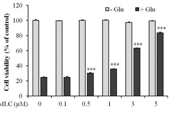
1. The protective effect of MLC on glutamate-induced neuronal cell death in HT-22 neurons.
To investigate whether MLC protects HT-22 neurons against glutamate-induced cell death
and MLC has the cytotoxicity in the cells, we examined the ctyotoxic effects of glutamate
(5 mM) on HT-22 cells with or without MLC pretreatment. The treatment of MLC did not
show any cytotoxic effect at concentrations less than 5 µM (Figure 1). Glutamate (5 mM)
decreased cell viability of HT-22 to 75.3 % compared to the non-treated group. Pretreatment
of MLC (0.5 - 5 µM) dose-dependently increased cell viability of HT-22 compared to the
glutamate-treated group. These results indicate that MLC protectedthe cells against
glutamate-induced cytotoxicity (Figure 1).
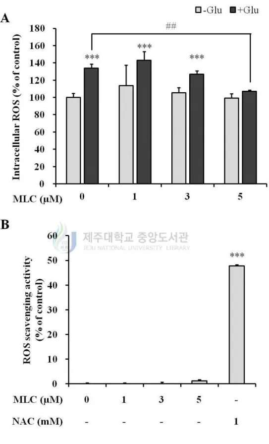
2. MLC inhibits intracellular ROS production by glutamate in HT-22 neurons.
To investigate whether MLC suppresses the glutamate-induced ROS in HT-22 cells, the
intracellular ROS levels were monitored by DCF-DA assay. Glutamate (5 mM) increased
intracellular ROS level up to 136 ± 4.3% (Figure 2A). The pretreatment of MLC (5 µM)
significantly reduced intracellular ROS compared to the glutamate-treated group. To
evaluate the antioxidant effect of MLC, DPPH assay was performed with MLC and
N-acetyl cysteine (NAC). MLC did not show the ROS scavenging effect (Figure 2B). These
results indicate that MLC plays a role to reduce the glutamate-induced ROS. It is estimated
that MLC is involved in the expression of antioxidant enzyme, albeit MLC is not
8
Figure 1. MLC protects HT-22 neurons against glutamate-induced neuronal cell death.
HT-22 cells were pretreated with MLC for 1 h. Then these cells were washed and incubated
with glutamate (5 mM) of fresh media for 12 h in the absence of MLC. The cell viability was
examined by MTT assay. The values were represented mean ±S.E.M. (n=3). ***p < 0.001 as
9
Figure 2. Glutamate-induced ROS generation was suppressed by MLC. (A) HT-22 cells
10
with glutamate (5 mM) for 12 h in the absence of MLC. Intracellular ROS levels were
measured using ROS-sensitive fluorescent DCF-DA. The values were represented mean ±
SEM (n=3). ***p < 0.001 as compared to the untreated control and ##p < 0.01 as compared
to the group treated with glutamate alone. (B) DPPH assay was perforemed to measure an
antioxidative effect of MLC. Free radical scavenging activities were indicated as %
inhibition. The values were represented mean ±S.E.M. (n=3). ***p < 0.001 as compared to
11
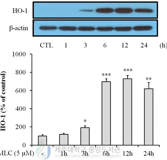
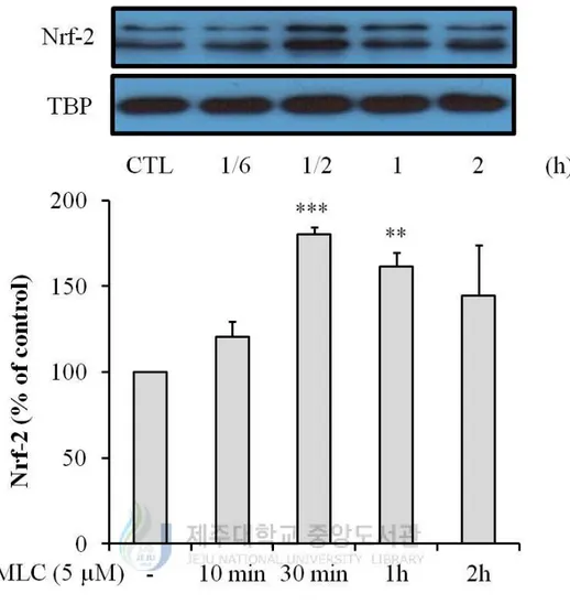
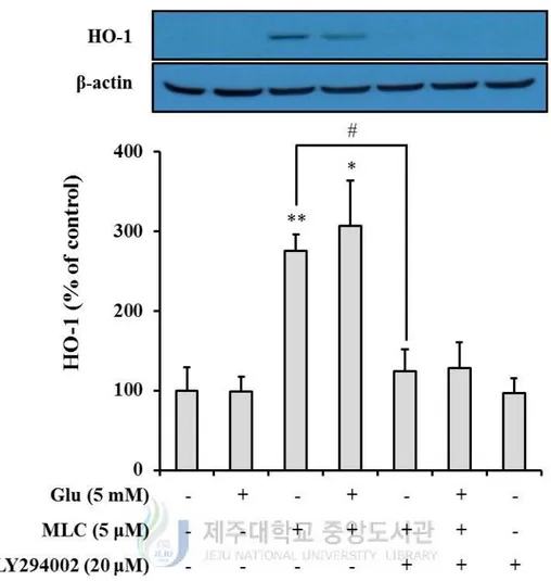
3. MLC activates Nrf-2/HO-1 signaling transduction in HT-22 neurons.
To determine that MLC regulate expression of HO-1, Western blot analysis was performed.
When MLC was applied in HT-22 cells, the induction of HO-1 was observed at 3 h and
highly increased at 6 h (Figure 3). As the translocation of Nrf-2 to nucleus can induce
expression of HO-1, we measured protein levels of Nrf-2 in nuclear fraction. Protein levels
of Nrf-2 were increased to a maximum at 30 min (Figure 4). These results indicate that
MLC induces the expression of HO-1 by Nrf-2 translocation. Accordingly, MLC has the
antioxidant effect on glutamate-induced oxidative stress.
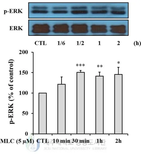
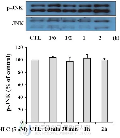
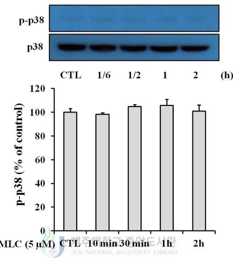
4. The role of MAPKs pathway in protective effects of MLC.
To evaluate which kinase of MAPKs was involved in Nrf-2 accumulation to nucleus,
phosphorylation rates of ERK, p38 and JNK were examined by Western blot analysis. The
treatment of MLC increased the phosphorylation of ERK compare to control group (Figure
5). In p38 and JNK, however, the significant increase of phosphorylation was not observed
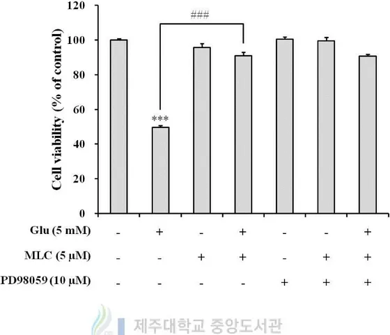
(Figure 6, 7). Thus, we confirmed the involvement of ERK activation in neuroprotection of
MLC. Cell viability that was increased by MLC on glutamate-induced cell death was not
affected by PD98059, an ERK inhibitor (Figure 8). These findings were suggested that the
activation of ERK was not involved in MLC effects, although the phosphorylation of ERK
12
Figure 3. MLC up-regulated HO-1 expression in HT-22 neurons. MLC was treated for 1
h in HT-22 cells. Then these cells were washed and further incubated in fresh media for 2 h,
5 h, 11 h and 23 h. The expression of HO-1 was examined by Western blot analysis. The
values were represented mean ± S.E.M. (n=3). *p < 0.05, **p < 0.01 or ***p < 0.001 as
13
Figure 4. MLC induced Nrf-2 translocation to nucleus in HT-22 neurons. MLC was
treated for 10 min, 30 min, 1 h and 2 h in HT-22 cells. The nuclei were fractionated from the
cytosol using NE-PER Nuclear and Cytoplasmic Extraction Reagets as described in
Materials and methods. Nrf-2 proteins were detected by Western blot analysis. The values
were represented mean ± S.E.M. (n=3). **p < 0.01 or ***p < 0.001 as compared to the
14
Figure 5. MLC regulated the phosphorylation of ERK. HT-22 cells were incubated with
MLC for the indicated times. The cells were lysed and the p-ERK and ERK proteins were
detected by Wetsern blot analysis. The values were represented mean ± S.E.M. (n=3). *p <
15
Figure 6. Effect of MLC on the phosphorylation of JNK. HT-22 cells were incubated with
MLC for the indicated times. These cells were lysed and the p-JNK and JNK proteins were
16
Figure 7. Effect of MLC on the phosphorylation of p38. HT-22 cells were incubated with
MLC for the indicated times. These cells were lysed and the p-p38 and p38 proteins were
17
Figure 8. Inhibition of ERK did not affect protective effects of MLC in HT-22 neurons.
PD98059 (10 μM) was treated for 1h prior to treatment of MLC. The cells were incubated
with MLC for 1 h. Then these cells were washed and incubated with glutamate (5 mM) for
12 h in the presence of PD98059. The values were represented mean ± S.E.M. (n=3). ***p <
0.001 as compared to the untreated control and ###p < 0.001 as compared to the group
18
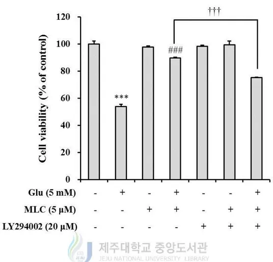
5. The PI3K/Akt pathway was involved in protective effects of MLC.
PI3K/Akt signaling pathway is involved in the activation of HO-1. Thus, we examined the
role of PI3K/Akt in protective effects of MLC. MLC-increased protein levels of HO-1 were
decreased by LY924002, a PI3K inhibitor (Figure 9). Also, the protective effect of MLC
was suppressed by LY294002 (Figure 10). These results indicate that PI3K/Akt signaling
19
Figure 9. The effect of PI3K inhibitor on MLC-induced HO-1 expression in HT-22 neurons. LY294002 (20 μM) was treated for 1h prior to treatment of MLC. These cells were
incubated with MLC (5 μM) for 1 h. Then these cells were washed and further incubated
with glutamate (5 mM) for 5 h in the absence of MLC and the presence of LY294002. The
cells were lysed and HO-1 proteins were detected by Western blot analysis. The values were
represented mean ± S.E.M. (n=3). *p < 0.05 or **p < 0.01 as compared to the untreated
20
Figure 10. PI3K involved in MLC-induced neuroprotection. LY294002 (20 μM) was
treated for 1h prior to treatment of MLC. These cells were incubated with MLC (5 μM) for 1
h. Then these cells were washed and incubated with glutamate (5 mM) for 12 h in the
absence of MLC and the presence of LY294002. The cell viability was measured by MTT
assay. The values were represented mean ± S.E.M. (n=3). ***p < 0.001 as compared to the
untreated control, ###p < 0.001 as compared to the group treated with glutamate alone and
21
Figure 11. A neuroprotective mechanism of MLC on glutamate-induced toxicity in HT-22 neurons.
22
ӀV. DISCUSSION
The excessive intracellular ROS causes various diseases by damaging cellular components,
such as DNA, lipids and proteins. In CNS, the high level of ROS results in neuronal cell
death, which contributes to neurodegenerative diseases. Oxidative glutamate toxicity has
been described in primary neuronal cell cultures (Murphy et al., 1989; Oka et al., 1993),
neuronal cell line (Davis and Maher, 1994; Miyamoto et al., 1989) and tissue slices (Vornov
and Coyle, 1991). Here I used the mouse hippocampal cell line, HT-22, because it is a
commonly used cell line for oxidative glutamate toxicity. The high level of extracellular
glutamate suppresses cystine uptake through the inhibition of glutamate/cystine antiporter,
which induces oxidative stress and neuronal cell death. In this process, HO-1 as antioxidant
enzyme is one of candidates to reduce oxidative stress. The present study investigated
whether MLC could inhibit oxidative glutamate toxicity.
Although MLC was not antioxidant, it could suppress the glutamate-induced ROS
generation (Figure 2). In some reports, it has been shown that phytochemicals had the
protective effect by activating some cytoprotective proteins including HO-1 (Hwang and
Jeong, 2008; Senthil Kumar et al., 2012; Zhang et al., 2012). The pretreatment of MLC
increases the expression of HO-1 and induces the nuclear translocation of Nrf-2 (Figure 3
and 4). Thus, it is suggested that the induction of HO-1 by MLC may inhibit the
glutamate-induced oxidative stress.
It was reported that the activation of Nrf-2 was controlled by its upstream regulators, such
as ERK, JNK, p38 and PI3K (Surh, 2003). First, MAPKs were targeted as on upstream of
Nrf-2 phosphorylation. It was observed that the treatment of MLC increased the
phosphorylation of ERK (Figure 5), but one inhibitor of ERK (PD98059) did not suppress
MLC-increased cell viability (Figure 8). So it was confirmed that MAPKs were not involved
23
are required. Next, it was examined that PI3K acts as an upstream regulator of Nrf-2. The
pretreatment of PI3K inhibitor (LY294002) reduced the expression of HO-1 that was
increased by MLC (Figure 9). Also MLC-induced neuroprotective effect was reduced by the
pretreatment of LY294002 (Figure 10). Therefore, it was suggested that MLC protected
HT-22 neurons against glutamate-induced oxidative stress via PI3K signaling pathway.
In conclusion, these findings demonstrated that MLC induced Nrf-2 activation through
PI3K signaling transduction and protected HT-22 neurons by suppressing glutamate-induced
24
V. REFERENCE
Beal, M.F., 1995. Aging, energy, and oxidative stress in neurodegenerative diseases. Annals
of neurology 38, 357-366.
Blokhina, O., Virolainen, E., Fagerstedt, K.V., 2003. Antioxidants, oxidative damage and
oxygen deprivation stress: a review. Annals of botany 91 Spec No, 179-194.
Coyle, J.T., Puttfarcken, P., 1993. Oxidative stress, glutamate, and neurodegenerative
disorders. Science (New York, N.Y 262, 689-695.
Cui, Y., Wu, J., Jung, S.C., Kim, G.O., Kyeong Ko, R., Lee, H.J., Yoo, E.S., Kang, H.K., Suk,
K., Eun, S.Y., 2012. Neuroprotective effect of methyl lucidone against microglia-mediated
neurotoxicity. European journal of pharmacology 690, 4-12.
Davis, J.B., Maher, P., 1994. Protein kinase C activation inhibits glutamate-induced
cytotoxicity in a neuronal cell line. Brain research 652, 169-173.
Hwang, Y.P., Jeong, H.G., 2008. The coffee diterpene kahweol induces heme oxygenase-1
via the PI3K and p38/Nrf2 pathway to protect human dopaminergic neurons from
6-hydroxydopamine-derived oxidative stress. FEBS letters 582, 2655-2662.
Ichino, K., Tanaka, H., Ito, K., Tanaka, T., Mizuno, M., 1988. Two New Dihydrochalcones
from Lindera erythrocarpa. Journal of natural products 51, 915-917.
Jenner, P., 1994. Oxidative damage in neurodegenerative disease. Lancet 344, 796-798.
Miyamoto, M., Murphy, T.H., Schnaar, R.L., Coyle, J.T., 1989. Antioxidants protect against
25
experimental therapeutics 250, 1132-1140.
Murphy, T.H., Miyamoto, M., Sastre, A., Schnaar, R.L., Coyle, J.T., 1989. Glutamate toxicity
in a neuronal cell line involves inhibition of cystine transport leading to oxidative stress.
Neuron 2, 1547-1558.
Oh, H.M., Choi, S.K., Lee, J.M., Lee, S.K., Kim, H.Y., Han, D.C., Kim, H.M., Son, K.H.,
Kwon, B.M., 2005. Cyclopentenediones, inhibitors of farnesyl protein transferase and
anti-tumor compounds, isolated from the fruit of Lindera erythrocarpa Makino. Bioorganic &
medicinal chemistry 13, 6182-6187.
Oka, A., Belliveau, M.J., Rosenberg, P.A., Volpe, J.J., 1993. Vulnerability of oligodendroglia
to glutamate: pharmacology, mechanisms, and prevention. J Neurosci 13, 1441-1453.
Ryter, S.W., Alam, J., Choi, A.M., 2006. Heme oxygenase-1/carbon monoxide: from basic
science to therapeutic applications. Physiological reviews 86, 583-650.
Senthil Kumar, K.J., Liao, J.W., Xiao, J.H., Gokila Vani, M., Wang, S.Y., 2012.
Hepatoprotective effect of lucidone against alcohol-induced oxidative stress in human
hepatic HepG2 cells through the up-regulation of HO-1/Nrf-2 antioxidant genes. Toxicol In
Vitro 26, 700-708.
Surh, Y.J., 2003. Cancer chemoprevention with dietary phytochemicals. Nature reviews 3,
768-780.
Thannickal, V.J., Fanburg, B.L., 2000. Reactive oxygen species in cell signaling. American
journal of physiology 279, L1005-1028.
Vornov, J.J., Coyle, J.T., 1991. Glutamate neurotoxicity and the inhibition of protein
26
Wang, S.Y., Lan, X.Y., Xiao, J.H., Yang, J.C., Kao, Y.T., Chang, S.T., 2008.
Antiinflammatory activity of Lindera erythrocarpa fruits. Phytother Res 22, 213-216.
Zhang, Z., Cui, W., Li, G., Yuan, S., Xu, D., Hoi, M.P., Lin, Z., Dou, J., Han, Y., Lee, S.M.,
2012. Baicalein protects against 6-OHDA-induced neurotoxicity through activation of
Keap1/Nrf2/HO-1 and involving PKCalpha and PI3K/AKT signaling pathways. Journal of
27
ABSTRACT IN KOREAN
산화적 스트레스는 신경세포 사멸을 유발시키는 주요한 원인 중의 하나로서 알쯔하이머 병, 파킨슨 병, 뇌졸중/허혈과 같은 신경 퇴행성 질환의 발병 및 진행과 관련이 있다고 알려져 있다. 그러므로 신경세포 내 활성산소가 급격하게 증가하는 병적 상태에서 이를 조절하여 신경세포의 사멸을 줄이는 것은 신경 퇴행성 질환에 있어서 매우 중요한 전략으로 평가되고 있다. 식물로부터 추출된 화합물이 여러 세포 종에서 세포 사멸에 대해 보호효과를 가진다는 연구가 많이 이루어 지고 있다. 비목나무 추출물에 포함된 화합물 중 하나인 methyl lucidone (MLC)이 미세신경아교세포(microglia)에서 신경염증을 억제하는 것이 이전에 보고되었다. 이번 연구에서는 신경세포에서 글루탐산염에 의해 유도된 산화적 스트레스에서 MLC의 신경보호 기전을 조사하였다. MLC (0.1~5 µM)를 처리하였을 때, 글루탐산염 신경독성 모델에서 글루탐산염에 의해 유도된 신경세포 사멸이 유의적으로 억제되는 것을 확인하였다. 산화적 글루탐산염 독성 상황에서 세포 내 활성산소의 양이 증가되는 것이 이미 알려져 있다. 이러한 상황에서 MLC는 글루탐산염에 의해 증가된 활성산소의 양을 억제하였다. 세포 내 활성산소의 감소가 MLC의 자체적인 항산화 활성에 의한 것인지 알아본 결과, MLC는 항산화 작용이 없는 것으로 확인 되었다. 따라서, MLC에 의한 세포 내 활성산소 생성의 억제는 세포 내 항산화 효소의 발현에 관여하는 것으로 생각되었고, 그 중 하나인 heme oxygenase (HO-1)를 조사하여 보았다. MLC는 HO-1의 발현을 증가시켰고 그의 상위 신호분자인 NF-E2-related factor 2 (Nrf-2)가 핵으로 이동하는 것을 증가시켰다. 이러한 과정에서 Nrf-2의 상위 신호전달을28
조절한다고 알려진 분자들 중 어떤 것이 관여하는 지 알아보았다. 각각의 억제제를 사용하여 실험한 결과, mitogen-activated protein kinases (MAPKs) 억제제는 MLC의 보호효과에 대해 아무런 변화를 보이지 않았지만 phosphatidylinositol 3-kinase (PI3K) 억제제는 MLC에 의해 유도된 HO-1의 발현과 보호효과를 감소시켰다. 이는 Nrf-2의 상위에 PI3K가 관여한다는 것을 의미한다. 결론적으로 MLC는 PI3K를 통해 Nrf-2의 핵으로 이동을 증가시키고 그에 따라 HO-1의 발현을 증가시킨다. 글루탐산염에 의해 증가된 세포 내 활성산소는 MLC에 의해 유도된 HO-1에 의해 억제되어 HT-22 신경세포에서 보호작용을 나타내는 것으로 확인되었다. 따라서 이번 연구는 MLC의 신경 퇴행성 질환에 대한 예방 및 치료제로의 활용 가능성에 대한 기초 자료를 제공하였다고 본다.
29
감사의 글
길게만 느껴졌던 2년이 정말 눈 깜짝할 사이 지나가고 이렇게 졸업논문을 쓰게 되었습니다. 짧지만 이 글을 빌어 그 동안 도움을 주셨던 분들께 감사인사를 드리고자 합니다. 학위과정 전후를 생각하면 너무나도 달라진 제 모습이 먼저 떠오릅니다. 이 분야에 대해선 무지했던 제가 실험을 하며 직면한 문제를 어떻게 해결해야 하는지 알게 되었습니다. 이렇게 달라질 수 있도록 2년의 시간 동안 제게 많은 가르침과 지도를 해주신 은수용 교수님께 먼저 감사의 말씀을 드립니다. 그리고 저의 논문 심사를 맡아 주시고 많은 조언을 해주신 정성철 교수님, 박주민 교수님께 진심으로 감사 드립니다. 저의 학위과정에 관심을 가져주시고 격려해 주시는 약리학교실의 강희경, 유은숙 교수님, 미생물학교실 고영상, 이근화 교수님, 생화학교실 조문제, 현진원 교수님, 조직학교실 박덕배, 이영기 교수님, 해부학교실 조사선, 윤상필, 김진우 교수님 및 모든 의과대학 교수님께 감사를 드립니다. 실험실 생활 하나도 모르던 저를 가르쳐 주고 이끌어 준 오금희 언니, 최연희 언니, 양윤실 언니, 강문석 오빠에게 진심으로 감사 드리고, 저와 같이 밤 늦게까지 실험실에 남아 공부하며 고생한 이지형 언니에게 감사 드립니다. 그리고 김선희 선생님, 실험실 후배 홀란, 유리에게도 감사의 마음을 전합니다. 그리고 대학원 동기 차지원 언니, 가영이, 새벽이에게도 우리 모두 수고했다는 말과 고맙다는 말을 전합니다. 세상에서 가장 사랑하는 우리 가족들에게 타지에서 고생한다고 걱정만 많이 끼쳤는데 이제 돌아가서 의젓한 딸, 누나 역할 하겠습니다. 2년 동안 믿고30
기다려주셔서 정말 감사합니다. 그리고 힘들다고 투정만 부렸는데도 이야기 잘 들어주고 다독여 준 내 친구들에게도 고맙다는 인사를 전합니다.