회춘양격산 물추출물의 항염증효과
이태호․조미정1․박숙자1․이종록․백영두․김종열2․권영규3․김상찬1*
대구한의대학교 한의과대학, 1 : 대구한의대학교 한방신약개발팀(BK21 Team), 2 : 한국한의학연구원, 3 : 부산대학교 한의전문대학원
Inhibitory Effects of Hoechunyanggyeok-san on Inflammation in Vivo and in Vitro
Tae Ho Lee, Mi Jeong Jo1, Sook Jarh Park1, Jong Rok Lee, Young Doo Back, Jong Reol Kim2, Young Kyu Kwon3, Sang Chan Kim1*
College of Oriental Medicine, 1 : BK21 Team, Daegu Haany University,
2 : Korea Institute of Oriental Medicine, 3 : School of Oriental Medicine, Pusan National University
Hoechunyanggyeok-san (HYS) is a traditional oriental herbal medicine widely used for treating inflammatory disorders. Although there are numerous clinical results of HYS reported in the literature of oriental hebal medicine, it has been rarely conducted to evaluate the immuno-biological activity. The present study was conducted to examine the anti-inflammatory effects of HYS extract (HYSE) in vivo and in vitro. To determine the cytotoxic concentration of HYSE, cell viability was tested by MTT assay. All four doses of HYSE (0.01, 0.03, 0.10 and 0.30 mg/ml) had no significant cytotoxicity during the entire experimental period. In order to measure NO levels in culture medium, the cells were treated with 1 µg/ml of LPS 1h before adding HYSE for 24 h and then culture medium were reacted with Griess reagent.
Increased NO production and iNOS expression were detected in LPS-activated cells compared to control. However, these increases were dose-dependently attenuated by treatment with HYSE. LPS plays a key role in leading to the massive production of pro-inflammatory cytokines such as TNF-α, IL-1β and IL-6 in macrophages. Thus, we next determined the levels of these cytokines. HYSE reduced the elevated production of TNF-α, IL-1β and IL-6 by LPS.
Moreover, the effects of HYSE were in a dose-dependent manner. In vivo, histopathological study, HYSE effectively inhe efed the increases of hind paw skin thicknesses and inflammatory cell infiltrations induced by carrageenan treatment. It, therefore, considered that HYSE will be favorably inhe efed the acute edematous inanner. In s. These findings showed that HYSE could have anti-inflammatory effects through the reduction of NO and inflammatory cytokines in macrophage.
Futhermore, the reduction of carrageenan-induced paw oedema by HYSE helps to understand its actions on inflammatory conditions.
Key words : Hoechunyanggyeok-san, inflammation, cytokine, edema
* 교신저자 : 김상찬, 대구시 수성구 상동 165 대구한의대학교 한의과대학
․E-mail : [email protected], ․Tel : 053-770-2247
․접수 : 2009/09/29 ․수정 : 2009/10/16 ․채택 : 2009/10/29
서 론
回春凉膈散은 萬病回春에 收載되어 있는 方劑로 連翹, 黃芩, 梔子, 桔梗, 黃蓮, 薄荷, 當歸, 生地黃, 枳實, 赤芍藥, 甘草 등의 11 種으로 構成되어, 三焦火盛으로 因한 口舌生瘡을 治療하는 方劑 이며, 淸熱解毒 凉血散火의 效能을 갖고 있다1).
回春凉膈散에 대한 연구로는, 김 등2)의 carrageenan 유도 부
종의 감소효과와, 강 등3)의 LPS로 활성화된 human monocyte의 IL-1β 생성억제 등에 대한 보고만 있으며, 回春凉膈散에 대한 연 구는 많은 부분에서 극히 제한적인 실정이다.
본 연구에서는 回春凉膈散이 淸熱解毒 凉血散火의 효능이
있음에 근거하여, 回春凉膈散 물추출물이 (HYSE;
Hoechunyanggyeok-san extract)이 LPS로 활성화된 Raw 264.7 cell에서 나타나는 염증매개물질들에 미치는 영향을 평가하였으 며, 또한 回春凉膈散이 동물모델에서도 유효한 항염증효과를 가 지는 지를 평가하기 위하여 carrageenan으로 유도된 rat의 발부 종 모델에서 부종의 정도 및 조직학적 평가를 실시하였다.
Carrageenan의 주입은 염증성 근육통을 유발하므로, 국소적 염증과, 통각과민의 연구에 널리 사용되고 있으며4), Raw 264.7 cell은 murine 대식세포의 cell line으로서, 대식세포는 여러 종류 의 숙주 반응에 관여하여 숙주의 방어와 숙주의 항상성 유지에 관여하는 것으로 알려져 있고, 염증 반응시에는 interleukin-1β (IL-1β), tumor necrosis factor-α (TNF-α) 및 interleukin-6 (IL-6) 와 같은 cytokine을 생산하여 감염초기에 생체 방어에 중요한 역 할을 하는 세포로 알려져 있다5).
Lipopolysaccharaide (LPS)는 인지질, 다당류 및 소량의 단 백질로 구성되며, 염증반응을 유발하는 유력한 인자로, 다양한 cytokine을 생성시키므로 염증반응을 연구하는 경우 빈용하는 실험모델6)로 확립되어 있다.
재료 및 방법
1. 回春凉膈散 추출물 (HYSE)의 제조
回春凉膈散의 재료는 대원약업사 (대구, 한국)에서 구입하여 관능평가를 한 후, <방약합편>7)의 용량을 근거로 回春凉膈散 4 첩 분량 122.8 g을 물 2 L에 넣고 3시간 전탕한 후 추출물을 거어 즈로 1차 여과하고 3000×g에서 3분간 원심분리하였다. 원심분리 후의 상층액만을 취하여 0.2 μm filter (Nalgene, New York, USA)로 여과하였다. 이 여과액을 rotary evaporator (EYELA, Tokyo, Japan)로 동결건조하여 21.58 g을 얻었으며, 사용 때까지 -20℃에서 보관하였다. 回春凉膈散 물추출물의 수율은 17.57 %였 으며 in vitro처치시에는 DMEM에 녹여 사용하였으며, in vivo 실험에서는 물에 녹여 사용하였다. 回春凉膈散의 구성 및 용량은 Table 1과 같다.
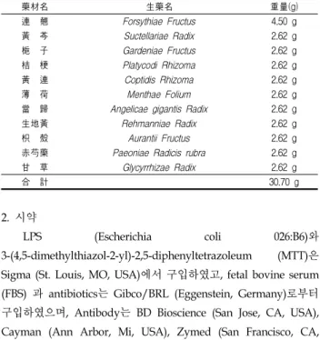
Table 1. Composition of Hoechunyanggyeok-san
藥材名 生藥名 重量(g)
連 翹 Forsythiae Fructus 4.50 g
黃 芩 Suctellariae Radix 2.62 g
梔 子 Gardeniae Fructus 2.62 g
桔 梗 Platycodi Rhizoma 2.62 g
黃 連 Coptidis Rhizoma 2.62 g
薄 荷 Menthae Folium 2.62 g
當 歸 Angelicae gigantis Radix 2.62 g
生地黃 Rehmanniae Radix 2.62 g
枳 殼 Aurantii Fructus 2.62 g
赤芍藥 Paeoniae Radicis rubra 2.62 g
甘 草 Glycyrrhizae Radix 2.62 g
合 計 30.70 g
2. 시약
LPS (Escherichia coli 026:B6)와 3-(4,5-dimethylthiazol-2-yl)-2,5-diphenyltetrazoleum (MTT)은 Sigma (St. Louis, MO, USA)에서 구입하였고, fetal bovine serum (FBS) 과 antibiotics는 Gibco/BRL (Eggenstein, Germany)로부터 구입하였으며, Antibody는 BD Bioscience (San Jose, CA, USA), Cayman (Ann Arbor, Mi, USA), Zymed (San Francisco, CA, USA)에서 구입하였고, NC paper는 Schleicher & Schuell (Dassel,
Germany)에서 구입하였다. TNF-α, IL-1β와 IL-6의 ELISA Kit는 Pierce endogen (Rockford, IL, USA)에서 구입하였다.
3. 세포배양
Murine macrophage cell line인 Raw 264.7 cells은 한국세포 주연구재단 (Seoul, Korea)에서 구입하였으며, Dulbecco’s modified Eagle’s medium (DMEM)에 10% fetal bovine serum (FBS), 100 U/ml penicillin 및100 μg/ml streptomycin을 혼합한 배지를 사용하여 37℃, 5% CO2 incubator에서 배양하였다. 실험 과정의 모든 cells은 80~90%의 confluence에서 실험하였고, 20 passages를 넘기지 않은 cell만 사용하였다.
4. 세포 생존율 측정
Raw 264.7 cell을 96-well plate에 5×10⁴ cells/well로 분주한 다음 回春凉膈散물추출물 (HYSE)을 농도별로 처치하여 세포의 생존율을 구하였다. 세포에 0.01, 0.03, 0.10, 0.30 mg/ml의 농도 로 HYSE를 처치한 후에 37℃, 5% CO2의 환경이 유지되는 배양 기에서 배양하였다. 배양 후 생존세포에 MTT (0.1 mg/ml)를 50
㎕넣고 4시간 배양한 후 배지를 조심스럽게 제거하고 생성된 formazan crystals을 DMSO에 녹여 Titertek Multiskan Automatic ELISA microplate reader (Model MCC/340, Huntsville, AL)를 사용하여 570 nm에서 흡광도를 측정하였다.
세포생존율은 control cell에 대한 백분율로 나타내었다.
i.e. viability(% control) =
100×/(absorbance of treated sample)/(absorbance of control)
5. NO생성량 측정
Raw 264.7 세포주로부터 생성된 nitric oxide (NO)의 양은 세포 배양액 중에 존재하는 NO2-의 형태로서 Griess 시약을 이 용하여 측정하였다. 간략하게 설명하면 세포배양 상등액 50 ㎕와 Griess시약 (1% sulfanilamide in 5% phosphoric acid + 1% α -naphthylamide in H2O) 50 ㎕를 96 well plates에 혼합하고 암 실에서 10분 동안 반응시킨 후 540 nm에서 Titertek Multiskan Automatic ELISA microplate reader (Model MCC/340, Huntsville, AL)로 흡광도를 측정하였다. NO2-의 농도는 sodium nitrate를 희석하여 흡광도를 측정하여 표준 곡선을 얻었다.
6. Immunoblot analysis
20 mM Tris Cl (pH 7.5), 1% Triton X-100, 137 mM sodium chloride, 10% glycerol, 2 mM EDTA, 1 mM sodium orthovanadate, 25 mM b-glycerophosphate, 2 mM sodium pyrophosphate, 1 mM phenylmethylsulfonylfluoride과 1 mg/ml leupeptin을 함유하는 buffer를 사용하여 cell을 lysis시켰 다. Cell lysates를 10,000×g로 10분간 원심 분리하여 debris를 제 거하였다. 각 protein의 발현은 각각의 antibody를 사용하여 면 역화학적 방법으로 분석하였으며, 2차 antibody는 alkaline phosphatase conjugated anti-rabbit을 사용하였다. 각 protein의
band는 ECL western blotting detection reagents (Amersham)를 사용하여 manufacturer's instruction에 따라 발색하였다. 발색 후 단백질의 발현량을 평가하기 위하여 image analyzing system (Ultra-Violet Products Ltd., Upland, CA, USA)을 이용하여 Densitometric analysis를 실시하였다.
7. Cytokine 측정
Cytokine을 측정하기 위하여 6-well plate에 cells (5×10⁵/ml) 을 분주하고 HYSE를 농도별로 처치한 다음, 1시간 후에 LPS를 처치하였다. LPS 처치 후 각 cytokine마다 특정 시간에 배지를 수거하여 cytokine을 측정하였다. 수거된 배지는 바로 측정하거 나, 측정전까지 -70℃에서 보관하였다. TNF-α, IL-1β와 IL-6는 ELISA Kit (Pierce endogen, Rockford, IL, USA)를 사용하여 측 정하였으며, 실험의 방법은 manufacturer's instruction에 따랐다.
8. 실험동물 및 처치
실험동물은 4주령 된 Sprague-Dawley계 수컷 흰쥐 (130-160 g)를 1주일 동안 환경에 적응시킨 후 실험에 사용하였으며, 사육 실 환경은 온도 20-23℃, 습도 60%, 12시간 light/dark cycle을 유 지하고, 사료 (Nestle Purina Petcare Korea, Seoul, Korea)와 음 료는 자유롭게 섭취하도록 하였다. 실험은 아무런 처치를 하지 않은 군을 Normal군으로 하고 기염제인 carrageenan (Sigma Chemical Co., St. Louis, USA; 100 μl/rat)만을 피하주사한 군을 carrageenan군으로 하였으며, carrageenan과 dexamethasone (1 mg/kg, P.O)을 투여한 dexamethasone군, carrageenan과 0.3 g/kg의 回春凉膈散을 투여한 0.3 g/kg HYSE군, carrageenan과 1 g/kg의 回春凉膈散을 투여한 1 g/kg HYSE군으로 나누었으 며, 각 군당 n수는 6마리로 하였다. dexamethasone과 HYSE는 4 일 동안 매일 1회 투여하였으며, 마지막 약물투여 1시간 후 carrageenan을 100 μl/rat로 rat의 오른쪽 발바닥에 주입하였다.
9. Paw edema의 유도 및 측정
4일째 약물을 투여하고 1시간 후 carrageenan을 투여하여 Paw edema를 유발시켰다. Paw edema의 측정은 carrageenan을 주입후 시간별로 (0, 1, 2, 3, 4시간) 부종측정기 (Plethysmometer, LE 7500; LETICA Scientific Instruments, Spain)를 이용하여 부 종 정도를 측정하였다.
10. 조직처리
Paw edema의 유발과 회복여부를 살펴보기 위해, carrageenan으로 염증이 유발된 rat의 오른쪽 하지의 발목아래를 절단하여 조직을 채취하여, 後肢의 발등 (dorsum pedis) 및 발바 닥 (ventrum pedis)의 피부 실질조직을 분리하여 10% 중성포르 말린에 6시간 이상 고정시킨 (d 탈수 및 파라핀 포매를 실시하 고, 3~4 μm의 longitudinal 절편을 제작하여 Hematoxylin-eosin 염색을 실시하고, 광학현미경 (Nikon, Japan)하에서 관찰하였다.
11. 조직학적 평가
Kim, et al8)의 방법을 변형시켜, 발등 및 발바닥 피부 (상피 에서 진피)의 두께를 mm 단위로 40배 현미경 시야에서 자동영 상분석장치 (DMI-300 Image Processing; DMI, Korea)를 이용하 여 각각 측정하였으며, 각각 1 mm2의 발등 및 발바닥 피부에 침 윤된 염증세포의 수 역시 자동영상분석장치를 이용하여, 200배 현미경 시야에서 측정하였다.
12. 통계적 검증
실험 결과는 mean ± S.D.로 나타내었으며, 처치군간의 유의 성은 one way analysis of varience (ANOVA)로 검정한 후 Newman-Kleuls test로 검정하였다. 통계적 유의성 검정은 p<0.05 또는 p<0.01로 하였다.
Fig. 1. Schedule of the in vivo study.
결 과
1. 回春凉膈散이 LPS로 유도된 Raw 264.7 cell의 NO production 에 미치는 영향
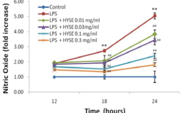
Raw 264.7 cell에서 HYSE의 NO 생성억제정도를 관찰하기 위하여 HYSE를 0.01, 0.03, 0.10, 0.30 mg/ml의 농도로 세포에 처 리하여 생성되는 NO양을 측정하였다. LPS군에서는 control군에 비교하여 NO의 생성량이 LPS처치 후 18 h, 24 h에서 각각 2.5 배, 5배 정도로 유의하게 증가하였으며, HYSE를 처치한 실험군 에서는 18 h, 24 h처치에서 전농도가 유의한 NO억제를 나타내 었다(Fig. 2).
Fig. 2. Effects of HYSE on the production of NO by LPS. Raw 264.7 cells were treated with 0.01, 0.03, 0.1, 0.3 mg/ml of HYSE dissolved in media for 1 h prior to the addition of LPS (1 μg/ml), and the cells were further incubated for 24 h. The concentrations of nitrite and nitrate in culture medium were monitored as described in the materials and methods section. Data represent the mean ± S.D. with eight separate experiments. (*, significant as compared to control. **P<0.01; #, significant as compared to LPS alone, ##P<0.01)
2. 回春凉膈散이 Raw 264.7 cell의 생존율에 미치는 영향 HYSE가 0.01, 0.03, 0.10, 0.30 mg/ml의 농도에서 LPS로 유 도된 Raw264.7 cell의 NO의 생성을 감소시킨 것이, HYSE의 세 포독성으로 인한 것인지를 관찰하기 위하여, HYSE를 0.01, 0.03, 0.10, 0.30 mg/ml로 처리하고 24시간 후에 MTT assay를 실시하 여 세포생존율을 측정하였다. 측정결과 12 h에서는 모든 경우 유 의한 세포독성이 나타나지 않았으나, LPS단독 처리 24 h에서는 Control에 비교하여 약 70% 정도의 세포독성을 나타내었다. 그 러나, LPS와 HYSE (0.01, 0.03, 0.10, 0.30 mg/ml)를 처리한 군에 서는 LPS 단독처치군에 비교하여 유의한 세포독성을 나타내지 않았으며, 오히려 HYSE는 농도의존적으로 유의한 세포독성을 억제함이 관찰되었다(Fig. 3).
Fig. 3. Effects of HYSE on the cell viability in LPS stimulated Raw264.7 cells. Raw264.7 cells were treated with 0.01, 0.03, 0.10, 0.30 mg/ml of HYSE dissolved in media for 1 h prior to the addition of LPS (1 μg/ml), and the cells were further incubated for 24 h. Data represent the mean ± S.D. with eight separate experiments. (*, significant as compared to control. **P<0.01)
A)
B)
Fig. 4. Effect of HYSE on the induction of iNOS by LPS. The levels of iNOS and actin protein were monitored 18h after treatment of cells with LPS (1μg/ml) with or without HYSE (0.01, 0.03, 0.10 and 0.30 mg/ml) pretreatment (i.e.
1h before LPS). Equal amounts of total protein were resolved by SDS-PAGE.
Expressions of iNOS protein were determined by immunoblot analysis using iNOS specific antibodies. The actin was used as a loading control (A). The relative density levels of protein bands were measured by scanning densitometry (B). The data represent the mean ± SD of three separate experiments.(*: significant compared with the control, **P<0.01, #: significant compared with the LPS alone,
##P<0.01).
4. 回春凉膈散이 LPS로 유도된 Raw 264.7 cell의 cytokine에 미 치는 영향
염증반응에 있어서 중요한 역할을 하는 TNF-α는 LPS반응의 주요 매개체로서 내재면역에 있어서도 중요한 역할을 한다9,10). 본 연구에서 LPS는 TNF-α의 분비를 유의성있게 증가시켰으며, HYSE는 0.10 및 0.30 mg/ml의 농도에서 TNF-α의 생성량을 유 의하게 감소시켰다(Fig. 5A). IL-1β는 NK cell의 활성, T-cell의 활성화, B-cell의 성숙을 활성화하는10) cytokine으로, 본 연구에서 도 LPS의 자극에 의하여 Raw264.7 cell의 IL-1β의 분비가 유의성 있게 증가하였으며, HYSE는 0.10 및 0.30 mg/ml의 농도에서 유 의하게 IL-1β의 생성량을 줄였다(Fig. 5B) IL-6는 B-cell이 활성화 되어 항체를 생산하는 plasma세포로 분화되도록 촉진하고, 항체 의 분비를 자극하는 cytokine으로 B-cell 분화단계의 후기에 주로 작용하여 imunoglobulin의 생성을 유도하고, T-cell의 증식에도 관련되어 있다11). 본 실험에서 LPS는 IL-6의 생성을 유의성 있게 증가시켰으며, HYSE는 0.03, 0.10, 0.30 mg/ml의 농도에서 LPS 로 유도된 IL-6를 유의성있게 감소시켰다(Fig. 5C).
A)
B)
C)
Fig. 5. The Effect of HYSE on LPS-stimulated cytokine production.
Production of cytokine was measured in the medium of Raw264.7 cells cultured with LPS (1 μg/ml) in the presence or absence of HYSE for 24 h. The amount of cytokine was measured by immunoassay as described in materials and methods. Data represent the mean ± S.D. with three separate experiments. (*, significant as compared to control. **P<0.01; #, significant as compared to LPS alone, #P<0.05, ##P<0.01)
5. 回春凉膈散이 carageenan으로 유도된 Rat의 paw edema에 미 치는 영향
부종측정기를 이용하여 paw edema를 측정한 결과, carrageenan을 주입한 군에서는 0, 1, 2, 3, 4 시간에 각각 1.00 ± 0.08, 1.68 ± 0.14, 2.21 ± 0.22, 3.02 ± 0.45, 3.02 ± 0.37을 나타내 어, 유의한 발 부종이 유발되었다. 그러나, 발부종유도 후 dexamethasone을 처치한 군에서는 1.03 ± 0.08, 1.02 ± 0.14, 1.01
± 0.17, 1.09 ± 0.16, 1.05 ± 0.17을 나타내어 실험 시간 동안 유의 한 발부종억제를 나타내었다. HYSE 0.3 g/kg를 투여한 군에서 는 0, 1, 2, 3, 4 시간에 1.12 ± 0.11, 1.22 ± 0.09, 1.74 ± 0.10, 2.02
± 0.24, 1.90 ± 0.15를 나타내어 1, 3, 4 시간에 유의한 발부종억제 를 나타내었으며, HYSE 1.0 g/kg를 투여한 군에서도 1.12 ± 0.12, 1.27 ± 0.12, 1.90 ± 0.23, 2.03 ± 0.09, 1.69 ± 0.15로 유의한 억제를 나타내었다(Fig. 6).
Fig. 6. Inhibition of Carrageenan-induced paw edema by HYSE.
HYSE was administered to rats at an oral dose of 0.3, 1.0 g/kg/day for 4 days before the induction of paw edema. Paw edema was induced by subcutaneously injecting 1% solution of carrageenan dissolved in saline (0.1 ml per animal) into the right hind paw. The swelling of the paw was measured 0~4 h after carrageenan injection. Dexamethasone (1 mg/kg p.o.) was used as a positive control. Data represent the mean ± S.D. of six animals. (## P<0.01, significant compared with carrageenan alone)
6. 回春凉膈散이 carageenan으로 유도된 Rat의 paw의 조직 변화 에 미치는 영향
HYSE의 carrageenan 유도 발부종에 대한 조직학적 영향을 평가하기 위하여 4 시간째의 발부종의 측정을 완료한 후 실험동 물을 희생하여 부종발의 발등 부분과 발바닥 부분에서 피부두께 및 침윤 염증세포의 수를 측정하였다. 본 실험의 결과, 발등피부 의 두께는 정상군에서 0.87 ± 0.11 mm, carrageenan군이 1.95 ± 0.35 mm로 유의성 있게 증가하였으며, 이러한 피부의 두께의 증 가는 dexamethasone 및 HYSE 0.3, 1.0 g/kg를 처치한 실험군에 서 0.75 ± 0.21, 1.17 ± 0.33, 1.08 ± 0.21 (mm)로 유의성있게 감소 하였다. 발바닥 부위의 피부두께에 있어서도 정상군은 0.70 ± 0.11 mm이었으나, carrageenan군에 있어서는 1.91 ± 0.24 mm로 유의성있게 증가하였으며, 이러한 발바닥의 피부두께의 증가 역 시 dexamethasone 및 HYSE 0.3, 1.0 g/kg를 처치한 실험군에서 0.84 ± 0.35, 1.37 ± 0.13 1.23 ± 0.31 (mm)으로 유의성있게 감소 하였다. 또한, 부종 발의 발등 조직에서 침윤 염증세포의 수 (cells/1 mm2)는 정상군에서는 6.40 ± 2.30개였으며, carrageenan군 은 28.40 ± 4.56개로 유의성있게 증가하였다. 이러한 염증세포의 증
가는 dexamethasone 및 HYSE 0.3, 1.0 g/kg의 처치에 의해 7.20 ± 2.28, 10.60 ± 2.61, 7.00 ± 2.70 (cells/1 mm2)을 나타내어 유의하게 감소되었다. 부종 발의 발바닥 조직에서 침윤염증세포의 수(cells/1 mm2)는 정상군이 6.40 ± 2.70개이었으며, carrageenan군은 831.80
± 124.99개로 유의한 증가를 나타내었다. 이러한 염증세포 침윤의 증가는 dexamethasone 및 HYSE 0.3, 1.0 g/kg의 처치에 의해 104.80 ± 64.10, 366.60 ± 110.50, 195.00 ± 80.10 (cells/1 mm2)로 유 의하게 염증세포의 침윤을 억제하였다(Table 2, Fig. 7, 8).
Table 2. Changes of histomorphometrical measurements in the present study
Groups
Dorsum pedis skin Ventrum pedis skin Thickness
(mm)
Infiltrated inflammatory cells
(cells/1 mm2)
Thickness (mm)
Infiltrated inflammatory cells
(cells/1 mm2) Normal 0.87±0.11 6.40±2.30 0.70±0.11 6.40±2.70 Carrageenan 1.95±0.35** 28.40±4.56** 1.91±0.24** 831.80±124.99**
Dexamethasone 0.75±0.21## 7.20±2.28## 0.84±0.35## 104.80±64.10##
HYSE 0.3 g/ml 1.17±0.33## 10.60±2.61## 1.37±0.13## 366.60±110.50##
HYSE 1.0 g/ml 1.08±0.21## 7.00±2.70## 1.23±0.31## 195.00±80.10##
Values are expressed as mean ± SD of 5 histological fields ** p<0.01 as compared with Normal control ## p<0.01 as compared with Carrageenan control.
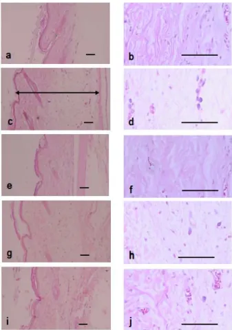
Fig. 7. Changes on histological profiles of the Dorsum Pedis skin in normal control (a, b), Carrageenan control (c, d), Dexamethasone (e, f), HYSE 0.3 g/kg (g, h) and HYSE 1.0 g/kg (i, j) treated groups.
Note that marked increases of skin thicknesses due to edematous changes were detected by carrageenan treatment with increases of inflammatory cell infiltrations.
However, these increases of skin thicknesses and inflammatory cell infiltrations were effectively inhibited by treatment of dexamethasone and two different dosages of HYSE, respectively. Arrow indicated total thicknesses measured. All HE stain;
Scale bars = 80 μm.
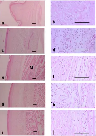
Fig. 8. Changes on histological profiles of the Ventrum Pedis skin in normal control (a, b), Carrageenan control (c, d), Dexamethasone (e, f), HYSE 0.3 g/kg (g, h) and HYSE 1.0 g/kg (i, j) treated groups. Note that marked increases of skin thicknesses due to edematous changes were detected by carrageenan treatment with marked increases of inflammatory cell infiltrations.
However, these increases of skin thicknesses and inflammatory cell infiltrations were effectively inhibited by treatment of dexamethasone and two different dosages of HYSE similar to that of dorsum pedis, respectively. M, muscle layers; Arrow indicated total thicknesses measured. All HE stain; Scale bars = 80 μm.
고 찰
回春凉膈散은 太平惠民和劑局方의 凉膈散에서 大黃, 芒硝, 竹葉, 蜂蜜을 제거하고, 黃連, 桔梗, 枳殼, 生地黃, 赤芍藥, 當歸를 加한 方劑이다1). 凉膈散의 구성은 크게 胸膈부위의 邪熱을 제거 하는 黃芩, 梔子, 連翹, 薄荷, 邪熱을 大小便으로 下하는 大黃, 芒 硝, 竹葉 그리고 淸熱潤燥, 調和諸藥하는 蜂蜜, 甘草의 3가지로 구성되어 있다.
回春凉膈散은 胸膈의 邪熱을 제거하기 위하여 凉膈散의 連 翹, 黃芩, 梔子, 薄荷에 黃連을 加하여 淸熱의 의미를 강화하였 고, 여기에 桔梗, 枳殼 (桔梗枳殼湯)을 배합하여, 理氣의 의미를 강화하였고, 또한 生地黃, 赤芍藥, 當歸, 甘草등을 加하여 邪熱로 인한 진액의 손상을 고려하고 있다.
回春凉膈散에 대한 연구로 김 등2)은 50.96 mg/20 g의 회춘 양격산추출물을 투여하여 carrageenan으로 유도된 paw edema 를 21%정도 억제함을 보고하였고, 강 등3)은 human monocyte에 회춘양격산추출물을 0.0001, 0.001, 0.01%처리하여 IL-1β가 8, 28, 43%가 감소함을 보고하였을 뿐 回春凉膈散에 대한 연구는 많은
부분에서 극히 제한적인 실정이다.
이러한 까닭에 본 연구에서는 回春凉膈散 물추출물의 NO 억제 기전 및 염증관련 cytokine의 발현에 대한 연구를 수행하기 위하여 열수추출된 回春凉膈散 (HYSE)이 LPS로 활성화된 Raw 264.7 cell에서 나타나는 염증매개물질들에 미치는 영향을 평가 하였으며, 또한 carrageenan 유발 발 부종에서의 부종의 정도 및 염증관련 지표에 대한 回春凉膈散의 효과를 평가하였다.
산화질소는 arginine과 O2로부터 nitric oxide synthase를 경 유하여 생성되는 radical로, 세포내에서 혈관조절, 숙주면역, 방 어, 신호전달 등에서 중요한 역할을 한다. septic shock, 뇌경색, 당뇨, 퇴행성신경질환 등이 NO의 과량생산에 연관되어 있다12). 대식세포가 이물질에 대응할 때 분비되는 IL-1β, TNF-α 및 nitric oxide (NO)는 숙주에 치명적인 결과를 초래 할 수 있는 것 으로 보고되고 있으므로13-15), NO 생성 저해제에 대하여 연구가 활발하게 이루어지고 있으며, 최근에는 靑黛, 苦楝皮, 當歸, 香附 子, 牡丹皮16-21) 등의 한약에서 이러한 조절제를 찾기 위해 많은 연구가 진행되고 있다.
Raw 264.7 cell에서 HYSE의 NO 생성억제정도를 관찰하기 위하여 HYSE를 0.01, 0.03, 0.10, 0.30 mg/ml의 농도로 세포에 처 리하여 생성되는 NO양을 측정하였다. LPS군에서는 control군에 비교하여 NO의 생성량이 유의성있게 증가하였으며, HYSE 0.01
~ 0.30 mg/ml을 처치한 실험군에서는 농도의존적으로 유의성있 는 NO의 생성억제를 나타내었다. 또한, HYSE는 12 h 및 24 h 에서 세포독성을 나타내지 않았으며, 오히려 농도의존적으로 LPS에 의한 세포독성을 억제하였다. Sharifi 등22)의 연구결과에 의하면, 납(Pb)이 PC-12 cell에서 유발하는 독성은, 납에 의한 NO의 과다생성으로 인해 세포독성이 유발됨을 밝혔으며, 박 등
23)은 감초성분 중의 하나인 liquiritigenin이 납에 의해 생성되는 NO의 생성을 억제하여 세포독성을 억제함을 보고한 바 있다.
HYSE가 LPS로 유도되는 세포독성을 억제한 것이 NO의 독성을 억제함에 기인한 것인지는 좀 더 연구가 진행되어야 할 것으로 생각된다.
NO는 iNOS를 경유하여 생성되는 것이므로24), HYSE와 iNOS단백질의 관련성을 조사하기 위하여 iNOS단백질의 발현량 을 조사하였다. LPS처치시에는 iNOS 단백질이 유의하게 발현이 증가되었으나, LPS에 HYSE 0.10, 0.30 mg/ml을 처치한 실험군 에서는 iNOS의 량이 농도의존적으로 유의하게 감소하였다. 이러 한 결과는 HYSE가 iNOS의 발현을 억제함으로서 NO의 생성을 억제함을 의미한다.
TNF-α는 pro-inflammatory cytokine으로서 macrophages, mast cells, endothelial cells 등으로부터 분비된다. 근래 TNF-α는 많은 자가면역질환에 있어서 염증의 개시 및 유지에 핵심적 역 할을 하는 것으로 알려져 있다9,10).
본 연구에서도 LPS는 Raw264.7 cell에서 TNF-α의 분비를 유의하게 촉진시켰으며, HYSE는 0.1 0및 0.30 mg/ml의 농도에 서 TNF-α의 생성량을 유의하게 감소시켰다. 이러한 결과는 HYSE가 여러 종류의 면역게재 염증성 질환을 유의하게 개선시 킬 수 있음을 의미한다.
염증관련 cytokine 중, IL-1β는 TNF-α, IL-2, IL-6와 함께 pro-inflammatory cytokine으로서 여러 면역학적 작용들과 연관 되어 있다. 특히 IL-1β는 T-cell의 활성화, B-cell의 성숙, NK cell 의 activity를 활성화한다10).
본 연구에서는 LPS의 자극에 의하여 IL-1β의 분비가 유의성 있게 증가하였으며, HYSE는 0.10, 0.30 mg/ml의 농도에서 모두 유의하게 IL-1β의 생성량을 줄였다.
단핵구나 대식세포에서 분비되는 IL-6는, 림프구를 활성화시 켜 항체생산을 증가시키는 것으로10), 본 실험에서 LPS는 IL-6의 분비를 유의성 있게 증가시켰으며, HYSE 0.10, 0.30 mg/ml은 LPS로 유도된 IL-6를 유의성 있게 감소시켰다.
Carrageenan의 국소적용은 염증세포의 침윤을 동반한 급성 부종을 초래하므로, 현재 여러 가지 항염증 물질의 급성 염증에 대한 효력평가에 널리 이용되고 있는 동물 모델 중 하나로25-27), 조직학적으로 투여부위에 국소적인 염증세포 침윤을 동반한 피 부 조직의 부종이 관찰되는 것으로 알려져 있다28-31).
기염제로는 김 등31)과 같이 carrageenan을 사용하였으며, HYSE는 0.3, 1.0 g/kg의 용량으로 투여하였다. 김 등2)은 HYSE 를 mouse에 2.55 g/kg를 투여하였으나, 본 연구에서는 HYSE의 수율이 17.57 %인 점을 고려하여 0.3 g/kg (건조약재로서 1.7 g/kg), 1.0 g/kg (건조약재로서 5.7 g/kg)로 투여하였다.
본 실험의 결과에서도 carrageenan은 발의 부종을 유도하였 으며, 0.3, 1.0 g/kg HYSE는 유의하게 발 부종을 억제하였다. 또 한 carrageenan은 정상 대조군에 비해 현저한 침윤 염증세포의 수적 증가 및 부종성 변화에 의한 발등 및 발바닥 피부 두께의 증가가 관찰되었다. 이러한 carrageenan 유발 급성 부종성 염증 소견은 HYSE의 투여에 의해 유의성 있게 억제되었다.
그러나, 본 연구는 連翹, 黃芩, 梔子, 桔梗, 黃連, 薄荷, 當歸, 生地黃, 枳殼, 赤芍藥, 甘草의 복합추출물로서, 이들 약물개별의 효과 및 약물간의 상호관계, 각 약물의 비율 등에 대해서는 아직 많은 부분이 밝혀져 있지 않으므로, 향후 이들의 효과 및 상호관 련성에 대하여 추가적 연구가 필요할 것으로 생각된다.
결 론
回春凉膈散물추출물 (HYSE)의 항염증효능을 평가하기 위하 여, Raw 264.7 cell을 LPS로 활성화시킨 후 nitric oxide의 생성 량, inducible nitric oxide synthase의 발현 및 interleukin-1β (IL-1β), IL-6, tumor necrosis factor-α (TNF-α) 등에 미치는 영향 및 carageenan으로 유도된 rat의 paw edema에 미치는 영향을 살펴본 바 다음과 같은 결론을 얻었다.
HYSE는 LPS에 의해 증가된 NO를 농도의존적으로 유의하 게 억제하였다. 또한 HYSE처치군은 LPS단독처리군에 비교하여 유의한 세포독성을 나타내지 않았다. HYSE는 LPS로 증가된 iNOS의 발현을 유의하게 감소시켰다. HYSE는 LPS에 의해 증가 된 TNF-α, IL-6, IL-1β를 유의성 있게 감소시켰다. HYSE는 carrageenan 으로 유도된 rat의 paw edema에 대하여 급성 부종 성 염증 소견을 유의성 있게 억제하였다.
감사의 글
본 연구는 지식경제부 지역혁신센터사업(대구한의대학교 한 방생명자원연구센터)의 지원에 의하여 이루어진 것입니다.
참고문헌
1. 허 준. 동의보감. 남산당, 서울, p 252, 420, 1987.
2. 김경준, 김중호, 채병윤. 회춘양격산이 항알레르기 및 소염 진 통 해열효과에 미치는 영향. 대한외관과학회지 7: 1-13, 1994.
3. 강승원, 노석선. 回春凉膈散과 龍石散이 抗炎作用에 미치는 影響. 대한외관과학회지 12(1):47-78, 1999.
4. 한유진, 이용태, 장경전. Carrageenan 유발염증에 대한 15Hz 전침의 효과에 대한 연구. 대한침구학회지 20(3):166-176, 2003.
5. Higuchi, M., Higashi, N., Taki, H., Osawa, T. Cytolytic mechanism of activated macrophages. Tumor necrosis factor and L-arginine-dependent mechanism acts as synergistically as the major cytolytic mechanism of activated macrophages. J Immunol. 144: 1425-1431, 1990.
6. 국윤범. 황련해독탕이 자발적 고혈압 백서의 혈압 및 신장 기 능에 미치는 영향. 대한한의방제학회지 10(1):113-129, 2002.
7. 黃道淵 原著. 南山堂編輯局 飜譯. 對譯證脈方藥合編. 南山堂.
서울, pp 229-230, 1985.
8. Kim, H.D., Cho, H.R., Moon, S.B., Shin, H.D., Yang, K.J., Park, B.R., Jang, H.J., Lim, L.S., Lee, H.S., Ku, S.K. Effect of exopolymers from Aureobasidum pullulans on formalin-induced chronic paw inflammation in mice. J Microbiol Biotechnol. 16: 1954-1960, 2006.
9. Lee, A.K., Sung, S.H., Kim, Y.C., Kim, S.G. Inhibition of lipopolysaccharide-inducible nitric oxide synthase, TNF-α and COX-2 expression by sauchinone effects on I-κBα phosphorylation, C/EBP and AP-1 activation. British journal of pharmacology 139: 11-20, 2003.
10. Delgado, A.V., McManus, A.T., Chambers, J.P. Production of tumor necrosis factor-alpha, interleukin 1-beta, interleukin 2, and interleukin 6 by rat leukocyte subpopulations after exposure to substance P. Neuropeptides. 37(6):355-361, 2003.
11. Jirik, F.R., Podor, T.J., Hirano, T., Kishimoto, T., Loskutoff, D.J., Carson, D.A., Lotz, M. Bacterial lipopolysaccharide and inflammatory mediators augment IL-6 secretion by human endothelial cells. J Immunol. 142(1):144-147, 1989.
12. Mori, M. Regulation of nitric oxide synthesis and apoptosis by arginase and arginine recycling. J Nutr. 137:
1616S-1620S, 2007.
13. McDaniel, M.L., Kwon, G., Hill, J.R., Marshall, C.A. and Corbett, J.A. Cytokines and nitric oxides in islet inflammation and diabetes. Proc. Soc. Exp. Biol. Med. 211:
24-32, 1996.
14. Corbett, J.A. and Mac, Daniel, M.L. Intraislet release of interleukin-1 inhibits beta cell function by inducing beta cell expression of inducible nitric oxide syntheses. J. Exp.
Med. 181: 559-568, 1995.
15. Cetkovic Cvrlje, M. and Eizirik, D.L. TNF and IFNγ potentiate the deleterious effects of IL-1β on mouse pancreatic islets mainly via generation of nitric oxide.
Cytokine. 6: 399-406, 1994.
16. Kawamata, H., Ochiai, H., Mantani, N., Terasawa, K.
Enhanced expression of inducible nitric oxide synthase by Juzen-taiho-to in LPS-activated RAW264.7 cells, a murine macrophage cell line. Am J Chin Med. 28: 217-226, 2000.
17. Lee, B.G., Kim, S.H., Zee, O.P., Lee, K.R., Lee, H.Y., Han, J.W., Lee, H.W. Suppression of inducible nitric oxide synthase expression in RAW 264.7 macrophages by two-carboline alkaloids extracted from Melia azedarach.
Eur J Pharmacol. 406: 301-309, 2000.
18. 장선일, 김형진, 황기명, 배현옥, 윤용갑, 정헌택, 김윤철. 활 성화된 설치류 RAW 264.7 대식세포에서 당귀에탄올 추출물 의 항염증 효과. 대한한의학방제학회지 10(2):189-197, 2002.
19. Seo, W.G., Pae, H.O., Oh, G.S., K.Y. Chai, Kwon, T.O., Y.G.
Yun, N.Y. Kim, H.T. Chung. Inhibitory effects of methanol extract of Cyperus rotundus rhizomes on nitric oxide and superoxide production by murine macrophage cell line, RAW 264.7 cells. J Ethnopharmacol. 76: 59-64, 2001.
20. 이영선, 한옥경, 신상우, 박종현, 권영규. 향부자 열수추출물 의 Nitric oxide 생성 및 iNOS 유전자 발현에 미치는 영향.
동의생리병리학회지 17(3):771-776, 2003.
21. Chun, S.C., Jee, S.Y., Lee, S.G., Park, S.J., Lee, J.R., Kim, S.C. Anti-Inflammatory Activity of the Methanol Extract of Moutan Cortex in LPS-Activated Raw264.7 Cells. Evid Based Complement Alternat Med. 4(3):327-333, 2007.
22. Sharifi, A.M., Mousavi, S.H., Bakhshayesh, M., Tehrani, F.K., Mahmoudian, M., Oryan, S. Study of correlation between lead-induced cytotoxicity and nitric oxide production in PC12 cells. Toxicol Lett. 160: 43-48, 2005.
23. 박은영, 박숙자, 이종록, 지선영, 변성희, 김상찬. PC-12 cell에 서 감초성분의 Liquiritigenin이 납에 의해 유도된 세포독성 과 nitric oxide production에 미치는 영향, 본초학회지 22(2):17-24, 2007.
24. Palmer, R.M., Ashton, D.S., Moncada, S. Vascular endothelial cells synthesize nitric oxide from L-arginine.
Nature. 333: 664-666, 1988.
25. Lee, J.H., Choi, Y.H., Choi, B.T. The anti-inflammatory effects of 2 Hz electroacupuncture with different intensities on acute carrageenan-induced inflammation in the rat paw.
Int J Mol Med. 16: 99-102, 2005.
26. Gupta, M., Mazumder, U.K., Gomathi, P., Selvan, V.T.
Antiinflammatory evaluation of leaves of Plumeria acuminata. BMC Complement Altern Med. 6: 36, 2006.
27. Rao, C.V., Verma, A.R., Gupta, P.K., Vijayakumar, M.
Anti-inflammatory and anti-nociceptive activities of Fumaria indica whole plant extract in experimental animals. Acta Pharm. 57: 491-498, 2007.
28. Holt, S., Comelli, F., Costa, B., Fowler, C.J. Inhibitors of fatty acid amide hydrolase reduce carrageenan-induced hind paw inflammation in pentobarbital-treated mice:
comparison with indomethacin and possible involvement of cannabinoid receptors. Br J Pharmacol. 146: 467-476, 2005.
29. Liu, J., Zhang, W., Zhou, L., Wang, X., Lian, Q.
Anti-inflammatory effect and mechanism of osthole in rats.
Zhong Yao Cai. 28: 1002-1006, 2005.
30. Beloeil, H., Ababneh, Z., Chung, R., Zurakowski, D., Mulkern, R.V., Berde, C.B. Effects of bupivacaine and tetrodotoxin on carrageenan-induced hind paw inflammation in rats (Part 1): hyperalgesia, edema, and systemic cytokines. Anesthesiology. 105: 128-138, 2006.
31. Kim, Y.W., Zhao, R.J., Park, S.J., Lee, J.R., Cho, I.J., Yang, C.H., Kim, S.G., Kim, S.C. Anti-inflammatory effects of liquiritigenin as a consequence of the inhibition of NF-kappaB-dependent iNOS and proinflammatory cytokines production. Br J Pharmacol. 154(1):165-173, 2008.