464
©The Korean Society of Food Science and Technology
큰비쑥
(
Artemisia fukudo
)
추출물의
murine macrophage RAW
264.7
세포에서
in vitro
항염효과
윤원종·이정아·김길남·김지영·박수영* (재)제주하이테크산업진흥원 제주생물종다양성연구소
In vitro
Anti-inflammatory Activity of the
Artemisia fukudo
Extracts in
Murine Macrophage RAW 264.7 Cells
Weon-Jong Yoon, Jung-A Lee, Kil-Nam Kim, Ji-Young Kim, and Soo-Yeong Park*
Jeju Biodiversity Research Institute, Jeju Hi-Tech Industry Development Institute
Abstract The present study describes the preliminary evaluation of the anti-inflammatory activities of Artemisia fukudo
extracts. The 80% ethanol extract of A. fukudo was sequentially fractionated with n-hexane, dichloromethane, ethylacetate,
and butanol. In order to effectively screen for anti-inflammatory agents, we first examined the extracts’ inhibitory effects on the production of pro-inflammatory cytokines activated with lipopolysaccharide. Moreover, we examined the inhibitory
effects of the A. fukudo extracts on pro-inflammatory factors (NO, iNOS, COX-2, and PGE2) in murine macrophage RAW
264.7 cells. The protein levels were determined by immunoblotting. Of the sequential solvent fractions, the n-hexane and
dichloromethane fractions inhibited the mRNA expression of pro-inflammatory cytokines (TNF-α, IL-1β, and IL-6),
production of NO and PGE2, and the protein levels of iNOS and COX-2. These results suggest that A. fukudo may have
significant effects on inflammatory factors, and may be a potential anti-inflammatory therapeutic plant.
Key words:Artemisia fukudo, pro-inflammatory cytokines, NO, PGE2, iNOS, COX-2
서 론
염증반응은생체나조직에 물리적작용이나화학적 물질, 세균 감염등의어떠한기질적변화를가져오는침습이가해질때그 손상부위를수복재생하려는기전이며(1), 일단자극이가해지면 국소적으로 histamine, serotonine, bradykinin, prostaglandins,
hydroxyeicosatetraenoic acid(HETE), leukotriene과같은혈관활성 물질이유리되어 혈관투과성이증대되면서염증을 유발한다. 그 러나지속적인 염증반응은도리어점막손상을촉진하고, 그결과 일부에서는 암발생 등의 질환을이끈다(2). 대식세포는 선천면 역뿐만아니라획득면역등다양한숙주반응에관여하여항상성 유지에 관여하는 것으로 알려져 있으며, 염증반응 시에는 nitric oxide(NO)와 cytokine을생산하여감염초기에 생체방어에중요한 역할을한다(3). 내독소로잘알려진 lipopolysaccharide(LPS)는그 람-음성균의세포외막에존재하며, RAW 264.7과같은 macrophage 또는 monocyte에서 tumor necrosis factor-α(TNF-α), interleukin-6
(IL-6), interleukin-1β(IL-1β)와같은 pro-inflammatory cytokine들을 증가시키는것으로 알려져있다(4,5). 또한이러한 염증매개물질 의형성은 phospholipase A2의활성으로 인해 arachidonic acid가
prostaglandin으로바뀌는과정및 NO형성과정으로이어지게된 다(6). 체내염증과정에서는과량의 NO 및 prostaglandin E2(PGE2)
등의염증인자가 inducible nitric oxide synthase(iNOS) 및
cycloox-ygenase (COX-2)에의해 형성된다. 이 중 NO는 체내방어기능, 신호전달기능, 신경독성, 혈관확장 등의 다양한생리 기능을 가 지고있다(7). 일반적인 NO의형성은박테리아를죽이거나종양 을 제거시키는 중요한 역할을 하지만(8), 염증상태에서 iNOS에 의해과잉생성된 NO는혈관투과성, 부종등의염증반응을 촉 진시킬뿐만아니라염증매개체의생합성을촉진하여염증을심 화시키는것으로알려져있다(9,10). PGE2는염증반응, 면역반응, 그리고 angiogenesis를 촉진하는 등암 발생에도 깊이 관여하고 있는것으로알려져있다(11). 쑥은국화과(Compositae) 쑥속(Artemisia)에속하며, 흔히쑥이라 불리울수있는식물로는지구북반구에 200여종이있고국내에 는 38 종이보고되고있으나, 흔히통칭하는쑥은종이명확하지 가않고쑥속(Artemisia)과유사종인떡쑥(Gnaphalium)과쑥부쟁이 (Aster)로구별할수있다. 이들 쑥에속하는식물들은독특한향 기와맛을가지고있어 종래로부터 식용또는약용으로널리사 용되어왔다. 쑥에는사람에게유익한성분이다수포함되어있어 예로부터널리이용되어왔으며, 쑥에포함된유효성분을생약학 적방법으로 연구한결과쑥에포함된다양한유효성분들이혈액 순환촉진, 이뇨작용, 강장작용, 지혈, 피부질환치료, 건위, 황달치 료, 항암작용등의 효과를나타내는것으로보고되고있다(12,13). 쑥의구성성분은생물학적으로나화학적으로다양하며단백질, 지 질, 당질, 비타민(A, B, C, D), 칼슘등이함유되어있어전통 의 학에 피부병, 위장병, 알레르기, 감기, 복통등거의쓰이지않는
*Corresponding author: Soo-Yeong Park, Jeju Biodiversity Research Institute, Jeju Hi-Tech Industry Development Institute, San 12-10 Sinrye, Namwon, Seogwipo, Jeju 697-943, Korea
Tel: 82-64-720-2820 Fax: 82-64-720-2801
E-mail: [email protected]
곳이없을정도로빈번하게사용되고있고풍부한식물이어서관 심이증가되고있다. 이식물에서 보고 된생물학적으로활성을 띄는다양한화합물들은 sesquiterpenes, flavonoids, coumarins,
iso-prenyl coumaric acid 유도체, caffeoylquinic acids, acetylenes,
ste-rols, phenoxychromene, anacetophenone glucoside, phenylpropene, methyl jasmonate, and γ-tocopherol이다(12).
본 연구에서는 쑥속 식물중 바닷가에 주로 서식하는 큰비쑥 (Artemisia fukudo)을 대상으로 그유효성분이 염증성 질환의 예 방및 치료제개발의 기초자료로 사용 될수 있는지 탐색하고, 식품산업에서 항염증 활성을 갖는 기능성식품으로의 가능성을 밝히고자 극성에따라순차적으로 용매 분획하여추출물 및분 획물을 가지고 LPS로 활성화된 RAW 264.7 세포에서 염증성
cytokine인 TNF-α, IL-1β 그리고 IL-6의억제 효과를 관찰하고,
NO의생성억제효과및 iNOS, COX-2 그리고 PGE2생성 및활
성억제를 조사하여기능성을탐색하고자 하였다. 실험방법 재료 제주도해안가일대에자생하고있는큰비쑥(A. fukudo)을 2006 년 9월경에채집하여제주대학교생명과학과에의뢰하여동정하 였으며, 채집된시료는증류수로 2-3회수세한뒤물기를제거한 뒤 2주간음건한후마쇄기로분쇄하여추출용시료로사용하였다. 용매 계통 분획 큰비쑥의에탄올 추출물및 순차적분획물의 제조는 80% 에 탄올(EtOH) 및분획용헥산(n-hexane), 디클로로메탄(CH2Cl2), 에 틸아세테이트(EtOAc) 그리고 부탄올(BuOH)을 사용하여 용매의 극성을이용한순차적추출법을 사용하였다. 즉분말건조된시 료에 80% 에탄올로 3회반복추출한 후여과하여얻어진 추출 액을감압 농축하여동결건조기로 완전히건조시켰다. 건조된에 탄올추출물에 10배량의증류수와동량의헥산을첨가하여분획 한후감압농축하여헥산 분획물을얻었다. 동일한방법으로디 클로로메탄, 에틸아세테이트, 부탄올그리고물층을분획하여각 각의순차분획물을얻었으며, 모든과정은 3회반복실시하였다. 세포 배양
대식세포 계열(murine macrophage cell line)인 RAW 264.7 세 포는 한국세포주은행(KCLB; Seoul, Korea)으로부터 분양 받아
penicillin-streptomycin 100 units/mL과 10% fetal bovine serum
(FBS)이함유된 DMEM 배지(GIBCO, Grand Island, NY, USA)를 사용하여 37oC, 5% CO
2 항온기에서배양하였으며, 3일에한번
씩계대배양을 시행하였다.
Nitric oxide(NO)
생성 평가RAW 264.7 세포를 10% FBS가첨가된 DMEM 배지를이용하 여 1.5×10⁵ cells/mL로조절한후 24 well plate 에접종하고, 추
출물시료와 LPS(1µg/mL)를동시에 처리하여 24시간 배양하였 다. 생성된 NO의 양은 Griess 시약 [1%(w/v) sulfanilamide,
0.1%(w/v) naphylethylenediamine in 2.5%(v/v) phosphoric acid]을 이용하여세포배양액중에존재하는 NO2−의형태로측정하였다.
세포배양 상등액 100µL와 Griess 시약 100µL를 혼합하여 96
well plates에서 10분동안반응시킨 후 540 nm에서흡광도를측 정하였다. 생성된 NO의양은 sodium nitrite(NaNO2)를 standard로
비교하였다.
RNA
분리 및RT-PCR
세포로부터의 total RNA 추출은 TRI-reagent(MRC, Cincinnati,
OH, USA)를이용하였으며, RNase-free한조건하에서이루어졌다.
1µg의 total RNA를 oligo(dT) 18 primer, dNTP(0.5µM), 1 unit
RNase inhibitor 그리고 M-MuLV reverse transcriptase(2U)로
70oC 5 min, 25oC 5 min, 37oC 60 min, 그리고 70oC에서 10 min heating 시킴으로서반응을중지시켰다.
Polymerase chain reaction(PCR)은합성된 cDNA로부터유전자를 증폭시키기 위하여 2µL cDNA, 4µM의 5'과 3' primer, 10×
buffer(10mM Tris-HCl, pH 8.3, 50 mM KCl, 0.1% Triton X-100), 250µM dNTP, 25 mM MgCl2, 1 unit Taq polymerase (Promega, Madison, WI, USA)를 섞고 distilled water로 전체를
25µL로맞춘다음 Perkin-Elmer Thermal Cycler(Perkin-Elmer Co.,
Norwalk, CT, USA)를 이용하여 PCR을 실시하였다. 이때 PCR
cycle은 94oC/45초, 55~60oC/45초, 72oC/60초, 30회이며, PCR에의
하여 생성된 산물은 1.5% agarose gel에서 전기영동을 실시하고
ethidium bromide로염색하여특정 band을확인하였다(Table 1).
Immunoblotting
배양이 끝난 세포를 수집하여 2-3회 PBS(phosphate buffered
saline)로세척한후 1 mL의 lysis buffer을첨가하여 30분간 lysis 시킨 후 12,000 rpm에서 20분간원심분리하여세포막성분 등을 제거하였다. 단백질농도는 BSA(bovine serum albumin)를표준화 하여 Bio-Rad Protein Assay Kit를 사용하여 정량하였다. 20-30 µg의 lysate를 8-12% mini gel SDS-PAGE로변성분리하여, 이를
PVDF(polyvinylidene difluoride) membrane(BIO-RAD, Richmond, CA, USA)에 200 mA로 2시간동안 transfer하였다. 그리고
mem-brane의 blocking은 5% skim milk가 함유된 TTBS(0.1% Tween
20 + TBS) 용액에서상온에서 2시간동안실시하였다. iNOS의발 현양을검토하기위한 항체로는 anti-mouse iNOS(1 : 1000)
(Cal-biochem, La Jolla, CA, USA)를 COX-2의발현양을검토하기위 한 항체로는 anti-mouse COX-2(1 : 1000)(BD Biosciences
Pharm-ingen, San Jose, CA, USA)을 TTBS 용액에서희석하여상온에서
2시간반응시킨 후 TTBS로 3회세정하였다. 2차항체로는 HRP
(horse radish peroxidase)가 결합된 anti-mouse IgG (Amersham
Pharmacia Biotech, Little Chalfont, UK)를 1 : 5000으로 희석하여 상온에서 30분간반응시킨 후, TTBS로 3회세정하여 ECL 기 질 (Amersham Biosciences, Piscataway, NJ, USA)과 1-3분간반 응후 X-ray 필름에감광하였다.
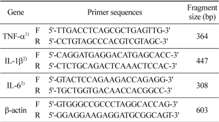
Table 1. Sequences of primers and fragment sizes of the investigated genes in RT-PCR analysis
Gene Primer sequences Fragment size (bp) TNF-α1) F 5'-TTGACCTCAGCGCTGAGTTG-3' 364 R 5'-CCTGTAGCCCACGTCGTAGC-3' IL-1β2) F 5'-CAGGATGAGGACATGAGCACC-3' 447 R 5'-CTCTGCAGACTCAAACTCCAC-3' IL-63) F 5'-GTACTCCAGAAGACCAGAGG-3' 308 R 5'-TGCTGGTGACAACCACGGCC-3'
β-actin F 5'-GTGGGCCGCCCTAGGCACCAG-3'R 5'-GGAGGAAGAGGATGCGGCAGT-3' 603
1)TNF-α: tumor necrosis factor-α, 2)IL-1β: interleukin-1β, 3)IL-6:
세포독성 평가
(LDH assay)
RAW 264.7 세포(1.5×10⁵ cells/mL)를 DMEM 배지에 추출물
시료와 LPS(1µg/mL)를동시 처리하여 24시간 배양한 후 배양 배지를얻어 3,000 rpm에서 5분간원심분리 하였다. LDH(lactate
dehydrogenase) assay는 non-radioactive cytotoxicity assay kit
(Promega)를이용하여측정했으며, 96 well plate에원심 분리하여 얻은배양배지 50µL와 reconstituted substrate mix를 50µL를넣 고, 실온에서 30분반응시킨후 50µL의 stop solution을넣은후
microplate reader(Bio-TEK Instruments Inc., Vermont, WI, USA)
를사용하여 490 nm에서흡광도를 측정하였다. 각시료군에 대
한평균 흡광도값을 구하였으며, 대조군 (LDH control, 1 : 5000) 의흡광도 값과비교하여 세포독성을평가하였다.
Prostaglandin E
2(PGE
2)
생성 평가RAW 264.7 세포를 DMEM 배지를이용하여 1.5×10⁵ cells/mL
로조절한후 24 well plate 에접종하고, 5% CO2항온기에서 18 시간전배양 하였다. 이후배지를제거하고 10배농도(1 mg/mL) 로조제된추출물시료 50µL와 450µL의 LPS(1µg/mL)를함유 한새로운 배지를동시에 처리하여전배양과 동일조건에서 배 양하였다. 24시간후 prostaglandin E2(PGE2)를측정하기위해배 양배지를원심분리(12,000 rpm, 3 min)하여상층액을얻었다. PGE2
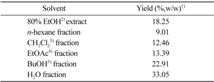
의 측정은 PGE2 ELISA kit(R&D Systems Inc., Minneapolis, MN, USA)를이용하여정량하였으며 standard 에대한표준곡선 의 r2값은 0.99 이상이었다. 통계분석 모든실험은 3회이상반복으로이루어졌으며, 실험결과는각 항목에 따라 평균치±표준편차 (SD)를 구하여 신뢰수준 95% (p< 0.05)에서통계적유의차를 평가하였다. 결과 및 고찰 에탄올 추출물및 순차분획물의수율 큰비쑥시료(1.2 kg)를 80% 에탄올로추출한후여과하여얻어 진추출액을감압 농축하여조추출물 219 g을얻었다. 그리고여 기서얻어진에탄올추출물(100 g)을 10배량의증류수로현탁시 킨 후에 헥산(n-hexane), 디클로로메탄(CH2Cl2), 에틸아세테이트 (EtOAc) 그리고 부탄올(BuOH) 등을순차적으로 분획하여 헥산 층에서 9.01 g, 디클로로메탄 층에서 12.46 g, 에틸아세테이트층 에서 13.39 g, 부탄올층에서 22.91 g 및잔사인물층에서 33.05 g 의분획물을얻었다. 각순차적분획물의수율은 Table 2에나타 내었으며, 추출에 사용한 큰비쑥 에탄올 추출물의 수율은 약 18.25%이었다. 에탄올추출물에대한각순차분획물 중헥산분 획물 수율이 9.01%로 가장 낮았고, 수용성 분획물이 33.05%로 가장높은수율을보였다.
Nitric oxide(NO)
생성억제 효과 NO는 NO 합성효소에 의해 L-arginine으로부터 생성되는무기 유리체로 면역반응, 세포독성, 신경전달계 및 혈관이완 등여러 가지생물학적인과정에관여하는것으로알려져있으며농도에 따라세포기능유지에중요한작용을하기도하고세포독성을일 으키기도한다(7,16). NO를생산하는 NOS는세포내에 존재하여calcium이나 calmodulin에 의존적인 형태인 constitutive NOS
(cNOS)와 calcium에비의존적으로대식세포나혈관내피세포가활 성화되거나 LPS와 같은세균의 내독소나 여러가지 cytokine에 의해 유도되는 형태인 iNOS의 형태가있다(17-22). LPS 자극에 의해발현된 iNOS는많은양의 NO를생성하게되며이에의한 세포독성은염증반응, 세포의돌연변이 및종양발생등에도 관 여하는 것으로 알려져 있다. 염증반응과 관련된 조직 손상에서 NO와 iNOS의발현이 증가되어있음이보고되어있다(8). 활성산소중하나이며, 최근염증유발에중요한역할을하는 것으로알려진 NO 생성에대한큰비쑥추출물과분획물의효과 를알아보았다. 생성된 NO 양을 Griess 시약을이용하여세포배 양액중에존재하는 NO2−의형태로측정하였다. 그결과헥산과 디클로로메탄 분획물에서대조군인 LPS 단독처리군에비해 높은 NO 생성억제효과를 관찰할수있었다(Table 3). 특히디클로로 메탄 분획물인경우 50µg/mL의농도에서도세포독성효과도보 이지않으면서아주우수한 NO 생성억제효과를 보여주었다.
Pro-inflammatory cytokine
생성 억제 효과 TNF-α와 같은다 기능성 cytokine은 정상조직에서 발현될 뿐 만아니라 병변 과정에서그 발현정도가 증가되며(23), 암촉진 과정에서 일어나는피부염증에중요한 역할을한다. TNF-α가 인 간의 염증성피부질환과 관련이 있음은이미많이 보고되어 왔 다(24,25). 또한여러 염증질환과 알러지 현상에 TNF-α에 대한 항체를 처리하였을 때증상이 완화되었다(26). 염증단계에 중추 적역할을하고 있는 cytokine인 TNF-α, IL-1β그리고 IL-6의발 현을저해시키거나 COX-2 활성저해에기인하는 PGE2의생성 억제를통해 pro-inflammatory factor의증가를수반하는병변과정 을조절할수있을가능성이높다.
대식세포인 RAW 264.7 세포로부터 염증성 사이토카인
(pro-inflammatory cytokine)의발현정도를 RT-PCR를통해알아본결
Table 2. Yield of each fraction extracted from A. fukudo
Solvent Yield (%,w/w)1) 80% EtOH2) extract 18.25 n-hexane fraction 09.01 CH2Cl23) fraction 12.46 EtOAc4) fraction 13.39 BuOH5) fraction 22.91 H2O fraction 33.05
1)Yield (%) = solid extract or fraction (g)/raw material (dry weight) or
ethanol extract×100, 2)EtOH: ethanol, 3)CH
2Cl2: dichloromethane, 4)EtOAc: ethylacetate, 5)BuOH: buthanol.
Table 3. Inhibitory effects of 80% ethanol extract and solvent
fractions of A. fukudo on the production of nitric oxide in RAW
264.7 cells Treatment 25 ( Inhibition (%) µg/mL) 50 (µg/mL) 80% EtOH extract 48.80* 64.41* n-hexane fraction 75.71** 87.87**1) CH2Cl2 fraction 85.45** 98.70** EtOAc fraction 61.30* 73.76** BuOH fraction - 02.59 H2O fraction -
-The production of nitric oxide was assayed from culture medium of RAW 264.7 cells stimulated with lipopolysaccharide (1µg/mL) in the
presence of 80% ethanol extract and solvent fractions of A. fukudo.
Significantly different from negative control (*p< 0.05, **p< 0.01).
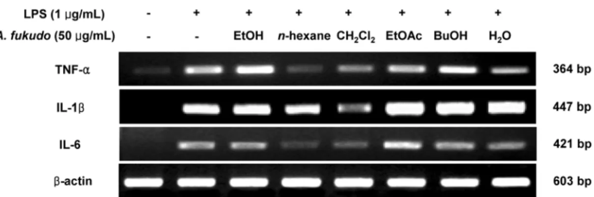
과, LPS 자극과함께처리하여 TNF-α, IL-1β 그리고 IL-6 생성 억제에 대한 큰비쑥 추출물 및 용매분획물을 50µg/mL 농도로 처리하였을때, TNF-α생성억제는 헥산과디클로로메탄 분획물 에서억제효과를 나타냈으며, IL-1β 생성억제는에틸아세테이 트, 부탄올그리고물분획물에서는 IL-1β의생성을오히려증가 시켰지만 디클로로메탄 분획물에서 억제효과를 보였으며, IL-6 생성억제는헥산과디클로로메탄분획물에서강한억제효과를 나타내었다(Fig. 1). iNOS 및 COX-2 생성 억제 효과 iNOS는평소에는 세포 내에 존재하지 않으나 일단 유도되면 장시간동안다량의 NO를생성하며, 생성된 NO는병리적인혈 관확장, 세포독성, 조직손상등과같은생체에유해한작용을나 타낸다. 그리고 염증상태에서 iNOS에 의해 생성된 NO는 혈관 투과성, 부종 등의염증반응을 촉진시킬뿐만아니라염증매개체 의 생합성을 촉진하여 염증을 심화시키는 것으로 알려져 있다 (27,28). RAW 264.7 세포에 LPS(1µg/mL)를사용하여 iNOS의생성을 유도한후큰비쑥추출물과분획물을 50µg/mL 농도로처리하여 iNOS 생성에대한억제정도를 immunoblotting를통해알아보았 다. 그결과 immunoblotting에의한 iNOS의단백질생성은헥산, 디클로로메탄 분획물에서대조군인 LPS 단독처리군에비해강 한억제효과를 나타내었다(Fig. 2). 본연구 결과, NO 생성억 제효과와초기-염증성 인자 (pro-inflammatory factor) 생성억제 효과가가장두드러지게나타났던디클로로메탄 분획물이 iNOS 의발현또한강하게억제시키는걸로보아 NO의생성억제기 전은 iNOS 발현억제를통해이루어진것으로여겨진다.
Cyclooxygenase(COX)는 arachidonic acid를 prostaglandins(PGs) 로 전환하는효소로 COX-1과 COX-2로분류된다. COX-1은 체 내에서 혈소판의 형성, 위벽보호, 신장기능의 유지 등정상적인 생체기능에 작용한다(29). 다수의 염증 억제 약물들의 작용기전 은 prostaglandin 합성억제를 나타내며 이는 COX-2의 생성 및 활성저해에 의한것이다. 따라서 COX-2에 의한 prostaglandin의 합성은염증반응을 매개하는것으로여겨진다. RAW 264.7 세포에 LPS(1µg/mL)를 사용하여 COX-2의생성 을유도한 후큰비쑥 추출물과분획물을 50µg/mL 농도로처리 하여 COX-2 생성에대한억제정도를 immunoblotting을통해 확 인하였다. 그결과, iNOS의단백질발현양상과마찬가지로 헥 산, 디클로로메탄분획물에서대조군인 LPS 단독처리군에비해 강한억제효과를나타내었다(Fig. 2). 세포 독성에 미치는 영향 LDH는모든 세포의 세포질 안에 존재하는 효소로서 pyruvic
acid와 lactic acid간의가역적전환에 관여하여촉매작용을 하며,
LDH를 내포한 조직이 파괴될 때 혈액 중으로 흘러나와 혈중 LDH가상승한다. RAW 264.7 세포(1.5×10⁵ cells/ mL)에시험약 물과 LPS(1µg/mL)를 동시 처리하여 24시간 배양한 후, LDH assay 방법을이용하여세포독성을확인한결과, 헥산분획물인 경우 50µg/mL 이상의농도에서다소 세포독성이나타났으나 다 른분획물에서는 세포독성이나타나지않았다(Fig. 3).
Fig. 1. Inhibitory effects of 80% ethanol extract and solvent fractions of A. fukudo on the mRNA expression of tumor necrosis factor-β,
interleukin-1β and interleukin-6 in RAW 264.7 cells. RAW 264.7 cells (1.0×10⁶ cells/mL) were pre-incubated for 18 hr, and the tumor
necrosis factor-α, interleukin-1β and interleukin-6 mRNA expressions were determined from 24 hr cultures of cells stimulated by
lipopolysaccharide (1µg/mL) in the presence of 80% ethanol extract and solvent fractions of A. fukudo (50 µg/mL).
Fig. 2. Inhibitory effects of 80% ethanol extract and solvent fractions of A. fukudo on the protein level of iNOS and COX-2 in RAW
264.7 cells. RAW 264.7 cells (1.0×10⁶ cells/mL) were pre-incubated for 18 hr, and the cells were stimulated with lipopolysaccharide (1µg/mL) in the presence of 80% ethanol extract and solvent fractions of A. fukudo samples (50 µg/mL) for 24 hr. iNOS and COX-2 protein levels were
Prostaglandin E2(PGE2) 생성 억제 효과
RAW 264.7 세포에서 염증성 PGE2 억제 효과를 ELISA kit를
이용하여정량하였다. RAW 264.7 세포에 LPS (1µg/mL)를사용 하여 PGE2의생성을유도한후큰비쑥추출물과분획물을 50µg/ mL 농도로처리하여확인한결과, PGE2 생성억제는디클로로메 탄분획물에서 다른분획물에 비해높은 억제효과를 나타내었 다(Fig. 3). 이는큰비쑥의디클로로메탄분획물에의해 LPS에의 해발현되는 PGE2 억제에영향을준다는것을의미하며, 이러한 결과는 COX-2의생성억제가 PGE2생성억제를통한것으로여 겨진다. 요 약 본연구는쑥추출물의 항염활성이 prostaglandins 합성의저 해및 pro-inflammatory cytokine의억제기전과관련이있을것으 로예상되어짐에따라, 큰비쑥(A. fukudo)을대상으로 80% EtOH 로추출하고추출물을극성에 따라용매분획을 실시하여, 큰비쑥 에탄올추출물및용매분획물들이염증반응의주체가되는대식 세포계열인 RAW 264.7 세포에서 LPS로유도된 TNF-α, IL-1β 그리고 IL-6와같은 pro-inflammatory cytokine과 NO의생성억제 효과, 그리고 iNOS와 COX-2의 단백질발현억제효과 및 PGE2
생성 억제효과 등을 통해 알아보았다. 대식세포 계열인 RAW 264.7 세포에 LPS로자극을주고큰비쑥 추출물을 처리하여확 인해본결과, 추출물및분획물들이다소 차이는있었지만 TNF-α, IL-1β 그리고 IL-6에서생성억제효과를나타났다. 또한헥산, 디클로로메탄및에틸아세테이트분획물에서 NO의생성억제효 과가강하게나타났으며, 헥산과디클로로메탄분획물에서는 iNOS, COX-2 및 PGE2 생성 억제효과가 다른분획물에 비해 강하게 나타났다. 이러한 결과는큰비쑥에서 유효성분추출을 통한항염 증물질의연구또는 예방하거나치료할 수있는염증억제성 분의분리및그작용기전 연구에중요한기초 자료가될것이 라사료된다. 또한큰비쑥 추출물로부터염증억제성분을도출하 고자 활성분획인 헥산과 디클로로메탄 분획물에 대하여 활성성 분의분리가진행중이다. 감사의 글 본 연구는 산업자원부 지방기술혁신사업(RTI04-02-07) 지원으 로수행되었으며, 지원에감사드립니다. 문 헌
1. Tizard IR. Immunology: An introduction inflammation. 2nd ed.,
Saunders College Publishing, New York, NY, USA pp. 423-441 (1986)
2. Willoughby DA. Heberden Oration, 1974. Human arthritis applied to animal models. Towards a better therapy. Ann. Rheum. Dis. 34: 471-478 (1975)
3. Higuchi M, Hisgahi N, Taki H, Osawa T. Cytolytic mechanisms of activated macrophages. Tumor necrosis factor and L-arginine-dependent mechanisms act synergistically as the major cytolytic mechanisms of activated macrophages. J. Immunol. 144: 1425-1431 (1990)
4. Willeaume V, Kruys V, Mijatovic T, Huez G. Tumor necrosis fac-tor-alpha production induced by viruses and by lipopolysaccha-rides in macrophages: similarities and differences. J. Inflamm. 46: 1-12 (1995-1996)
5. Funk CD, Frunk LB, Kennedy ME, Pong AS. Fitzgerald GA. Human platelet/erythroleukemia cell prostaglandin G/H synthase: cDNA cloning, expression, and gene chromosomal assignment. FASEB J. 5: 2304-2312 (1991)
6. McDaniel ML, Kwon G, Hill JR, Marshall CA, Corbett JA. Cytokines and nitric oxide in islet inflammation and diabetes. P. Soc. Exp. Biol. Med. 211: 24-32 (1996)
7. Moncada S, Palmer RM, Higgs EA. Nitric oxide: physiology, pathophysiology, and pharmacology. Pharmacol. Rev. 43: 109-142 (1991)
8. Weisz A, Cicatiello L, Esumi H. Regulation of the mouse induc-ible-type nitric oxide synthase gene promoter by interferon-γ,
bac-terial lipopolysaccharide, and NG-monomethyl-L-arginine. Biochem.
J. 316: 209-215 (1996)
9. Ryu JH, Ahn H, Kim JY, Kim YK. Inhibitory activity of plant extracts on nitric oxide synthesis in LPS-activated macrophage. Phytother. Res. 17: 485-489 (2003)
10. Mu MM, Chakravortty D, Sugiyama T, Koide N, Takahashi K, Mori I, Yoshida T, Yokochi T. The inhibitory action of quercetin on lipopolysaccharide-induced nitric oxide production in RAW 264.7 macrophage cells. J. Endotoxin. Res. 7: 431-438 (2001) 11. Kim JY, Jung KS, Jeong HG. Suppressive effects of the kahweol
and cafestol on cyclooxygenase-2 expression in macrophages. FEBS Lett. 569: 321-326 (2004)
12. Tan RX, Zheng WF, Tang HQ. Biologically active substances from the genus Artemisia. Planta Med. 64: 295-302 (1998) 13. Yoon WJ, Lee JA, Kim JY, Oh DJ, Jung YH, Lee WJ, Park SY.
Anti-oxidant activities and anti-inflammatory effects on Artemisia scoparia. Korean J. Pharmacogn. 37: 235-240 (2006)
14. Lee TB. llustrated Flora of Korea. Hyangmoon Publishing Co., Seoul, Korea p.757 (1979)
15. Wakefield PE, James WD, Samlaska CP, Mettzer MS. Tumor necrosis factor. J. Am. Acad. Dermatol. 24: 675-685 (1991) 16. Nathan C, Xie QW. Nitric oxide synthases: roles, tolls and
con-trols. Cell 78: 915-918 (1994)
17. Liu RH, Hotchkiss JH. Potential genotoxicity of chronically ele-vated nitric oxide. Mutat. Res. 339: 73-89 (1995)
18. Rockey DC, Chung JJ, McKee CM, Noble PW. Stimulation of inducible nitric oxide synthase in rat liver by hyaluronan frag-ments. Hepatology 27: 86-92. (1998)
19. Nunokawa Y, Ishida N, Tanaka S. Cloning of inducible nitric oxide synthase in rat vascular smooth muscle cells. Biochem. Bioph. Res. Co. 191: 89-94 (1993)
20. Lyons CR, Orloff GJ, Cunningham JM. Molecular cloning and
Fig. 3. Inhibitory effects of 80% ethanol extract and solvent fractions of A. fukudo extracts on cell cytotoxicity and the prostaglandin E2 production in RAW 264.7 cells. RAW 264.7 cells (1.5×10⁵ cells/mL) were stimulated by lipopolysaccharide (1
µg/mL) with 80% ethanol extract and solvent fractions (50 µg/mL)
from A. fukudo for 24 hr. Supernatants were then collected after 24
hr and prostaglandin E2 concentration from supernatants was determined by ELISA method. Cell viability was determined using lactate dehydrogenase method. The data represent the mean ± standard deviation of triplicate experiments.
functional expression of an inducible nitric oxide synthase from a murine macrophage cell line. J. Biol. Chem. 267: 6370-6374 (1992)
21. Geller DA, Lowenstein CJ, Shapiro RA, Nussler AK, Disilvio M, Wang SC, Nakayama DK, Simmons RL, Snyder SH, Billiar TR. Molecular cloning and expression of inducible nitric oxide syn-thase from human hepatocytes. P. Natl. Acad. Sci. USA 90: 3491-3495 (1993)
22. Santos-Gomes PC, Seabra RM, Andrade PB, Fernandes-Ferreira M. Determination of phenolic antioxidant compounds produced by calli and cell suspensions of sage (Salvia officinalis L.). J. Plant Physiol. 160: 1025-1032 (2003)
23. Dinarello CA. Pro-inflammatory cytokines. Chest 118: 503-508 (2000)
24. Piguet PF, Grau GE, Houser C, Vassalli P. Tumor necrosis factor is a critical mediators in hapten induced irritant and contact hypersensitivity reaction. J. Exp. Med. 173: 673-679 (1991) 25. Burrell R. Human responses to bacterial endotoxin. Circ. Shock
43: 137-153 (1994)
26. Galea E, Feinstein DL, Reis DJ. Induction of calcium-indepen-dent nitric oxide synthase activity in primary rat glial cultures. P. Natl. Acad. Sci. USA 89: 10945-10949 (1992)
27. Tezuka Y, Irikawa S, Kaneko T, Banskota AH, Nagaoka T, Xiong Q, Hase K, Kadota S. Screening of Chinese herbal drug extracts for inhibitory activity on nitric oxide production and identification of an active compound of Zanthoxylum bugeanum. J. Ethnophar-macol. 77: 209-217 (2001)
28. Kim RG, Shin KM, Chun SK, Ji SY, Seo SH, Park HJ, Choi JW, Lee KT. In vitro antiinflammatory activity of the essential oil from Ligularia fischeri var. spiciformis in murine macrophage RAW 264.7 cells. Yakhak Hoeji 46: 343-347 (2002)
29. Seibert K, Zhang Y, Leahy K, Hauser S, Masferrer J, Perkins W, Lee L, Isakson P. Pharmacological and biochemical demonstration of the role of cyclooxygenase-2 in inflammation and pain. P. Natl. Acad. Sci. USA 91: 12013-12017 (1994)