Otolaryngology & Dermatology 2009;22(2) : 92-103
葎草가 항염 효과에 미치는 영향
황순이
1)*·조미정
2,3)·김상찬
2,3)·지선영
1)1) 대구한의대학교 안이비인후피부과교실
2) 한의과대학
3) 한방신약개발팀 (BK21 Team)
Anti-inflammaory effects of the MeOH extract of Humulus japonicus in vivo
Sun-Yi Hwang·Mi-Jeong Jo·Sang-Chan Kim·Seon-Young Jee
Objectives : The present study was examined to evaluate the anti-inflammatory effects of the Humulus japonicus MeOH extracts (HJE) in vivo.
Methods : The effects of HJE on anti-inflammation were measured by production of NO, iNOS (inducible Nitric Oxide Synthase), COX-2, IκBα (Inhibitor kappa B alpha), NFκB (Nuclear Factor kappa B), TNF-α (Tumor Necrosis Factor-alpha) and IL-1β (Interleukin-1β), IL-6 in Raw 264.7 macrophage cells stimulated with LPS.
Results : 1. All concentrations of HJE(0.03 and 0.10 ㎎/㎖) had no significant cytotoxicity in Raw 264.7 cell during the entire experimental period.
2. The level of NO and iNOS in culture medium was dramatically increased by LPS application.
However, these increases were dose-dependently(0.03 and 0.10 ㎎/㎖) attenuated by treatment with HJE.
3. HJE extract reduced PGE
2levels in a dose-dependent manner as a consequence of inhibition of COX-2 protein expression in Raw 264.7 macrophage cells stimulated with LPS.
4. 0.10 ㎎/㎖ HJE significantly inhibited the phosphorylation of IκBα indicating the suppression of NF-κB pathway in Raw 264.7 macrophage cells stimulated with LPS.
5. 0.10 ㎎/㎖ HJE significantly inhibited the production of TNF-α in Raw 264.7 macrophage cells stimulated with LPS.
6. All concentrations of HJE significantly inhibited the production of IL-1β, IL-6 in Raw 264.7 macrophage cells stimulated with LPS.
Conclusions : These results provide evidences that therapeutic effect of HJE on heat syndrome, especially due to the acute inflammation, are partly due to the reduction of some of inflammatory factors by inhibiting iNOS and COX-2 through the suppression of p-IκBα. Moreover, it suggests that the mechanism of action of HJE comes from the suppression of inflammatory mediators, such as NO, PGE
2and pro-inflammatory cytokines.
Key words : Humulus japonicus, Anti-inflammaory effects, NO, iNOS, COX-2, IκBα , NFκB, TNF-α, IL-1β, IL-6
교신저자 : 황순이, 대구 수성구 상동 165
대구한의대학교 부속 대구한방병원 안이비인후피부과교실 (Tel : 053-770-2178, E-mail : [email protected])
∙접수 2009/06/22 ∙수정 2009/07/22 ∙채택 2009/08/03
І . 서 론
염증 반응 물질 중 NO (Nitric Oxide) 및 COX-2 (Cyclooxygenase-2)의 생성 저해제는 염증 반응조절제로서의 가능성 때문에 연구가 활발하게 이루어지고 있으며, 최근에는 牡丹皮, 靑黛, 苦楝 皮, 款冬花, 當歸, 香附子 등의 한약에서 이러한 조 절제를 찾기 위해 많은 연구가 진행되고 있다
1-7). 濕熱壅塞之實證을 主하고 外瘍과 陽毒에 外敷할 수 있는 약으로 주변에서 쉽게 구할 수 있는 율초 (葎草, Humulus japonicus )는 梁代 陶弘景의 《名 醫別錄》에 勒草로 처음 수록되어 있으나 현재 임 상에서 많이 사용되지 못하고 있는 실정이다
8,9). 현재까지 율초에 대한 연구로는 세포보호작용, 항 돌연변이효과, 항산화작용 등이 보고되고 있으나 습열을 치료하는 기전과 관련된 연구는 드물다
10-2). 이에 율초의 항염증효과의 기전에 관한 연구로 저자는 MeOH로 추출된 율초 (HJE)가 LPS로 활 성된 Raw 264.7 cells에서의 NO 생성, iNOS (inducible Nitric Oxide Synthase), COX-2 발현 및 IκBα (Inhibitor kappa B alpha), NFκB (Nuclear Factor kappa B), TNF-α (Tumor Necrosis Factor-alpha), IL-1β (Interleukin-1β), IL-6 등의 염증매개물질 발현에 어떠한 영향을 미 치는지 살펴보았다.
Ⅱ. 재료 및 실험방법
1. 율초 추출물 (HJE)의 제조
율초는 2008년 7월 대구광역시 수성구 외환동
원장지 주변에서 채취하였으며, 율초 400 g을 MeOH 6L에 넣고 3일간 추출한 후, 추출물을 거 즈로 1차 여과하고 3000× g 에서 3분간 원심분리 하였다. 원심분리 후의 상층액 만을 취하여 0.2 μ m filter (Nalgene, New York, USA)로 여과하였 다. 이 여과액을 Rotary evaporator (EYELA, Tokyo, Japan)로 동결 건조하여 28.81 g을 얻었으 며, 사용 때까지 -20℃에서 보관하였다. HJE의 수 율은 7.20%였으며 DMSO에 녹여 사용하였다.
2. 시약 및 기구
LPS (Difco, Detroit, MI, USA)와 MTT는 Sigma (St. Louis, MO, USA)에서 구입하였고, FBS와 Antibiotics는 Gibco/BRL (Eggenstein, Germany)로부터 구입하였으며, antibody는 BD Bioscience (San Jose, CA, USA), Cayman (Ann Arbor, Mi, USA), Zymed (San Francisco, CA, USA)에서 구입하였고, NC paper는 Schleicher &
Schuell (Dassel, Germany)에서 구입하였다.
TNF-α, IL-1β, IL-6, COX-2의 ELISA Kit는 Pierce endogen (Rockford, IL, USA)에서 구입하 였다.
3. 세포 배양
Murine macrophage cell line인 Raw 264.7 cells는 한국세포주연구재단 (Seoul, Korea)에서 구 입하였으며, DMEM에 10% FBS, 100 U/㎖
penicillin 및 100 ㎍/㎖ streptomycin을 혼합한
배지를 사용하여 37℃, 5% CO
2incubator에서 배
양하였다. 실험과정의 모든 cell은 80~90%의
confluence에서 실험하였고, 20 passages를 넘기지
않은 cell만 사용하였다.
4. 세포 생존율 측정
Raw 264.7 cells를 96 well plates에 5×10
4cell/well로 분주한 다음 HJE를 농도별로 처치하여 세포의 생존율을 구하였다. 세포에 0.03 및 0.10
㎎/㎖의 농도로 HJE를 처치한 후에 37℃, 5%
CO
2의 환경이 유지되는 배양기에서 배양하였다.
배양 후 생존세포에 MTT (0.1 ㎎/㎖)를 50 ㎕넣 고 4시간 배양한 후 배지를 조심스럽게 제거하고 생성된 Formazan crystals을 DMSO에 녹여 Titertek Multiskan Automatic ELISA microplate reader (Model MCC/340, Huntsville, AL, Austria)를 사용하여 570 ㎚에서 흡광도를 측정하 였다. 세포생존율은 control cell에 대한 백분율로 나타내었다. [i.e. viability (% control) = 100×/
(absorbance of treated sample)/(absorbance of control)].
5. NO 생성량 측정
Raw 264.7 cells로부터 생성된 NO의 양은 세 포 배양액 중에 존재하는 NO
2-의 형태로서 Griess 시약을 이용하여 측정하였다. 세포배양 상등액 50
㎕와 Griess시약 (1% sulfanilamide in 5%
phosphoric acid + 1% α-naphthylamide in H
2O) 50 ㎕를 96 well plates에 혼합하고 암실에 서 10분 동안 반응시킨 후 540 ㎚에서 Titertek Multiskan Automatic ELISA microplate reader 로 흡광도를 측정하였다. NO
2-의 농도는 sodium nitrate를 희석하여 흡광도를 측정하여 표준 곡선 을 얻었다.
6. Immunoblot analysis
20 mM Tris Cl (pH 7.5), 1% Triton X-100, 137 mM sodium chloride, 10% glycerol, 2 mM EDTA, 1 mM sodium orthovanadate, 25 mM
b-glycerophosphate, 2 mM sodium pyrophosphate, 1 mM phenylmethylsulfonylfluoride과 1 ㎎/㎖
leupeptin을 함유하는 buffer를 사용하여 cell을 lysis시켰다. Cell lysates를 10,000× g 로 10분간 원 심 분리하여 debris를 제거하였다. 각 단백질의 발 현은 각 단백질에 특이적 항체를 사용하여 면역화 학적 방법으로 분석하였으며, 2차 antibody는 Alkaline phosphatase conjugated anti-rabbit을 사용하였다. 단백질의 발현은 ECL western blotting detection reagents (Amersham, UK)를 사용하여 manufacturer's instruction에 따라 발색 하였다. 발색 후 각 단백질의 발현량을 평가하기 위하여 Image analyzing system (Ultra-Violet Products Ltd., Upland, CA, USA)을 이용하여 Densitometric analysis를 실시하였다.
7. Cytokine과 PGE2 측정
Cytokine을 측정하기 위하여 6 well plates에 5×10
5cell/㎖를 분주하고 HJE를 농도별로 처치한 다음, 1시간 후에 LPS를 처치하였다. LPS 처치 후 각 cytokine마다 특정 시간에 배지를 수거하여 cytokine을 측정하였다. 수거된 배지는 바로 측정 하거나, 측정 전까지 -70℃에서 보관하였다. TNF- α, IL-1β와 IL-6는 ELISA Kit를 사용하여 측정 하였으며, PGE
2는 R&D Systems Inc.
(Minneapolis, MN, USA)를 사용하였으며, 실험의 방법은 manufacturer's instruction에 따랐다.
8. 통계적 검증
실험 결과는 mean±SD로 나타내었으며, 처치군
간의 유의성은 one way analysis of varience
(ANOVA)로 검정한 후 Newman - Keuls test로
검정하였다. 통계적 유의성 검정은 p <0.05 또는
p <0.01로 하였다.
Ⅲ. 결 과 1. NO production에 미치는 영향
Fig. 1. Effects of HJE on the production of NO by LPS.
Raw 264.7 cells were treated with 0.03, 0.10 ㎎/㎖
of HJE dissolved in DMSO for 1 h prior to the addition of LPS (1 ㎍/㎖), and the cell was further incubated for 24 h. The concentration of nitrite and nitrate in culture medium was monitored as described in materials and methods section. Data represent the mean±SD with eight separate experiments.
*: significant as compared to control, ** p <0.01 #: significant as compared to LPS alone, ## p <0.01
2. Raw 264.7 cells의 생존율에 미치는 영향
A) B)
Fig. 2. Effects of HJE on the cell viability (A) and NO/cell viability (B) in LPS stimulated Raw 264.7 cells.
Raw 264.7 cells were treated with 0.03, 0.10 ㎎/㎖ of HJE dissolved in DMSO for 1 h prior to the addition of LPS (1 ㎍/㎖), and the cell was further incubated for 24 h. Data represent the mean±SD with eight separate experiments.
*: significant as compared to control, ** p <0.01 #: significant as compared to LPS alone, ## p <0.01
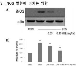
3. iNOS 발현에 미치는 영향
A)
B)
Fig. 3. Effect of HJE on the induction of iNOS expression by LPS.
The level of iNOS protein was monitored 24 h after treatment of cells with LPS (1 ㎍/㎖) with or without HJE (0.03, 0.10 ㎎/㎖) pretreatment (i.e. 1 h before LPS). Equal amounts of total protein were resolved by SDS-PAGE. Expressions of iNOS protein were determined by immunoblot analysis using specific iNOS antibodies. The actin was used as a loading control (A). The relative density level of protein bands was measured by scanning densitometry (B). Data represent the mean±SD of three separate experiments. One-way ANOVA was used to compare the multiple group means followed by Newman - Keuls test.
*: significant compared with the control, ** p <0.01 #: significant compared with the LPS alone, ##
p <0.01
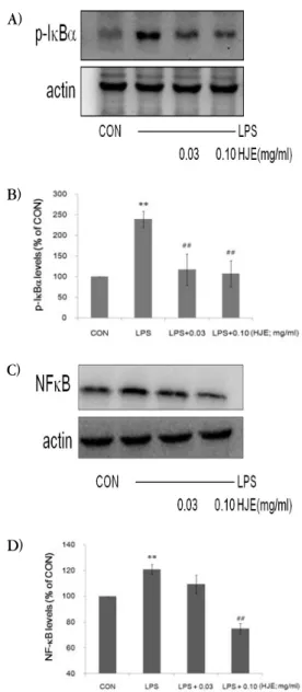
4. Iκ Bα , NFκB 발현에 미치는 영향
A)
B)
C)
D)
Fig. 4. Effects of HJE on the expression of IκBα and NFκB by LPS.
The level of IκBα and NFκB protein was monitored 15 min and 60 min after treatment of cells with LPS (1 ㎍/㎖) with or without HJE pretreatment (i.e. 1 h before LPS). The actin was used as a loading control (A, C). The relative density level of the bands was measured by scanning densitometry (B, D).
*: significant as compared to control, ** p <0.01 #: significant as compared to LPS alone, ## p <0.01
5. TNF-α 에 미치는 영향
Fig. 5. The effect of HJE on LPS-stimulated TNF- α production.
Production of TNF-α was measured in the medium of Raw 264.7 cells cultured with LPS (1 ㎍/㎖) in the presence or absence of HJE for 12 h. The amount of TNF-α was measured by immunoassay as described in materials and methods. Data represent the mean±SD with three separate experiments. One-way ANOVA was used for comparisons of multiple group means followed by Newman - Keuls test.
*: significant as compared to control, ** p <0.01 #: significant as compared to LPS alone, # p <0.05,
## p <0.01
6. COX-2의 발현에 미치는 영향
A)
B)
Fig. 6. Effect of HJE on the induction of COX-2 by
LPS.
The level of COX-2 protein was monitored 24 h after treatment of cells with LPS (1 ㎍/㎖) with or without HJE (0.03, 0.10 ㎎/㎖) pretreatment (i.e. 1 h before LPS). Equal amounts of total protein were resolved by SDS-PAGE. Expressions of COX-2 protein were determined by immunoblot analysis using COX-2 specific antibodies. The actin was used as a loading control (A). The relative density level of protein bands was measured by scanning densitometry (B). Data represent the mean±SD of three separate experiments. One-way ANOVA was used to compare the multiple group means followed by Newman - Keuls test.
*: significant compared with the control, ** p <0.01 #: significant compared with the LPS alone, ##
p <0.01
7. PGE
2에 미치는 영향
Fig. 7. Inhibition of LPS-activated PGE
2production by HJE.
Raw 264.7 cells were cultured with LPS (1 ㎍/㎖) in the presence or absence of HJE for 24 h to determine the level of PGE
2. The cultured medium was collected and directly assayed for PGE
2. The data represent the mean±SD of three separate experiments. One-way ANOVA was used to compare the multiple group means followed by Newman - Keuls test.
*: significant compared with the control, ** p <0.01 #: significant compared with the LPS alone, ##
p <0.01
8. IL-1β, IL-6에 미치는 영향
A)
B)
Fig. 8. The effect of HJE on LPS stimulated IL-1β, IL-6 production.
Production of IL-1β, IL-6 were measured in the medium of Raw 264.7 cells cultured with LPS (1 ㎍/
㎖) in the presence or absence of HJE for 12 h, 6h.
The amount of IL-1β, IL-6 were measured by immunoassay as described in materials and methods. Data represent the mean±SD with three separate experiments. One-way ANOVA was used for comparisons of multiple group means followed by Newman - Keuls test.
*: significant as compared to control, ** p <0.01 #: significant as compared to LPS alone, ## p <
0.01
Ⅳ. 고 찰
外瘍과 陽毒의 外敷에 사용되는 율초는 우리 주
변에서 쉽게 구할 수 있음에도 불구하고 우리나라
임상에서 많이 응용되지 못하는 실정이다
8). 율초
(葎草, Humulus japonicus )는 삼과 (Cannabinaceae)
에 속한 1년생 혹은 다년생 덩굴식물인 환삼덩굴
의 전초로서, 잎은 대생하며 줄기와 엽병에 가시가
있고 7~8월에 담황색의 꽃이 피는 자웅이주의 식
물로서, 우리나라 전역에 분포하고 있다
11,13). 勒草,
黑草, 割人藤, 鋸鋸藤, 拉拉藤, 五瓜龍, 大葉五瓜龍 등의 別名으로 불리며
8,9,14), 性味는 苦甘寒無毒하고 肝肺大腸膀胱의 4經에 작용하며, 淸退虛熱, 淸熱解 毒, 利尿通淋, 止泄痢, 驅瘀血 등의 효능이 있어, 虛熱不退, 瘧疾, 潮熱盜汗, 肺癆咳嗽, 廱腫, 瘰癧, 濕疹, 瘙痒, 熱淋澁痛, 小便不利, 下痢, 痔疾, 肺結 核, 肺膿瘍, 肺炎, 泄瀉 등의 병증을 치료한다
14,15). 율초의 성분 연구에 따르면 全草에는 luteolin, glucoside, choline, asparagine, tannin, 정유, 수 지가, 열매에는 humulone, lupulone 등이, 잎에는 0.015%의 cosmosin과 vitexin 등이 들어 있다
16). 또한 이 등, 박 등은 율초에 관한 세포 독성완화, 항산화효과를 보고하였다
10,12). 그러나 항염증효과 의 기전에 관한 연구는 부족하여 저자는 율초의 iNOS 억제 기전 및 염증관련 cytokine의 발현에 대한 연구를 수행하였다.
염증(inflammation)은 전통적으로 “균의 감염, 열, 외상, 항원항체반응 등 생체조직의 기질변화를 초래하는 침습에 대한 생체의 방어 기전”라고 정의 되지만, 현재에는 염증을 신체 국소에서 일어나는 상해에 대한 생체조직의 방어반응으로 인식하고 있다. 즉, 각종 유해한 자극에 응답하여, 자극에 대한 상해를 제거하여 원래의 상태로 회복하려는 생체방어반응이 염증반응이다. 염증이 발생한 부위 는 발적, 발열, 동통, 종창과 같은 징후가 발생된
다
17,18). 수많은 원인에 의하여 일어나는 염증반응
은 그 원인과 반응조직의 차이에 상관없이 거의 유사한 변화를 보인다. 이 변화는 조직 손상 후에 일어나는 변화로서 손상에 의하여 생체내 국소부 위에 유리되는 공통적인 물질요인의 존재를 추정 케 한다. 이러한 화학적 매개체로는 활성산소, NO, PG, 염증을 유발시키는 여러 cytokine 등이 있다
19).
염증 반응에서의 유해자극은 직접 국소에 작용 해 손상을 주기도 하지만, 대부분 내인성 화학전달 물질을 통해 간접적으로 국소의 혈관이나 세포에
전달된다. 염증반응의 주요 화학 전달 매개물질로 는 크게 즉시형 혈관투과성 항진에 관여하는 amine류 (histamine, serotonin 등)와 kinin류 (bradykinin 등), 지연형 반응에 주로 작용하는 cytokine류와 PG와 IL류 등의 4군으로 분류된다
16)
. 면역과 염증에 관련된 여러 cytokine중 IL-1
β, IL-6 및 TNF-α는 대식세포에서 생산되는 대
표적인 염증성 cytokine으로 각종 염증질환의 발생
과 진행에 중요한 작용을 하는 것으로 보고되고
있다. 특히 염증반응에 관여하는 세포 중에서 대식
세포는 각종 cytokine을 분비하여 대식세포의 유
주, T 세포의 활성화와 증식억제, 혈관신생작용 등
을 나타내어 염증반응을 조절한다고 인식되고 있
다
20). 대식세포는 염증 반응시에 IL, TNF-α와 같
은 cytokine을 생산하고, COX-2를 활성화시켜
PG를 생산하여 감염초기의 생체 방어에 중요한
역할을 하는 세포로 알려져 있다
21,22). 또한
COX-2에 의해 생성되는 PGE
2는 세포자멸사의 억
제, 세포분열 및 암세포 전이, 혈관신생을 유도하
여 종양의 형성에 기여하는 것으로 알려져 있다
23).
Gram-negative 박테리아의 세포벽 구성성분인
LPS는 인지질, 다당류 및 소량의 단백질로 구성되
며, 염증반응을 유발하는 유력한 인자로 대식세포
의 TLR과 결합하여 다양한 cytokine을 생성시키
므로 염증반응 연구에서 빈용되는 실험모델이다
24).
일반적으로 LPS에 의해 활성화된 대식세포의 염증
반응에는 다량의 Proinflammatory cytokines,
NO, PGE
2가 iNOS와 COX-2에 의해 생성된다
25).
NO는 NOS효소에 의해 만들어지며, 체내의 염
증과정에서는 과량의 NO가 만들어져 관절염을 비
롯한 각종 급성 혹은 만성 염증 질환에서 중요한
역할을 하는 것으로 알려져 있다. NOS는 I형, II
형, III형의 3종류가 있는데, 이 중 생체에서 항상
성과 관련해 중요한 역할을 담당하는 I형이나 III
형과 달리 II형은 iNOS로 cytokine이나 세균 등에
서 분비되는 LPS나 calcium ionophore에 의해 일
부 세포에서 생성되며, 생성된 iNOS는 과량의 NO를 생성해 각종 염증질환에 작용하는 것으로 알려져 있다. 생산된 과량의 NO는 그 자체로도 유전자 및 단백질에 독성을 나타내지만 활성산소 의 하나인 superoxide anion(O
2-)과 반응해 맹독성 을 가진 peroxynitrite (ONOO-)를 생성하므로 더 욱 강력한 독성물질로 변화되어 암 형성과 진행에 중요한 역할을 하는 것으로 보고되고 있다. 따라서 각종 염증의 발생억제와 치료를 위해서는 NO의 발생을 억제시켜 주는 것이 중요하다
26).
본 연구에서는 LPS로 활성화된 Raw 264.7 cells에서 HJE의 NO 생성억제를 평가하기 위하여 HJE를 0.03 및 0.10 ㎎/㎖의 농도로 LPS 처치 전 1시간에 Raw 264.7 cells에 처리하여 생성되는 NO의 양을 측정하였다. LPS군에서는 control군에 비교하여 12, 18, 24 시간에서 NO의 생성량이 다 량 증가하였으며, 0.03 및 0.10 ㎎/㎖ HJE를 처치 한 실험군에서는 시간 및 농도 의존적으로 유의성 있는 NO의 생성억제를 나타내었다.
HJE가 0.03 및 0.10 ㎎/㎖의 농도에서 LPS로 유도된 NO의 생성을 억제시킨 이유가 HJE의 세 포독성으로 인한 것인지를 평가하기 위하여 HJE의 농도에 따라 MTT assay를 실시하여 cell viability 를 측정하였다. 실험결과 실험에 사용된 HJE는 LPS 단독처치군에 비교하여 유의한 세포독성을 나 타내지 않았다. HJE는 오히려 LPS에 의한 세포독 성을 억제하였다. Sharifi 등은 PC-12 cell (Rat Pheochromocytoma)에서의 납의 독성은 납에 의 한 NO의 과다생성이 세포독성을 유발함을 밝히고, 그 세포독성은 iNOS inhibitor인 L-NAME에 의해 억제됨을 밝혔다
27). 이러한 결과로 볼 때 본 연구 에서도 LPS에 의한 과량의 NO가 Raw 264.7 cells의 세포생존율을 저하시켰으며, HJE는 NO의 생성을 억제함으로서 세포생존율을 증가시킨 것으 로 판단된다.
NO는 L-arginine으로부터 iNOS에 의하여 생성
되므로, HJE가 LPS로 활성화된 대식세포에서 iNOS의 발현에 미치는 영향을 평가하였다
28). LPS 처치 시에는 iNOS의 발현이 유의하게 증가되었으 나, LPS에 0.03 및 0.10 ㎎/㎖ HJE를 전처치한 실험군에서는 iNOS의 양이 농도 의존적으로 유의 하게 감소하였다. 이러한 HJE에 의한 iNOS의 발 현의 억제는 NO의 생성 억제를 유도함을 의미한다.
COX는 I형과 II형의 2가지 isoform이 존재한 다. I형 효소인 COX-1은 위장관 보호, 신장의 혈 류조절, 혈소판 응집 등 인체의 정상적인 기능을 유지하는데 중요한 작용을 하는 house keeping enzyme인 반면, 외부 자극에 의해 발현이 유도되 는 II형인 COX-2는 염증과 암 등의 각종 퇴행성 질환에 중요한 역할을 한다
29,30).
한편, 염증에 관련되는 iNOS와 COX-2의 발현 에 관여하는 것으로 NFκB가 대표적인 전사조절 인자로 알려져 있다. 대식세포는 LPS 등의 염증유 발 자극에 의해 NFκB pathway가 활성화되어 iNOS나 TNF-α 등의 유전자 발현에 관련한다
31,32)