Received: 1 March, 2016 Revised: 14 March, 2016 Accepted: 14 March, 2016 Corresponding author: Byoung-Hee Lee
Department of Physical Therapy, College of Health and Welfare, Sahmyook University, 815 Hwarang-ro, Nowon-gu, Seoul 01795, Republic of Korea Tel: 82-2-3399-1634 Fax: 82-2-3399-1639 E-mail: [email protected]
This is an Open-Access article distributed under the terms of the Creative Commons Attribution Non-Commercial License (http://creativecommons.org/licens es/by-nc/4.0) which permits unrestricted non-commercial use, distribution, and reproduction in any medium, provided the original work is properly cited.
Copyright © 2016 Korean Academy of Physical Therapy Rehabilitation Science
http://dx.doi.org/10.14474/ptrs.2016.5.1.29 pISSN 2287-7576
eISSN 2287-7584
Phys Ther Rehabil Sci 2016, 5 (1), 29-33 www.jptrs.org
Comparison of three different surface plank exercises on core muscle activity
Jin Lee
a, Kwanghyun Jeong
b, Hyuna Lee
b, Jaeyeon Shin
b, Jaelim Choi
b, Seungbeom Kang
b, Byoung-Hee Lee
baDepartment of Physical Therapy, Hyun-Myoung Medical Center, Seoul, Republic of Korea
bDepartment of Physical Therapy, College of Health and Welfare, Sahmyook University, Seoul, Republic of Korea
Objective: This study compared the muscle activities of the erector spinae (ES), the external oblique (EO), and the rectus abdom- inis (RA) on three different surfaces. The purpose of this study was to determine which surface induces the highest muscle activity during the plank exercises. The information from this study can be used to recommend plank exercises to athletes and patients with weak core muscles.
Design: Cross-sectional study.
Methods: The subjects include 20 adult males attending S University in Seoul. Participants completed each plank exercise on three different surfaces. To measure muscle activities, researchers used the values from electromyography. The measurement ex- cluded the initial two and final two seconds and collected information on the RA, EO, and ES in each posture of each subject.
Results: The left external oblique showed significant differences between the plank position on stable ground (ST) and the plank position using a suspension device (SL) (p<0.05) and between the plank position on the unstable ground (US) and SL (p<0.05).
The right rectus abdominis and left rectus abdominis displayed statistically significant differences between the ST and the US (p<0.05) and between the ST and the SL (p<0.05). The right erector spinae had a statistically significant difference between ST and US (p<0.05).
Conclusions: The plank exercise strengthens the core muscles effectively, and muscle activity is related to the posture of the ex- ercise and the location of the muscle. These results suggest that plank exercises improve muscle activities. Additionally, plank ex- ercises can be applied to general medical care.
Key Words: Electromyography, Isometric exercise, Posture
Introduction
Core muscles are the deep and shallow muscles of the trunk; they stabilize the spinal column, align the body, and enhance performance when the extremities move [1]. Weak- nesses in core muscles can cause changes in body config- uration and compress the posterior joints of the lumbar spine. Additionally, excessive anterior or posterior leaning of the pelvis accompanied by the tension of the thor- acolumbar fascia may result in increased shocks on the flanks and lower extremities [2].
Core exercises can prevent injuries in sports and rehabili-
tation treatment by maximizing muscle power and endur- ance [3]. Exercises to strengthen core muscles include the crunch exercise, the bridge exercise, and the plank exercise [4]. Among them, the plank exercise estimates and strength- ens the stability of the core muscles. Core muscle stability is crucial for preventing injuries to the knees, hip joints, and the lumbar spine. Moreover, core muscle stability relieves back pain by maintaining proper alignment for posture and gait [5].
The plank exercise is a posture designed to use body
weight to resist gravity; it can be performed on various surfa-
ces, in variable positions, with devices, and impacts multiple
Figure 1. The plank position on stable ground. Figure 3. The plank position using a suspension device.
Figure 2. The plank position on unstable ground.
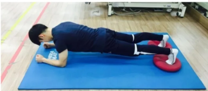
joints. Plank exercises on the unstable surface using dynam- ic cushions for the lower extremities can induce greater ac- tivities than plank exercises of the upper extremities on the unstable or on the stable surface [6]. Plank exercises using a suspension device generates greater core muscle activities compared to plank exercises performed on stable or unstable surfaces [5].
Few studies have comprehensively examined core mus- cle activity in the plank posture on stable and unstable surfa- ces, or while using a suspension device. This study com- pared the muscle activities of the erector spinae (ES), ex- ternal oblique (EO), and rectus abdominis (RA) on three dif- ferent surfaces while subjects performed plank exercises.
The purpose of this study is to determine which surface in- duces the highest muscle activity during the plank exercises.
The information from this study can be used to recommend plank exercises to athletes and patients with weak core muscles.
Methods Subjects
The subjects include 20 adult males attending Sahmyook University in Seoul. The present study was approved by Sahmyook University Institutional Review Board and each subject was able to follow instructions and gave informed consent by signing an approved consent form; thus, the rights of human subjects were protected. The selection cri- teria included individuals who: understood the study, a body mass index between 18.5 and 25 kg/m
2, volunteered to join, and had exercised less than three times a week. The ex- clusion criteria included individuals who had: exercise limi- tations from a physician, used steroid or protein supple- ments, drank more than two bottles of alcohol a week, or could not maintain the plank posture.
Procedures
Twenty subjects practiced the plank position and warmed up before the researchers measured core muscle activity.
After the warm-up, five subjects were withdrawn from the study due to immature posturing. The order of the three ex- ercises was randomly assigned to the fifteen subjects.
The plank position utilized had the same initial posture.
Both forearms maintained contact with the ground while the hands made fists and the elbows maintained a distance of 30 cm apart. The researchers instructed the subjects to protract the scapulas and maintain 90-degree angles at the ankles.
The abdomen was contracted using the abdominal draw- ing-in maneuver method; the heights of the shoulders and hips from the ground were keep at 25 cm. The plank exercise on the stable ground required that the feet touch the ground (Figure 1) [7]. The plank exercise on the unstable ground uti- lized a dynamic cushion at the bottom of each foot; the air pressure was the same in the two cushions. The subjects were required to maintain the posture [8] for 15 seconds (Figure 2). In the plank exercise using a suspension device, each ankle was tied to the straps of the device. The heights of the ankles from the ground were maintained at 40-50 cm.
Support was provided as needed so that the subject did not
excessively tilt towards the front or swing from side to side
(Figure 3) [5]. All subjects were advised to breathe regularly
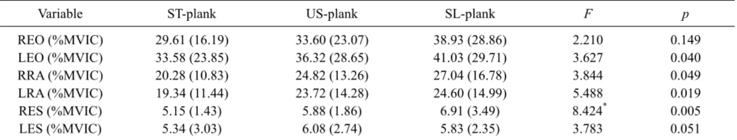
Table 1. Comparison of muscle activities between groups (N=15)
Variable ST-plank US-plank SL-plank F p
REO (%MVIC) 29.61 (16.19) 33.60 (23.07) 38.93 (28.86) 2.210 0.149
LEO (%MVIC) 33.58 (23.85) 36.32 (28.65) 41.03 (29.71) 3.627 0.040
RRA (%MVIC) 20.28 (10.83) 24.82 (13.26) 27.04 (16.78) 3.844 0.049
LRA (%MVIC) 19.34 (11.44) 23.72 (14.28) 24.60 (14.99) 5.488 0.019
RES (%MVIC) 5.15 (1.43) 5.88 (1.86) 6.91 (3.49) 8.424* 0.005
LES (%MVIC) 5.34 (3.03) 6.08 (2.74) 5.83 (2.35) 3.783 0.051
Values are presented as n (%) or mean (SD).
ST-plank: the plank position on stable ground, US-plank: the plank position on unstable ground, SL-plank: the plank position using a sus- pension device, REO: right external oblique muscle, LEO: left external oblique muscle, RRA: right rectus abdominal muscle, LRA: left rectus abdominal muscle, RES: right erector spinal muscle, LES: left erector spinal muscle, MVIC: maximum voluntary isometric contraction.
while performing plank exercises.
Each experiment consisted of a three minute warm up and three sets of a plank exercise maintained for fifteen seconds.
A break was given for 30 seconds between each set; a three- minute break was given between performing each different plank exercise. To measure muscle activities, researchers used the values from electromyography (TELEMYO 2400T G2
Ⓡ; NORAXON, Scottsdale, AZ, USA). The measure- ment excluded the initial two and final two seconds and col- lected information on the RA, EO, and ES in each posture of each subject. The greatest value of electromyography when each muscle was contracted to its maximum was regarded as the maximal voluntary isometric contraction (MVIC). The MVIC was measured three times for five seconds each by having the participant resist the researcher’s manual resis- tance. To help assess manual resistance to the RA, the sub- ject lifted their upper body while flexing the hip joints and the knees; the evaluator applied resistance on both shoul- ders. For the EO, the subject rotated the erected upper body to the left and right; the evaluator applied resistance on both shoulders. For the ES, the subject laid down on their abdo- men, placed their hands with locked fingers on the back of the head, and lifted the upper body up as much as possible while an evaluator helped to fix the lower body and another evaluator applied resistance on both shoulders [5].
The electromyography leads were placed on 2 cm lateral points from the umbilicus with 3 cm longitudinal intervals for the RA. For the EO, the leads were placed between the 12th rib and the iliac crest, along the fibers of the EO just above the anterior superior iliac spine with 2 cm intervals.
For the ES, the leads were placed on 2 cm lateral points from the spinous process of the level of the iliac crest with 2 cm longitudinal intervals [9].
Analysis
The average and maximum values were calculated using PASW ver. 18.0 (IBM Co., Armonk, NY, USA). Resear- chers used Mauchly’s Test of Sphericityto determine if the variances of the differences between all possible pairs of groups were equal. Researchers analyzed the data using re- peated measures analysis of variance to evaluate the differ- ences between the groups. The threshold of the statistical significance was 0.05.
Results
The general characteristics of the subjects revealed no significant difference between the samples. The average weight and height of the subjects were 67 kg and 174.5 cm, respectively.
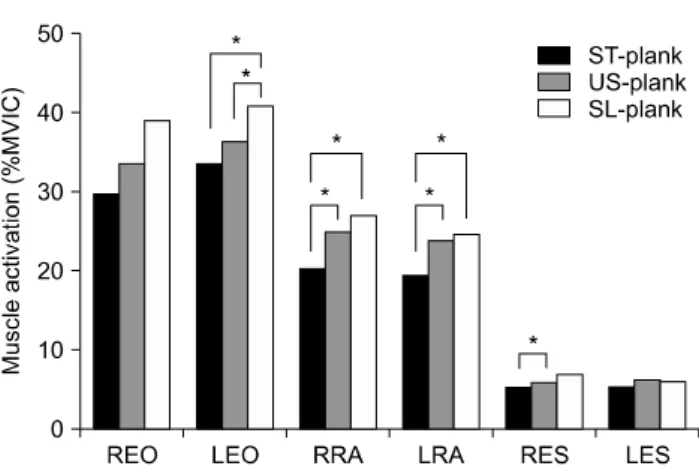
Performing a plank exercise on three different surfaces re- vealed statistically significant differences in the muscle ac- tivities of the left external oblique (LEO), right rectus ab- dominis (RRA), left rectus abdominis (LRA), and right erec- tor spinae (RES) (p<0.05); there were no significant muscle activity differences in the right external oblique (REO) and left erector spinae (LES) (Table 1, Figure 4). The LEO showed significant differences between the plank position on stable ground (ST) and the plank position using a suspen- sion device (SL) (p=0.028) and between the plank position on the unstable ground (US) and SL (p=0.047); however, there were no significant differences between ST and US.
The RRA displayed statistically significant differences be-
tween the ST and the US (p=0.012) and between the ST and
the SL (p=0.034); however, there were no significant differ-
ences between the US and the SL. The LRA exhibited stat-
istically significant differences between ST and US
Figure 4. Comparison of muscle activities between groups.
ST-plank: the plank position on stable ground, US-plank: the plank position on unstable ground, SL-plank: the plank position using a suspension device, REO: right external oblique muscle, LEO: left external oblique muscle, RRA: right rectus abdominal muscle, LRA: left rectus abdominal muscle, RES: right erector spinal mus- cle, LES: left erector spinal muscle, MVIC: maximum voluntary iso- metric contraction (*p<0.05).
(p=0.004) and between ST and SL (p=0.028); however, there were no significant differences between US and SL.
The RES had a statistically significant difference between ST and US (p=0.005).
Discussion
The purpose of this study was to compare core muscle ac- tivities in plank exercises performed on three different surfa- ces among adult men. Exercises on an unstable surface in- tensify the activities of the muscles and the cooperation pat- tern among the stabilizing muscles [10]. Additionally, the exercises provide changed sensory inputs to the muscles and activate the proprioceptors and neuroadaptive mechanisms.
In this study, US and SL showed significant muscle activ- ities in the RA, EO, and ES compared to the ST (p<0.05).
Tong et al. [7] measured muscle activities of the RA, EO, and ES in 36 healthy adults with a program consisting of the primary plank position (60 seconds), plank position with one upper extremity lifted (15 seconds each side), plank po- sition with one lower extremity lifted (15 seconds each side), plank position with contralateral extremities lifted together (15 seconds each side), and back to the primary plank posi- tion (30 seconds) without rest between cycles. Tong et al. [7]
reported significant differences between the plank exercise requiring one upper extremity lifted and the basic plank po- sition (p<0.05), between the plank exercise with one lower extremity lifted and the plank exercise with one upper ex-
tremity lifted (p<0.05), and between the plank exercise with contralateral extremities lifted together and the plank ex- ercise with one lower extremity lifted (p<0.05). Czaprowski et
al. [11] measured muscle activities in the right RA, EO, andinternal oblique (IO) of 33 healthy adults in plank exercises supporting the forearms on the stable ground, using a BOSU ball, and gym balls. The order of the exercises was des- ignated by a random ballot; a one minute break was given between each exercise. As a result, among all of the muscles measured (RA, EO, and IO), significant changes were ob- served between the plank exercise on the BOSU ball and the plank exercise on stable ground, between the plank exercise on the gym ball and the plank exercise on stable ground, and between the plank exercise on the gym ball and the plank ex- ercise on the BOSU ball (p<0.05). The current study demon- strates a consistent result with Tong et al. [7] and Czaprowski et
al. [11] The consistency between all three studies may be be-cause the plank exercise on the unstable surface requires more muscle demands to maintain the elevated position compared to stable surface [5].
In this study, LES showed no significant differences in muscle activity while RES showed significant differences in muscle activity between ST and US and between ST and SL.
Tong et al. [7] reported significant differences in the RA and the EO among plank positions with different bases of support. However, the muscle activity of the ES was weak and not included in the timeline measuring the values (10 seconds). Even though the ES is used before and after the plank exercises, it demonstrates very low muscle activity during the plank exercise. The role of the ES in the plank ex- ercise is limited as the plank exercise resists body weight and gravity with the anterior surface of the trunk. Despite the weak muscle activity of the ES, the plank exercise can be used to evaluate and strengthen the core muscles, mainly fo- cusing on the transversus abdominis (TrA). Mok et al. [5]
measured and compared the muscle activities of the RA, EO, IO, TrA, and lumbar multifidus (LMF) in 18 healthy adults in the hip addition plank (HAP) using a suspension device, chest press (CP), 45
orow (ROW), and hamstring curl (HC).
The EO, IO, and TrA showed the most significant muscle ac- tivity in the HAP compared to the CP, ROW, or HC (p<0.05); interestingly, the HC induced the greatest muscle activity of the LMF than any other exercise (p<0.05).
Czapowski et al. [11] measured and compared the muscle
activities of the RA, EO, IO, and TrA in the supine bridge,
side bridge, and plank exercise on different surfaces. The
greatest muscle activity occurred in the plank exercise
(p<0.05). Based on the previous studies, the plank exercise strengthens the core muscles effectively, and muscle activity is related to the posture of the exercise and the location of the muscle.
Conflict of Interest
The authors declared no potential conflicts of interest with respect to the authorship and/or publication of this article.
References
1. Key J. ‘The core’: understanding it, and retraining its dysfunction.
J Bodyw Mov Ther 2013;17:541-59.
2. Kline JB, Krauss JR, Maher SF, Qu X. Core strength training us- ing a combination of home exercises and a dynamic sling system for the management of low back pain in pre-professional ballet dancers: a case series. J Dance Med Sci 2013;17:24-33.
3. Wilk BR, Stenback JT, Gonzalez C, Jagessar C, Nau S, Muniz A.
Core muscle activation during Swiss ball and traditional abdomi- nal exercises. J Orthop Sports Phys Ther 2010;40:538-9; author reply 539-41.
4. Ekstrom RA, Donatelli RA, Carp KC. Electromyographic analy-
sis of core trunk, hip, and thigh muscles during 9 rehabilitation exercises. J Orthop Sports Phys Ther 2007;37:754-62.
5. Mok NW, Yeung EW, Cho JC, Hui SC, Liu KC, Pang CH. Core muscle activity during suspension exercises. J Sci Med Sport 2015;18:189-94.
6. Do YC, Yoo WG. Comparison of the thicknesses of the trans- versus abdominis and internal abdominal obliques during plank exercises on different support surfaces. J Phys Ther Sci 2015;27:
169-70.
7. Tong TK, Wu S, Nie J. Sport-specific endurance plank test for evaluation of global core muscle function. Phys Ther Sport 2014;
15:58-63.
8. Schoffstall JE, Titcomb DA, Kilbourne BF. Electromyographic response of the abdominal musculature to varying abdominal exercises. J Strength Cond Res 2010;24:3422-6.
9. Youdas JW, Boor MM, Darfler AL, Koenig MK, Mills KM, Hollman JH. Surface electromyographic analysis of core trunk and hip muscles during selected rehabilitation exercises in the side-bridge to neutral spine position. Sports Health 2014;6:416- 21.
10. Vera-Garcia FJ, Grenier SG, McGill SM. Abdominal muscle re- sponse during curl-ups on both stable and labile surfaces. Phys Ther 2000;80:564-9.
11. Czaprowski D, Afeltowicz A, Gębicka A, Pawłowska P, Kędra A, Barrios C, et al. Abdominal muscle EMG-activity during bridge exercises on stable and unstable surfaces. Phys Ther Sport 2014;15:162-8.