Article Info
Received October 1, 2019 Revised November 2, 2019 Accepted January 7, 2020 Corresponding Author Jong-duk Choi E-mail: [email protected]
https://orcid.org/0000-0002-9663-4790
Key Words Exercise therapy Stability Sling
Ultrasonography
Background: Bridging exercises are used to enhance the functional stability of the lum- bopelvic region in clinical settings. Although most of the studies on bridging exercises have compared the complete activation of the trunk muscles, some recent studies have examined the functional stability of the trunk and the lumbopelvic region and assessed the appropriate recruitment of the local and global muscles during different task levels.
Objects: The purpose of this study was to investigate the changes in muscle thickness in the transverse abdominis (TrA), internal oblique (IO), and external oblique (EO) muscles during a common bridging exercise on an unstable surface and to determine whether these changes differ based on the surface used.
Methods: Twenty-five healthy young adults (8 males, 17 females) were recruited. The sub- jects were randomly assigned to either the exercise progression with a sling bridge group or the ball bridging exercise progression group, each with three stages of increasing difficulty.
Each position was measured three times with an ultrasonic diagnostic imaging system, and the mean values were recorded for analysis.
Results: No significant differences were observed between the TrA, IO, or EO muscle thick- ness ratios between the sling and ball exercise groups (p > 0.05). There were also no signifi- cant differences in the EO muscle thickness ratios between the tasks irrespective of whether the sling or ball was used. However, the TrA and IO thickness ratios in both groups were significantly greater during stages 2 and 3 compared to stage 1.
Conclusion: The results suggest that the use of slings and balls during bridging exercises is effective in activating the deep abdominal muscles.
Copyright ⓒ Korean Research Society of Physical Therapy
This is an Open Access article distributed under the terms of the Creative Commons Attribution Non-Commercial License (http://creativecommons.org/licenses/by-nc/4.0) which permits unrestricted non-commercial use, distribution, and reproduction in any medium, provided the original work is properly cited.
INTRODUCTION
The stability of the lumbopelvic region and neuromuscular control of the trunk not only provide flexibility of body parts but also prevent musculoskeletal injuries [1]. Abnormal pos- tures, inappropriate muscle activation, and abnormal move- ments of local muscles lead to loss of control of the lumbopel- vic region and serious problems in trunk stability [2,3].
The trunk musculature consists of local muscles and global muscles that play important roles in many functional and ath- letic activities. Larger and superficially located muscles in the abdominal and back region include global muscles, such as the rectus abdominis, the paraspinalis, and the external oblique [4,5]. These muscles are mainly used for trunk or hip flexion,
extension, and abduction and contribute to overall trunk sta- bility. Local muscles, such as the transverse abdominis, the internal oblique, and the multifidus, are smaller and deeply located in the abdominal and back region. These are directly connected to spinal segments and involved in fine spinal con- trol and the stability of spinal segmental motions [5,6].
Bridging exercises are used to enhance the functional sta- bility of the lumbopelvic region in clinical settings. Bridging exercises can relieve lumbopelvic pain, make the patient feel more comfortable, and retrain the local muscles and the global muscles to ensure that these muscles can be recruited at ap- propriate rates [7]. Many studies have examined the effects of bridging exercises on lumbar stability. García-Vaquero et al.
[8] compared abdominal muscle activation during bridging
Physical Therapy Korea
PTK https://doi.org/10.12674/ptk.2020.27.1.87 pISSN: 1225-8962 eISSN: 2287-982X Phys Ther Korea. 2020;27(1):87-92
Original Article
Comparison of Abdominal Muscle Thickness Using Ultrasound Imaging During Bridging Exercises With a Sling and Ball in Healthy Young Adults
Young Moon
1,2, MSc, PT, Jong-duk Choi
2, PhD, PT
1
Department of Movement Development, The ERUM Child Development Center,
2Department of Physical Therapy, College of Health and
Medical Science, Daejeon University, Daejeon, Korea
exercises performed with one leg raised with that during tra- ditional bridging exercises, and Bjerkefors et al. [9] compared EMG activation of the back and the abdominal muscles during bridging exercise in a series of postures.
Unstable surfaces such as Swiss balls, air cushions, and sling suspension systems were used to increase muscle activation and coactivation to enhance the stability of the lumbopelvic region in theprogressing stage [10,11]. The results showed that the unstable surfaces were more effective in activating trunk- stabilizing muscles than the stable surfaces.
Most of the studies of bridging exercises conducted thus far have compared complete activation of the trunk muscles. Few studies have assessed the appropriate recruitment of the local muscles and the global muscles during different levels of diffi- culties of tasks to examine the functional stability of the trunk and the lumbopelvic region. In addition, the levels of muscle activation on a series of surfaces and the activation rates of the deep muscles and the superficial muscles have not been measured.
Thus, the purpose of the present study was to examine the thicknesses of the transverse abdominal muscle, the internal oblique muscle, and the external oblique muscle when bridg- ing exercises of different degrees of difficulties were performed on unstable surfaces using sling suspension systems and balls.
MATERIALS AND METHODS
1. Subjects
Twenty-five healthy young persons (8 males and 17 females) who understood the content of the study and voluntarily agreed to participate were recruited. The inclusion criteria were as follows: no experience of low back pain during the last six months, no orthopedic surgery, no neurological illness affecting the lower extremities or the lumbopelvic region, and sufficient muscle strength and range of motion to perform the tasks. The mean age of the subjects was 22.28 ± 2.82 years, the mean height was 165.92 ± 8.63 cm, and the mean weight was 58.84 ± 9.84 kg.
2. Ultrasound Imaging
Prior to enrolling subjects, examiners underwent a super- vised training program for the ultrasonic diagnostic imaging system by an experienced physical therapist. The center of the transducer was placed on the transverse plane across the
abdominal wall on the axillar line in the middle of the area be- tween the no. Twelve costal cartilage on the right side and the iliac crest to obtain clear images of the deep abdominal layer [12]. To standardize the location of the transducer, the space where the transverse abdominal and the thoracolumbar fascia meet each other was shown at the end of the right side of the images [13]. The angle of the transducer was adjusted to opti- mize the visualization of the images.
In the present study, the ultrasound images were obtained in B (brightness) mode. To control the effects of breathing cycles on the abdominal muscle thicknesses and ensure that the mea- surement times were identical among the subjects, the subject maintained the position of each exercise for 5 seconds at the last expiration, which was determined by visual observation of the abdominal region. Three repeated ultrasound images were used to determine the abdominal muscle thicknesses [14].
To determine the abdominal muscle thicknesses from the ultrasound images, the distances between the superior and in- ferior hyperechoic muscle fascias of the transverse abdominal muscle and the distances between the internal oblique muscle and the external oblique muscle were measured. The measure- ments were made at the same position in areas (1 cm away from the center of the muscles) where the muscles are sepa- rated from their muscle fascias [15]. The measurement of the muscle thicknesses is presented in Figure 1.
The ratios of changes in the muscle thicknesses of the three abdominal muscles were determined by dividing the average thickness value measured in contracted states during each ex- ercise by the average thickness measured in resting states.
Figure 1. Ultrasound image of the abdominal muscles. EO, external
oblique; IO, internal oblique; TrA, transverse abdominis.
EO
IO
TrA
3. Measurement Procedure
In the present study, an ultrasonic diagnostic imaging system was used to measure the thickness of the abdominal muscle during the bridging exercises using the suspense sling system and balls with and without air cushions, which were used in addition to the slings and balls to provide different degrees of difficulties of tasks.
At that start of the bridging exercises, the subjects maximally bent their knee joints and placed both their arms on their normal anatomical position, with the palms of their hands facing down. Their knees and both feet were close together, and the soles of the feet were placed parallel on the ground.
The subjects were instructed not to press the floor hard with their heads to ensure that their heads did not bear the weight.
A blanket of the same height as the air cushion was placed between the scapular and C7. In the bridging exercises us- ing a suspension sling system, the heights of the straps of the slings were determined so that the straps were at the knee level of individual subjects, and the straps were placed at points perpendicular to the suspension sling system. In the bridging exercises using balls, a 55 cm high ball was used.
The bridging exercises using slings and balls were divided into three stages (Figure 2). In the stage 1 bridging exercises, the sling straps and the ball were placed under the knee joints of each subject, and the subject was instructed to perform ab- dominal draw-in maneuvers. The subject was then instructed
to perform a bridging exercise to place the knees, hips, and shoulders along a straight line and hold the posture for 5 sec- onds. In the stage 2 bridging exercises, in the starting posi- tion, the slings and the ball were placed below the ankle joint to lengthen the lever arm, and the same bridging exercise as performed in stage 1 was performed. In stage 3, in the starting position, the straps of the sling and the ball were placed un- der the ankle joints as in stage 2, and an air cushion was used instead of a blanket. Then, the subject performed the same bridging exercise as performed in stage 1. All the subjects were educated about abdominal draw-in maneuvers and bridging exercises in each stage in advance. The order of the indepen- dent variables (two exercises and three stages) was determined via randomized control trials. A rest time of 1 minute was al- lowed between each experiment, and each experiment was repeated three times.
4. Data Analysis
In the present study, statistical analyses were conducted us- ing the SPSS ver. 18.0 program (IBM Corp., Armonk, NY, USA).
The ultrasound image show the means and standard devia- tions. Ratio of abdominal muscle thickness under independent variables (stages) were examined using One-way ANOVAs with repeated measures. The least significant difference was used as post hoc analyses. The level of statistical significance α was set to 0.05.
Figure 2. Bridging exercises using slings and balls. (A) Stage 1, (B) stage 2, (C) stage 3 of bridging exercises using slings and (D) stage 1, (E) stage 2, (F)
stage 3 of bridging exercises using balls.
A B C
D E F
RESULTS
1. Thicknesses of the Abdominal Muscles at Different Levels of Difficulties during Bridging Exercises Using Slings And balls
The thicknesses of the transverse abdominal muscle, the in- ternal oblique muscle, and the external oblique muscle in each stage of the bridging exercises using slings and balls are shown in Table 1.
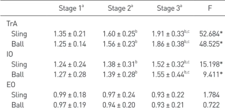
2. Changes in the Thicknesses of Abdominal Muscles during Bridging Exercises Using Slings and Balls The muscle thickness ratios (mean contracted-rest thickness ratios) of the transverse abdominal muscle, the internal oblique muscle, and the external oblique muscle in each stage of the bridging exercises performed at different lever arm lengths and degrees of instability are shown in Table 2. Changes in the thicknesses of the abdominal muscles in each stage of the bridging exercises performed using slings and those performed using balls were compared. According to the results, there were no significant differences (p > 0.05). In the muscle thickness ratios of the transverse abdominal muscle, the internal oblique muscle, and the external oblique muscle. Changes in the thick- nesses of the abdominal muscles throughout individual stages of the bridging exercises performed using the slings and balls were compared. According to the results, the thicknesses of the transverse abdominal muscle and the internal oblique muscle were significantly higher in stage 2 compared to stage 1 and significantly higher in stage 3 compared to stages 1 and 2 (p <
0.05). However, the external oblique muscle did not show any significant differences between the stages (p > 0.05).
DISCUSSION
In clinical settings, rather than rehabilitation training on stable surfaces, rehabilitation training on unstable surfaces is recommended for therapeutic exercises for re-education of damaged muscles. In particular, to improve lumbar stabiliza- tion, loads imposed on the abdominal muscles are increased by appropriately adjusting the level of difficulty during bridg- ing exercises performed on unstable surfaces. However, studies of the degrees of contraction of the abdominal muscles during tasks of different degrees of difficulties due to the type of un- stable surface are rare. Therefore, the present study aimed to examine change in the thicknesses of the abdominal muscles when the lever arm length and the degree of instability were changed. The results of the present study indicated that chang- es in the thicknesses of the deep abdominal muscles increased as the level arm length and the degree of instability increased, regardless of the type of unstable surfaces.
Bridging exercises are an efficient exercising method that can optimally contract the local muscles, such as the trans- verse abdominal muscle and the internal oblique muscle, in relation to the global muscles, such as the external oblique muscle. They are one type of lumbar stabilization exercise that can relieve lumbar instability and improve the trunk muscles’
coactivation functions [16]. It has been reported that bridg- ing exercises performed on unstable surfaces such as balls and slings stimulate proprioception receptors and increase muscle recruitment more so than the same exercises performed on stable surfaces. To increase the degree of difficulty, increas- ing the length of the lever arm or the degree of instability is
Table 1. Thickness of the abdominal muscle at different levels during
bridging exercise (N = 25)
Rest Stage 1 Stage 2 Stage 3
Sling
TrA (mm) 2.52 ± 0.39 3.41 ± 0.80 4.02 ± 0.76 4.79 ± 1.00 IO (mm) 5.91 ± 1.42 7.51 ± 2.32 8.13 ± 2.49 8.88 ± 2.38 EO (mm) 4.02 ± 1.23 3.92 ± 1.26 3.81 ± 1.25 3.68 ± 1.25 Ball
TrA (mm) 2.52 ± 0.39 3.14 ± 0.52 3.90 ± 0.64 4.66 ± 0.99 IO (mm) 5.91 ± 1.42 7.43 ± 2.31 8.13 ± 2.24 9.12 ± 2.96 EO (mm) 4.02 ± 1.23 3.85 ± 1.17 3.69 ± 1.12 3.67 ± 1.12 Values are presented as mean±standard deviation. TrA, transverse ab- dominis muscle; IO, internal oblique muscle, EO,external oblique muscle.
Table 2. Ratio of abdominal muscle thickness to resting condition at dif-
ferent exercise method (N = 25)
Stage 1
aStage 2
aStage 3
aF TrA
Sling 1.35 ± 0.21 1.60 ± 0.25
b1.91 ± 0.33
b,c52.684*
Ball 1.25 ± 0.14 1.56 ± 0.23
b1.86 ± 0.38
b,c48.525*
IO
Sling 1.24 ± 0.24 1.38 ± 0.31
b1.52 ± 0.32
b,c15.198*
Ball 1.27 ± 0.28 1.39 ± 0.28
b1.55 ± 0.44
b,c9.411*
EO
Sling 0.99 ± 0.18 0.97 ± 0.24 0.93 ± 0.22 1.784 Ball 0.97 ± 0.19 0.94 ± 0.20 0.93 ± 0.21 0.722 Values are presented as mean±standard deviation. TrA, transverse ab- dominis muscle; IO, internal oblique muscle, EO,external oblique muscle.
a