Comparison of the Muscle Activity of Lumbar Stabilizers Between Stoop and Semi-Squat Lifting Techniques at Different Lifting Loads
Hoe-song Yang1, MSc, PT, Oh-yun Kwon2, PhD, PT, Yeon-seop Lee1, PhD, PT
1Dept. of Physical Therapy, College of Cheongam,
2Dept. of Physical Therapy, College of Health Science, Yonsei University,
Dept. of Ergonomic Therapy, The Graduate School of Health and Environment, Yonsei University
Abstract
1)This study was performed to compare the muscle activity of lumbar stabilizers between stoop and semi-squat lifting techniques at different lifting loads. Twenty healthy subjects (9 males, 11 females) were recruited for this study. Muscle activity of external obliques (EO), internal obliques (IO) and lumbar multifidus (LM) muscle was measured by surface electromyography during stoop and semi-squat lifting at different lifting loads (10%, 20%, and 30% of the subject’s body weight). A one-way repeated measure ANOVA was applied. The results showed that EMG activity of EO was significantly increased with a load of 30% of body weight compared to 10% and 20% of body weight in both lifting techniques (p<.05).
Muscle activity of LM was significantly increased in 20% compared to 10% and 30% compared to 10% of subject’s body weight in stoop lifting and the muscle activity of LM was significantly increased in 20%
compared to 10%, 30% compared to 20%, and 30% compared to 10% of the subject’s body weight in semi-squat lifting (p<.05). However, there was no significant difference in activity of IO according to lifting loads in both lifting techniques. There were no significant differences in muscle activity of EO, IO, and LM between stoop and semi-squat technique (p>.05). Therefore, the results of this study suggested that the EO can contribute to increase the lumbar stability during stoop and semi-squat lifting at 30% of body weight rather than at lower loads, and the LM seems to act as counteractor to imposed loads during stoop and semi-squat lifting with increasing loads.
[Hoe-song Yang, Oh-yun Kwon, Yeon-seop Lee. Comparison of the Muscle Activity of Lumbar Stabilizers Between Stoop and Semi-Squat Lifting Techniques at Different Lifting Loads. Phys Ther Kor.
2012;19(3):105-114.]
Key Words: Lifting loads; Lifting techniques; Lumbar stabilizer; Surface electromyography.
Introduction
It has been reported that nearly 60% of industrial workers experience low back pain in their lifetime.
Low back pain not only generates economic, pro- ductive losses in the work site, such as increased nursing rate and insurance costs for occupational ac- cidents, but also negatively influences the quality of life in individuals. Thus, measures for preventing low back pain are being sought from various angles
(Murtezani et al, 2011). Recently, although manual material handling activities have been decreased with developing automatic equipment, lifting of objects is often used in industrial sites. Among manual han- dling activities, lifting is reported as the etiology with the highest risk factor among the various caus- es of low back pain (Granada and Marras, 1995;
Ferguson, et al, 1997). Musculoskeletal diseases were caused by lifting activities in 48.3% of cases and by transport or movement in 33.1% (KOSHA, 2010).
Corresponding author: Oh-yun Kwon [email protected]
There are several lifting techniques in common use, such as stoop (back lift), semi-squat, and squat.
Straker (2003) stated that a squat lifting is charac- terized with a start position of deep knee flexion with the trunk close to erect. Quantitatively this can be described as knee flexion around 45° and trunk flexion < 30° for most workers when lifting from floor level. Stoop lifting techniques involve the in- clined trunk and nearly-extended knees. Semi-squat technique uses a posture midway between the squat and stoop lifts.
Many studies have been conducted to compare various parameters among lifting techniques. In a study conducted on lifting postures, the squat lifting posture was found a proper lifting posture for re- ducing the burden on low back ligaments by de- creasing pressure on the 5th lumbar segment and the 1st sacrum (Lariviere et al, 2002; Faber et al, 2009).
However, although squat lifting was a safer posture for lifting objects, many workers prefer stoop lifting as it consumes less energy (Grag and Herrin, 1979) and is easier to keep one’s balance (Hagen et al, 1993). Hwang et al (2009) reported that the knee ex- tension that is the prominent kinematic feature dur- ing the squat lifting was produced by the con- tributions of the kinetic factors from the hip and an- kle joints (extensor moment and power generation), and that the lumbar extension that is the prominent kinematic feature during the stoop lifting could be produced by the contributions of the knee joint ki- netic factors (flexor moment, power absorption).
Lifting from semi-squat postures, involving a moder- ate range of flexion at both knees and trunk, allows a pattern of inter-joint coordination that appears to be functional in reducing muscular effort (Burgess-Limerick, 2001).
Lifting capacity can be enhanced through spinal stabilization, which is achieved by cooperative co-ac- tivation of abdominal muscles and intra-abdominal pressure (Cholewicki et al, 1999). The co-activation of abdominal muscles and intra-abdominal pressure (IAP) are important factors that contribute to trunk
stabilization. By increasing intra-abdominal pressure, the compressive force of the spine is decreased (Cholewicki et al, 2000). Lumbar spinal stability is essential to reduce the risk of tissue overload or damage in carrying out various functions (Ferreira and Hodges, 2004). Various studies reported the im- portance of transverse abdominis (TrA) and local muscle system in lumbar spinal segmental stabiliza- tion (Davidson and Hubley-Kozey, 2005; Urquhart et al, 2005). Delitto and Lose (1992) demonstrated that the heavy load presented greater activity of erector spinae and oblique muscles than both the moderate and the light loads, and that the moderate load showed greater activity than the light loads during squat lifting. Allison and Henry (2001) reported that abdominal activation was increased during sustained sub-maximal trunk extension efforts.
However, there has been no study to find the muscle activity of lumbar stabilizers during lifting at different loads. Therefore, the present study was performed to compare of the muscle activity of lum- bar stabilizers (external oblique; EO, internal oblique;
IO, and lumbar multifidus; LM) between stoop and semi-squat lifting techniques at different lifting loads.
We hypothesized that the muscle activity of lumbar stabilizers would increase as increased lifting load, And muscle activity of lumbar stabilizers would be different between stoop and semi-squat lifting techniques.
Methods
Subjects
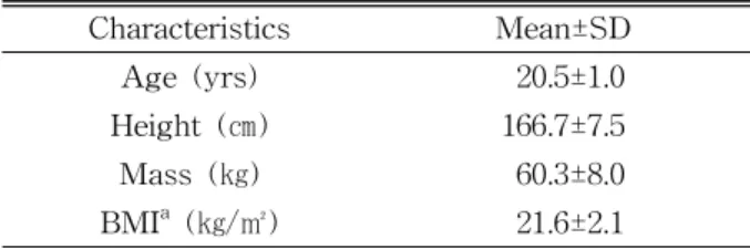
Twenty healthy subjects (9 males, 11 females) participated in this study. This study selected sub- jects without congenital deformity or medical history in the muscular and neurological system of the back and legs and excluded subjects that had experienced trauma or pain in the back or legs but had regularly participated in exercise for the past 6 months. The general characteristics of subjects were showed in
Characteristics Mean±SD
Age (yrs) 20.5±1.0
Height (㎝) 166.7±7.5
Mass (㎏) 60.3±8.0
BMIa (㎏/㎡) 21.6±2.1
aBody Mass Index.
Table 1. General characteristics of subjects (N=20) Table 1. The subjects were informed of the inves- tigational protocol and possible risks and gave writ- ten consent prior to their participation in this study.
Instruments and Measurements.
The lifting load determined by the strain gauge 2) The strain gauge with the Noraxon Myoresearch 1.06 XP software was used to control the lifting load. The strain gauge was attached to the steel wire-connected steel bar (15 W 315 D × 328 H (㎜)) on the back strength dynamometer1) Strain gauge was connected with Noraxon software. The subject was asked to watch a computer monitor for main- taining the predetermined lifting load. The lifting or- der was randomly determined to reduce variables re- lated with the order effect.
Surface electromyography3)
To measure the muscle activity of EO, IO, and LM in right side, a surface EMG system2) was used.
Surface electrodes (Ag-Ag/Cl; Biopac, diameter 2 ㎝, interelectrode distance 2 ㎝) were used for collecting EMG signals. Sampling rate was 1,000 ㎐ and band pass (30∼500 ㎐) and notch filter (60 ㎐) were applied. EMG signals were processed into the root mean square (RMS) using the software (Myolab-2.12 version). The surface electrode was attached on the lateral abdominal area, the part with the highest muscle activity. To minimize skin resistance before attachment, the electrode area was shaved and wiped with alcohol. After the area was completely dried, electrodes were attached to the muscle belly. Ground
electrode was attached on the C7 spinous process.
Among abdominal muscles, electrodes for EO were attached over approximately 15 ㎝ lateral to the um- bilicus, electrodes for IO were attached over halfway between the anterior superior iliac spine of the pelvis and the midline, just superior to the inguinal liga- ment; and electrodes for LM were attached at 2 ㎝ lateral to L5 of the spinous process (Silfies et al, 2005). To normalize the muscle activity measurement, maximum voluntary isometric contraction (MVIC) measurement of EO and IO were achieved according to the previous literature (Sánchez-Zuriaga, 2009).
The subject was in a sit-up posture positioned on a bench with the legs bent and feet strapped down with a belt. The subject was asked to twist his/her upper trunk to right, and left while his/her thorax was manually braced by the principal investigator.
MVIC measurement of LM was performed in prone position. The subject was strapped in a prone posi- tion, with the torso horizontally cantilevered over the end of the bench (Biering-Sorensen position). The subject was asked to extend the trunk in the sagittal plane while manual resistance was applied on the shoulders by the principal investigator. EMG activity was collected for 50 milliseconds for measuring the MVIC of each muscle. For normalization, RMS of the middle three second MVIC was measured three times for each muscle. The average RMS of the measurement was used to determine the MVIC of each muscle.
Procedures
Lifting loads were determined according to sub- ject’s body weight. 10%, 20%, and 30% of their in- dividual body weights were calculated. Body weight was measured by digital weight scale. Two lifting techniques (stoop and semi-squat lifting) were ap- plied at different lifting loads. Stoop lifting technique was defined as the lifting position with maintaining lumbar lordotic curve and hip flexed 30 degrees without knee flexion. Semi-squat lifting technique 1) Takei TKK 5402, Takei Scientific Inst. Co. Ltd, Tokyo, Japan.
2) BTS Pocket EMG, Garbagnate Milanese, Milano, Italy.
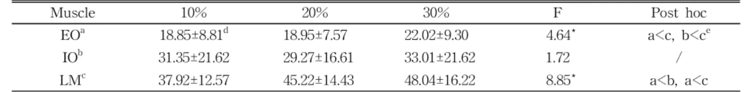
Muscle 10% 20% 30% F Post hoc
EOa 18.85±8.81d 18.95±7.57 22.02±9.30 4.64* a<c, b<ce
IOb 31.35±21.62 29.27±16.61 33.01±21.62 1.72 /
LMc 37.92±12.57 45.22±14.43 48.04±16.22 8.85* a<b, a<c
aexternal oblique, binternal oblique, clumbar multifidus,dmean±standard deviation, ea: 10%, b: 20%, c: 30%, *p<.05.
Table 2. Comparison of muscle activity of EO, IO, and LM at different loads in stoop lifting (Unit: %MVIC)
Muscle 10% 20% 30% F Post-hoc
EOa 18.86±8.81d 19.82±7.95 22.29±8.08 8.18* a<c, b<ce
IOb 28.41±16.17 30.56±16.38 37.47±20.49 2.22 /
LMc 37.29±12.20 43.94±14.13 49.20±14.48 40.64* a<b, b<c, a<c,
aexternal oblique, binternal oblique, clumbar multifidus,dmean±standard deviation, ea: 10%, b: 20%, c: 30%, *p<.05.
Table 3. Comparison of muscle activity (%MVIC) of EO, IO, and LM at different loads in squat lifting (Unit: %MVIC) was defined as the lifting position with maintaining
lumbar lordotic curve and hip and knee flexed 30 degrees. During lifting, the pelvis is aligned in an anterior tilt. Each subject practiced the two lifting techniques for familiarization with the testing procedure. Subjects were asked to hold the lifting loads of 10%, 20%, and 30% of their body weight and hold the posture for 50 milliseconds. Subjects were asked to not elevate scapula, and to extend the trunk during the lifting test. Measurement was per- formed three times and 3 minute resting periods were allowed between each trial. EMG signals were collected 50 milliseconds and the middle 30 milli- seconds used for data analysis. EMG data of the ini- tial one second and final one millisecond were excluded. The normalized muscle activity of EO, IO, and LM was expressed as percent of MVIC.
Statistical Analysis
Descriptive statistics were used to analyze the general characteristics of subjects. A one-way re- peated measure ANOVA was used to compare the activity of the lumbar stabilizer (EO, IO, and LM) at different loads in each lifting technique and paired t-test was performed to determine the difference in muscle activity between two lifting techniques.
Post-hoc multiple comparison test was performed.
This study was analyzed using PASW Statistics ver- sion 18.0 software. Significance level was set as α=.05.
Results
The results of a one-way repeated ANOVA re- vealed a significant difference for the activity of the EO and LM at different loads in both lifting techni- ques (p<.05), and a paired t-test revealed no sig- nificant difference between the lifting techniques at each muscle according to lifting loads (p>.05).
Comparison of muscle activity at different loads in stoop lifting
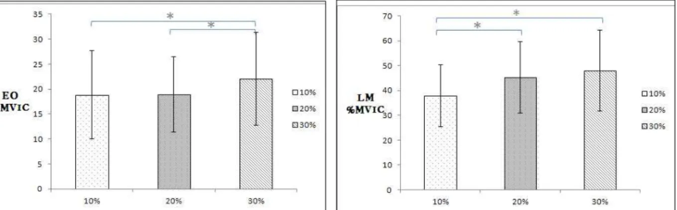
The muscle activity of EO and LM were statisti- cally significant difference according to increased loads from the results of a one-way repeated meas- ures ANOVA in stoop lifting (p<.05). However, the muscle activity of IO was not significantly different (p>.05). The results of the post-hoc analysis showed that the muscle activity of EO was significantly in- creased in 30% compared to 10% and 20%, whereas the muscle activity of LM was significantly in- creased in 20% compared to 10% and 30% compared
Figure 2. Comparison of muscle activity of (%MVIC) EO and LM at different loads in semi-squat lifting (EO: external oblique, LM: lumbar multifidus, %MVIC: %maximum voluntary isometric contraction, *p<.05).
Figure 1. Comparison of muscle activity (%MVIC) of EO and LM at different loads in stoop lifting (EO: external oblique, LM: lumbar multifidus, %MVIC: %maximum voluntary isometric contraction,
*p<.05).
to 10% of subject’s body weight in stoop lifting (Table 2) (Figure 1).
Comparison of muscle activity at different loads in semi-squat lifting
The muscle activity of EO and LM were statisti- cally significantly different according to increased load from the results of a one-way repeated meas- ures ANOVA in semi-squat lifting (p<.05). The muscle activity of IO was not significantly different (p>.05). The results of the post-hoc analysis showed that EO was significantly increased in 30% compared to in 10% and 20%, whereas the muscle activity of LM was significantly increased in 20% compared to 10%, 30% compared to 20%, and 30% compared to 10% of the subject’s body weight in semi-squat lift- ing (Table 3) (Figure 2).
Discussion
The purpose of the present study was to compare of the muscle activity of lumbar stabilizers (EO, IO, and LM) between stoop and semi-squat lifting tech- niques at different lifting loads. The results of this study showed that muscle activity of EO and LM increased significantly as increased lifting load both stoop and semi-squat lifting. However, there was no difference in muscle activity of IO according to lift- ing loads in both stoop lifting and semi-squat lifting.
In addition there was no significant difference in muscle activity of lumbar stabilizers between the two lifting techniques.
Tan et al (1993) reported that maximum extension torque was significantly increased at 35 degrees of trunk flexion and many activities of daily living and
trunk activities such as manual material handling and lifting are performed in the sagittal plane with the trunk slight flexed to about 30~60 degrees.
Therefore, we selected 30 degrees of trunk flexion for stoop and semi-squat lifting techniques (Nordin et al, 1984). During lifting, the lumbar spine is main- tained in its normal lordosis and the pelvis is aligned in an anterior tilt. It minimizes stretch on the poste- rior elements of the lumbar spine and thereby de- creases the stress on these structures (Kisner and Colby, 2007). Delitto and Rose (1992) recommended that the greater trunk muscle activity occur with the anterior tilt position for optimal muscular support for the spine while lifting loads, thereby reducing the risk for low back injury. Therefore, we asked sub- jects to maintain the lumbar natural lordotic curve for minimizing lumbar injury during lifting.
The activity of abdominal muscle plays a vital role in determining the trunk’s ability to maintain the normal posture and effectively function to do daily activities (Granata and Orishimo, 2001; Lavender et al, 1992) and is deeply related with the trunk stabili- zation of lumbar spinal disorder patients (Silfies et al, 2005; van Dieen et al, 2003). However, Tan et al (1993) reported that there was no co-activation of abdominal muscles at sub-maximal extension efforts at 0, 15, and 35 degrees of trunk flexion, whereas, Lavender et al (1992) stated that abdominal muscles are activated during submaximal lifting. Sitilertpisan et al (2011) stated that the lateral abdominal muscle group (LAM) including the IO, EO, and TrA shared a role in controlling the lumbar spine during weight-lifting. The EO and IO may control the ro- tary torque and balance the external loads on the lumbar segment (Hodges and Gandevia, 2000;
Richardson et al, 2004; Teyhen et al, 2005).
Therefore, the present study sought to determine whether IO and EO muscles are activated for lumbar stabilization during lifting at different lifting loads. In the present study, the muscle activity of EO was statistically significantly different according to in- creased loads However, the muscle activity of IO
was not significantly different (p>.05). The results of the post-hoc analysis showed that the muscle activ- ity of EO was significantly increased in 30%
(22.02±9.30) compared to 10% (18.85±8.81) and 20%
(18.95±7.57) in stoop lifting. And the EMG activity of EO was also significantly increased in 30%
(22.29±8.08) compared to 10% (18.86±8.81) and 20%
(19.82±7.95) in semi-squat lifting. Tan et al (1993) found that an activity of abdominal oblique muscles did not appear at submaximal levels (MVIC 20, 40, 60, 80%), but there was a activity of abdominal obli- ques at maximal level, Because of an increase in the need of trunk stabilization through coactivation of the abdominal oblique muscles.
Three major mechanisms of lifting have been introduced. There are the intra-abdominal pressure (IAP) (Cholewicki et al, 1999; Hodges et al, 2005;
McGill 2002), the lumbodorsal fascia mechanism (Tesh et al, 1987), and the hydraulic amplifier (McGill et al, 1996). The stability of spine can be achieved by two mechanisms: antagonistic muscle co-activation (Gardner-Morse and Stokes, 1998) and/or an increase in IAP. IAP can increase by co-activation of the abdominal musculature, dia- phragm, and pelvic floor muscles (Cresswell and Thorstensson, 1989; Cresswell et al, 1994). Norris (1995) stated that contraction of the transverse ab- dominis and internal and external oblique cause an incresed IAP when the glottis is closed. The result of the study showed that muscle activity of EO was significantly increased with increasing lifting loads in either technique. These results suggest that EO con- tributes to increased IAP during stoop and semi-squat lifting techniques.
The rigidity of the trunk segment must be main- tained during daily activities to allow for sufficient loading and energy transfer during loading.
Paraspinal muscles, in particular the erector spinae, act as the primary agonist in lifting activities, whereas abdominal muscles such as rectus abdomi- nis, IO, EO, and TrA act as the antagonist (Chow et al, 2005; Kingma and van Dieen, 2004). Previous lit-
eratures stated that the LM can contribute to stabili- zation of the lumbar spine (Barr et al. 2005; Beneck and Kulig, 2012; Freeman et al. 2010; Hides et al, 2008; Lee et al, 2011). Biomechanical studies showed that the LM has a high capacity to stabilize the spine when spinal stability is challenged (Panjabi, 1992; Wilke et al, 1995), and control the spinal seg- ment’s neutral zone (Panjabi et al, 1989). The LM may contribute to sub-maximal extension efforts at greater exertion intensity (Olson, 2010). Although, LM has been considered as an important lumbar stabilizer during lifting objects, there has been no study to determine the muscle activity of LM during lifting. In the present study, the muscle activity of LM was significantly increased in 20% (45.22±14.43) compared to 10% (37.92±12.57) and 30% (48.04±16.22) compared to 10% (37.92±12.57) of subject’s body weight in stoop lifting. And the muscle activity of LM was significantly more increased in 20%
(43.94±14.13) compared to 10% (37.29±12.20), 30%
(49.20±14.48) compared to 20% (43.94±14.13), and 30% (49.20±14.48) compared to 10% (37.29±12.20) of the subject’s body weight in semi-squat lifting.
Previous studies reported that the activity of para- spinal muscles significantly increased as lifting load increased (Dolan et al, 1993; Lariviere et al. 2002;
Olson, 2010; Tan et al, 1993; Yoon et al, 2012). LM muscle works together with TrA, diaphragm, and pelvic floor muscle to increase lumbar stability dur- ing lifting. Increased muscle activity of LM during lifting with 30% of body weight resulted in counter- balancing to the EO muscle contraction for maintain- ing the neutral position of lumbar spine.
Straker (2003) reported that there were different advantages and disadvantages according to various situations as the squat posture reduced shear force exerted on lumbar spine, whereas the stoop posture decreased muscular fatigue. Van Dieen et al (1999) found that reasonable evidence was not found for verifying the kinetic advantage of the squat rather than stoop posture in lifting activities. Kingma et al (2004) reported that squat lifting results in higher
compression forces on the spine compared to stoop lifting when the load is not positioned between the legs. In the present study, the EMG activity of EO, IO, and LM was not significantly different between stoop and semi-squat lifting technique. These result do not support the hypothesis of this study.
This study has some limitations. First, subjects were young and healthy. Therefore, the results of this study cannot be generalized to all populations.
Second, deep muscles such as diaphragm, TrA, and pelvic floor contribute to lumbar stabilization; how- ever, the muscle activity of theses muscles was not measured in this study. Further study is needed to compare the muscle activity of TrA, diaphragm, and pelvic floor muscle according to lifting loads and lift- ing techniques.
Conclusion
The purpose of this study was to compare the muscle activity of lumbar stabilizers (EO, IO, and LM) between stoop and semi-squat lifting techniques at different lifting loads. Twenty healthy subjects participated. The muscle activity of EO was sig- nificantly increased at increased loads (30% of body weight) in stoop and squat lifting techniques and LM was significantly increased with increased loads in stoop and squat lifting techniques. However, the ac- tivity of IO was not significantly different at differ- ent loads in either lifting technique. Therefore, we suggest that the EO and LM can contribute to in- creased lumbar stability as increased loads in stoop and semi-squat lifting techniques.
References
Barr KP, Griggs M, Cadby T. Lumbar stabilization:
Core concepts and current literature, part 1. Am J Phys Med Rehabil. 2005;84(6):473-480.
Beneck GJ, Kulig K. Multifidus atrophy is localized
and bilateral in active persons with chronic uni- lateral low back pain. Arch Phys Med Rehabil.
2012;93(2):300-306.
Burgess-Limerick R. Squat, stoop, or something in between? Int J Ind Ergon. 2003;31(3):143-148.
Cholewicki J, Juluru K, McGill SM. Intra-abdominal pressure mechanism for stabilizing the lumbar spine. J Biomech. 1999;32(1):13-17.
Cholewicki J, Simons AP, Radebold A. Effects of ex- ternal trunk loads on lumbar spine stability. J Biomech. 2000;33(11):1377-1385.
Chow DH, Cheng IY, Holmes AD, et al. Muscular and centre of pressure response to sudden re- lease of load in symmetric and asymmetric stoop lifting tasks. Appl Ergon. 2005;36(1):13-24.
Cresswell AG. Thorstensson A. The role of the ab- dominal musculature in the elevation of the in- tra-abdominal pressure during specified tasks.
Ergonomics. 1989;32:1237-1246.
Cresswell AG, Oddsson L, Thorstensson A, The in- fluence of sudden perturbations on trunk muscle activity and intraabdominal pressure while standing. Exp Brain Res. 1994;98:336-341.
Davidson KL, Hubley-Kozey C. Trunk muscle re- sponses to demands of an exercise progression to improve dynamic spinal stability. Arch Phys Med Rehabil. 2005;86(2):216-223.
Delitto RS, Rose SJ. An electromyographic analysis of two techiques for squat lifting and lowering.
Phys Ther. 1992;72(6):438-448.
Dolan P, Adams MA. The relationship between EMG activity and extensor moment generation in the erector spinae muscles during bending and lift- ing activities. J Biomech. 1993;26(4-5):513-522.
Faber GS, Kingma I, Bruijn SM, et al. Optimal in- ertial sensor location for ambulatory measure- ment of trunk inclination. J Biomech.
2009;42(14):2406-2409.
Ferguson SA, Marras WS. A literature review of low back disorder surveillance measures and risk factors. Clin Biomech (Bristol, Avon).
1997;12(4):211-226.
Ferreira PH, Ferreira ML, Hodges PW. Changes in recruitment of the abdominal muscles in people with low back pain. Spine (Phila Pa 1976).
2004;29(22):2560-2566.
Freeman MD, Woodham MA, Woodham AW. The role of the lumbar multifidus in chronic low back pain: A review. PM R. 2010;2(2):142-146.
Gardner-Morse MG, Stokes IA. The effects of ab- dominal muscle coactivation on lumbar spine stability. Spine (Phila Pa 1976). 1998;23(1):86-91.
Granata KP, Marras WS. The influence of trunk muscle coactivity on dynamic spinal loads. Spine (Phila Pa 1976). 1995;20:913-919.
Granata KP, Orishimo KF. Response of trunk muscle coactivation to changes in spinal stability. J.
Biomech. 2001;34(9):1117-1123.
Hagen KB, Hallén J, Harms-Ringdahl K.
Physiological and subjective responses to max- imal repetitive lifting employing stoop and squat technique. Eur J Appl Physiol Occup Physiol.
1993;67(4):291-297.
Hides JA, Stanton WR, McMahon S, et al. Effect of stabilization training on multifidus muscle cross-sectional area among young elite crick- eters with low back pain. J Orthop Sports Phys Ther. 2008;38(3):101-108.
Hodges PW. Gandervia SC. Activation of the human diaphragm during a repetitive postural task. J physiol. 2000;522:165-175.
Hodges PW, Eriksson AE, Shirley D, et al. Intra-ab- dominal pressure increases stiffness of the lum- bar spine. J Biomech. 2005;38(9):1873-1880.
Hwang S, Kim Y, Kim Y. H. Lower extremity joint kinetics and lumbar curvature during squat and stoop lifting. BMC Musculoskelet Disord.
2009;2:10-15.
Kingma I, Bosch T, Bruins L, et al. Foot positioning instruction, initial vertical load position and lift- ing technique: Effects on low back loading.
Ergonomics. 2004;47(13):1365-1385.
Kisner C, Colby LA. Therapeutic Exercise:
Foundations and techniques 5th. Philadelphia,
F.A. Davis Company. 2007;256-258.
KOSHA. Industrial Accident Cause Investigation.
2010. http://www.kosha.or.kr
Lariviere C, Gagnon D, Loisel PA. Biomechanical comparison of lifting techniques between sub- jects with and without chronic low back pain during freestyle lifting and lowering task. Clin Biomech (Bristol, Avon). 2002;17(2):89-98.
Lavender SA, Tsuang YH, Andersson GB, et al.
Trunk muscle cocontraction: The effects of mo- ment direction and moment magnitude. J Orthop Res. 1992;10(5):691-700.
Lee HI, Song J, Lee HS, et al. Association between cross-sectional areas of lumbar muscles on magnetic resonance imaging and chronicity of low back pain. Ann Rehabil Med. 2011;35(6):
852-859.
McGill SM. A revised anatomical model of the ab- dominal musculature for torso flexion efforts. J Biomech. 1996;29(7):973-977.
McGill SM. Low Back Disorders: Evidence based prevention and rehabilitation. Human kinetics publishers, Champaign, IL. 2002:230-239.
Murtezani A, Ibraimi Z, Sllamniku S, et al.
Prevalence and risk factors for low back pain in industrial workers. Folia Med (Plovdiv). 2011;53 (3):68-74.
Nordin M, Ortengren R, Andersson GB.
Measurements of trunk movements during work.
Spine (Phila Pa 1976). 1984;9(5):465-469.
Norris CM. Spinal stabilisation: 3. Satabilisation mechanisms of the lumbar spine. Physiotherapy.
1995;81(2):72-79.
Olson MW. Trunk muscle activation during sub-maximal extension efforts. Man Ther. 2010;
15(1):105-110.
Panjabi MM, Abumi K, Duranceau J, et al. Spinal stability and intersegmental muscle forces. A bi- oimechanical model. Spine (Phila Pa 1976).
1989;14(2);194-200.
Panjabi MM. The stabilizing system of the spine.
Part 1. Function, dysfunction, adaptation, and
enhancement. J Spinal Disord. 1992;5(4):383-389.
Richardson CA, Hodges PW, Hides J. Therapeutic Exercise for Lumbopelvic Stabilization. In: A motor control approach for the treatment and prevention of low back pain. 2nd ed. Edinburgh, Churchill Livingstone. 2004:30-40.
Silfies SP, Squillante D, Maurer P, et al. Trunk muscle recruitment patterns in specific chronic low back pain populations. Clin Biomech (Bristol, Avon). 2005;20(5):465-473.
Sitilertpisan P, Pirunsan U, Puangmali A, et al.
Comparison of lateral abdominal muscle thick- ness between weightlifters and matched controls.
Phys Ther Sport. 2011;12(4):171-174.
Straker L. Evidence to support using squat, semi-squat and stoop techniques to lift low-ly- ing objects. Int J Ind Ergon. 2003;31(3):149-160.
Tan JC, Parnianpour M, Nordin M, et al. Isometric maximal and submaximal trunk extension at different flexed positions in standing: Triaxial torque output and EMG. Spine (Phila Pa 1976).
1993;18(16):2480-2490.
Tesh KM, Dunn JS, Evans JH. The abdominal mus- cles and vertebral stability. Spine (Phila Pa 1976). 1987;12:501-508.
Teyhen DS, Miltenberger CE, Deiters HM, et al. The use of ultrasound imaging of the abdominal drawing-in maneuver in subjects with low back pain. J Orthop Sports Phys Ther. 2005;35(6):
346-355.
Urquhart DM, Hodges PW, Allen TJ, et al.
Abdominal muscle recruitment during a range of voluntary exercises. Man Ther. 2005;10(2):
144-153.
van Dieën JH, Hoozemans MJ, Toussaint HM. Stoop or squat: A review of biomechanical studies on lifting technique. Clin Biomech (Bristol, Avon).
1999;14(10):685-696.
van Dieën JH, Kingma I, van der Bug P. Evidence for a role of antagonistic cocontraction in con- trolling trunk stiffness during lifting. J Biomech.
2003;36(12):1829-1836.
This article was received June 21, 2012, was re- viewed June 21, 2012, and was accepted August 29, 2012.
Sánchez-Zuriaga D, Vera-Garcia FJ, Moreside JM, et al. Trunk muscle activation patterns and spine kinematics when using an oscillating blade:
Influence of different postures and blade orientations. Arch Phys Med Rehabil. 2009;90(6):
1055-1060.
Wilke HJ, Wolf S, Claes LE, et al. Stability increase of the lumbar spine with different muscle groups. A biomechanical in vitro study. Spine
(Phila Pa 1976). 1995;20(2):192-198.
Yoon J, Shiekhzadeh A, Nordin M. The effect of load weight vs. pace on muscle recruitment during lifting. Appl Ergon. 2012;43(6):1044-1050.