http://crossmark.crossref.org/dialog/?doi=10.14474/ptrs.2018.7.2.72&domain=pdf&date_stamp=2018-06-25
Received: 20 April, 2018 Revised: 24 May, 2018 Accepted: 24 May, 2018
Corresponding author: Changho Song (ORCID http://orcid.org/0000-0002-5709-3100)
Department of Physical Therapy, College of Health Science and Social Welfare, Sahmyook University, 815 Hwarang-ro, Nowon-gu, Seoul 01795, Republic of Korea
Tel: 82-2-3399-1631 Fax: 82-2-3399-1639 E-mail: [email protected]
This is an Open-Access article distributed under the terms of the Creative Commons Attribution Non-Commercial License (http://creativecommons.org/licenses/
by-nc/4.0) which permits unrestricted non-commercial use, distribution, and reproduction in any medium, provided the original work is properly cited.
Copyright © 2018 Korean Academy of Physical Therapy Rehabilitation Science
https://doi.org/10.14474/ptrs.2018.7.2.72 pISSN 2287-7576
eISSN 2287-7584
Phys Ther Rehabil Sci 2018, 7 (2), 72-77 www.jptrs.org
Comparison of the effects of different core exercise on muscle activity and thickness in healthy young adults
Mingyun Ko
a, Changho Song
baDepartment of Physical Therapy, Chunnam Techno University, Gokseong, Republic of Korea
bDepartment of Physical Therapy, College of Health Science and Social Welfare, Sahmyook University, Seoul, Republic of Korea
Objective: This study aimed to compare the effects of core exercise methods on muscle activation and muscle thickness in healthy young adults and to propose effective core exercise methods.
Design: Three-group pretest-posttest design.
Methods: A total of 30 healthy young adults (14 males, 16 females) voluntarily participated in the study. Subjects were random- ized to the prone plank exercise (n=10), reverse plank exercise (n=10), or bridge exercise (n=10) groups. Muscle activity and thickness of the rectus abdominis (RA), multifidus (MF), external oblique (EO), and internal oblique (IO) muscles were measured using surface electromyography and ultrasound. Subjects from each group participated in the exercises five times a week, with five 20-second sets during week 1. The set time was increased by 10 seconds per week.
Results: Muscle activity and thickness in the prone plank, reverse plank, and bridge exercise group were statistically significant different for RA, MF, EO, and IO changes over time, and interaction between time and groups were also significantly different (p<0.05). We analyzed statistically significant differences between groups using a one-way analysis of variance for each period. A significant difference was observed after 4 weeks of exercise (p<0.05).
Conclusions: The results suggest that the prone plank exercise is a beneficial method for enhancing muscle activation and thick- ness of the RA, EO, and IO compared to the reverse plank and bridge exercises. On the other hand, the reverse plank and bridge ex- ercises are effective methods for enhancing the MF compared to the prone plank exercise.
Key Words: Abdominal oblique muscles, Electromyography, Exercise, Paraspinal muscles, Rectus abdominis
Introduction
Physical activity has decreased due to automation and mechanization in the 21st century. It has caused increased body fat and weakened muscle strength and function.
Muscle weakness is a major cause of lower back disease, and the need for exercise is emerging. A recent emphasis on ac- tive movements has emerged, such as strengthening ex- ercises for the lower back or strengthening muscles around the spine and stabilizing the spine. In particular, core ex- ercises are being used to improve the ability to control the body by securing its center of gravity through strengthening
the core muscles and promoting movement and stability [1,2].
Core exercises are used to strengthen the spinal, abdomi- nal, pelvic floor, and hip muscles, which maintain the core of the body. It contributes to the maintenance of the right body center of pressure, allows for efficient body movement, as well as improves the ability to maintain a stable posture and balance control by stabilizing and providing neutral control of the trunk and the spine, and reduces pain [3,4].
The core of the body is composed of abdominal muscles
at the front of the trunk, paraspinal and gluteal muscles on
the back, the diaphragm at the upper part of the core, and pel-
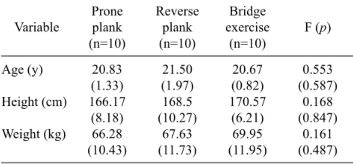
Table 1. General characteristics of the subjects (N=30) Variable
Prone plank (n=10)
Reverse plank (n=10)
Bridge exercise
(n=10)
F (p)
Age (y) Height (cm) Weight (kg)
20.83 (1.33) 166.17
(8.18) 66.28 (10.43)
21.50 (1.97) 168.5 (10.27) 67.63 (11.73)
20.67 (0.82) 170.57
(6.21) 69.95 (11.95)
0.553 (0.587)
0.168 (0.847)
0.161 (0.487) Values are expressed as mean (SD).
vic floor and hip girdle at the lower core. Core muscles are classified into large and local muscles. The former are all segmental muscles that maintain balance against external loads on the body, while the latter are important in maintain- ing stability of the anterior, posterior, and lateral body [5].
The large muscles include the external oblique (EO), rectus abdominis (RA), paraspinal muscles, and the local muscles include the interspinales, intertransversarii, internal oblique (IO), multifidus (MF), and transverse abdominis muscle.
Because core muscle activity plays an important role at the center of the trunk in whole-body movements and postural adjustments, positive changes in the core muscles help maintain body posture alignment and maintain a dynamic balance during functional activity [6-8]. The core muscles also play an important role in the performance of all physical activities and sports [9].
Many studies have been conducted to investigate the ef- fectiveness of core exercises. These predominantly include the treatment of patients with low back pain and exercise performance in athletes. However, there is a lack of studies comparing different core exercises and their effect on phys- ical activity and back pain prevention in the general population.
Thus, the purpose of this study was to compare the effects of prone plank, reverse plank, and bridge exercises on mus- cle activity and thickness in healthy young adults and pro- pose effective core exercises.
Methods Subjects
Thirty healthy young adults (14 males, 16 females), from Chunnam Techno University located in Jeonnam, Korea, participated in this study. Subjects were fully informed about the purpose and method of the study before participat- ing and written informed consent was voluntarily provided.
The Institutional Review Board of Sahmyook University approved all protocols and procedures (IRB No.
2-1040781-AB-N-01- 2018004HR). The characteristics of the subjects are shown in Table 1.
Subjects were recruited according to the following ex- clusion criteria: (1) having neurological or orthopedic dis- eases, (2) taking medication for muscle relaxation, (3) those who participated in a similar study within a year, and (4) those with a skin problem that would be affected by the at- tachment of the surface electrode and the application of ul- trasound gel.
Exercise procedures
Sex, age, height, weight, and other general information were recorded for all subjects. The selected subjects were divided into 3 groups by random sampling to minimize se- lection bias and were randomly assigned to one of 3 groups:
prone plank, reverse plank, and bridge exercise. The ran- domization process was performed using computer-gene- rated numbers produced by a basic random number gen- erator (Random Allocation Software ver. 1.0; Isfahan University of Medical Sciences, Isfahan, Iran) [10].
Before the intervention, all subjects were assessed for maximum voluntary isometric contraction (MVIC) and muscle thickness of the RA, MF, EO, and IO. The inter- vention was performed in each group for 4 weeks and out- come measures were re-measured at 2 weeks and 4 weeks as in the pre-test. Each group performed 5 sets of 20 seconds in the first week, 5 sets of 30 seconds in the second week, 5 sets of 40 seconds the third week, and 5 sets of 40 seconds in the fourth week. Exercises were performed 5 times a week. A 30-second break was given between each set to reduce mus- cle fatigue.
Exercise procedures
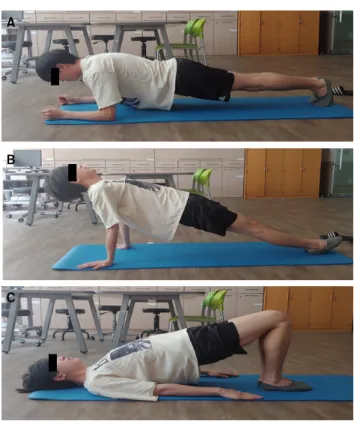
For the prone plank exercise, study participants were asked to maintain their elbows flexed at 90° in the push-up position, with the forearms and toes on the floor. The width between both arms was as wide as the shoulders, and the width between the feet was as wide as the pelvis. The sub- jects’ hip, pelvis, and lumbar spine were aligned (Figure 1A).
In the reverse plank exercise, subjects lifted their pelvis off the floor in a supine position with elbows extended, and the knees and trunk aligned. Both arms were separated at shoulder width (Figure 1B).
Subjects in the bridge exercise group were asked to bend
A
B
C
Figure 1. The position according to the exercise methods. (A) Prone plank exercise method. (B) Reverse plank exercise method. (C) Bridge exercise method.
their knees to 90° with the heel of their feet on the floor. Both arms were at shoulder-width apart, and their palms were faced down. The subject lifted their hips up to keep their pel- vis in line with the lumbar spine at 0° according to the verbal instructions of the evaluator (Figure 1C).
Experimental equipment
Muscle activity measure and data processing
We used a surface electromyography (EMG) device (Trigno Wireless EMG; Delsys Inc., Natick, MA, USA) to measure muscle activation of the RA, MF, EO, and IO ac- cording to the different intervention methods. The skin was wiped with rubbing alcohol and dried off to minimize skin resistance. Two surface electrodes were use applied with one electrode placed on the muscle belly of the RA, MF, EO, and IO muscles and the other placed within a distance of 2 cm. The MVIC of each muscle was measured after adequate practice and rest period. The electrode placement and MVIC measurements used were similar to those described by Cram
et al. [11]. To normalize the EMG value of each muscle, theMVIC was measured for 7 seconds in a manual muscle test- ing position. A 5-minute break was given between each of the 3 repetitions.
Measured electromyographic signals were processed and analyzed using the Delsys EMGworks 4.1.1 (Delsys Inc.).
The EMG signals were sampled at a frequency of 1,000 Hz, and a band-pass filter of between 20 to 450 Hz. After under- going the full-wave rectification process, it was processed with the root mean square and was analyzed. Excluding the first and last 2 seconds of the 7-second measurement period, the mean values from the middle 5 seconds during the max- imal contraction data of each muscle were represented as the
%MVIC values for normalization.
Muscle thickness measurement
An ultrasound imaging device (MyLab One; Esaote, Genova, Italy) was used to measure changes in muscle thickness. The subjects were prevented from receiving visu- al biofeedback during the measurement process. In the pre- and post-test, the measurement area was marked with an ex- perimental pen to measure at the same location. The same evaluator performed the measurements, and the maximum value was used after 3 repeated measurements.
Measurements for the RA were performed using the probe transversally while the subject was in a supine position. It was measured inwardly and upward 2 cm at the umbilicus [12]. For the MF measurement, the subject in was prone po- sition, and a pillow supported his or her hip and ankle joints.
After palpating the transverse process of the lumbar spine 4 to 5 levels, the probe was positioned lengthwise by vertically placing it on the waist spine center line. The shape of the spi- nous process was checked on the monitor screen, and the probe was measured with inclination until the facet joint was clearly visible [13]. The EO and IO were measured in the area of the right lower quadrant of the subject, aligned with the anterior superior iliac spine, which was inward and downward 2 cm [12].
Statistical analysis
All statistical analyses were performed using IBM SPSS
Statistics ver. 19.0 (IBM Co., Armonk, NY, USA). The gen-
eral characteristics of the subjects were analyzed using de-
scriptive statistics, and results were presented as the
mean±standard deviation. The Shapiro-Wilks testing was
performed to test for normality, and the normal distribution
was satisfied. The one-way placement variance analysis was
performed for the group-to-group homogeneity test and
group comparison. The repeated measures analysis of var-
iance was performed to compare the difference between
groups, the changes between the different time points, and
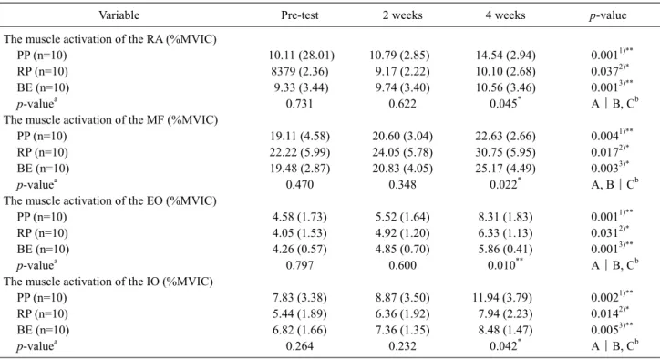
Table 2. Changes in muscle activation according to the exercise methods (N=30)
Variable Pre-test 2 weeks 4 weeks p-value
The muscle activation of the RA (%MVIC)
PP (n=10) 10.11 (28.01) 10.79 (2.85) 14.54 (2.94) 0.0011)**
RP (n=10) 8379 (2.36) 9.17 (2.22) 10.10 (2.68) 0.0372)*
BE (n=10) 9.33 (3.44) 9.74 (3.40) 10.56 (3.46) 0.0013)**
p-valuea 0.731 0.622 0.045* A|B, Cb
The muscle activation of the MF (%MVIC)
PP (n=10) 19.11 (4.58) 20.60 (3.04) 22.63 (2.66) 0.0041)**
RP (n=10) 22.22 (5.99) 24.05 (5.78) 30.75 (5.95) 0.0172)*
BE (n=10) 19.48 (2.87) 20.83 (4.05) 25.17 (4.49) 0.0033)*
p-valuea 0.470 0.348 0.022* A, B|Cb
The muscle activation of the EO (%MVIC)
PP (n=10) 4.58 (1.73) 5.52 (1.64) 8.31 (1.83) 0.0011)**
RP (n=10) 4.05 (1.53) 4.92 (1.20) 6.33 (1.13) 0.0312)*
BE (n=10) 4.26 (0.57) 4.85 (0.70) 5.86 (0.41) 0.0013)**
p-valuea 0.797 0.600 0.010** A|B, Cb
The muscle activation of the IO (%MVIC)
PP (n=10) 7.83 (3.38) 8.87 (3.50) 11.94 (3.79) 0.0021)**
RP (n=10) 5.44 (1.89) 6.36 (1.92) 7.94 (2.23) 0.0142)*
BE (n=10) 6.82 (1.66) 7.36 (1.35) 8.48 (1.47) 0.0053)**
p-valuea 0.264 0.232 0.042* A|B, Cb
Values are expressed as mean (SD).
Repeated measure analysis of variance: 1)within-subjects factors, 2)between-subjects factors, 3)interaction.
RA: rectus abdominis, MVIC: maximum voluntary isometric contraction, PP: prone plank exercise group, RP: reverse plank exercise group, BE: bridge exercise group, MF: multifidus, EO: external oblique, IO: internal oblique.
aOne-way analysis of variance, bpost hoc.
*p<0.05, **p<0.01.
the interaction between the time and groups. A one-way anal- ysis of variance was performed to compare the groups at each time point. The post-test was conducted using the Duncan test. The statistical significance level was set to α= 0.05.
Results
Homogeneity between groups was examined among the 30 subjects (16 males, 14 females). The prone plank, reverse plank and bridge exercise groups were identified as the same group with no difference. The general characteristics of the subjects are listed in (Table 1).
The differences in muscle activity based on the exercise method is listed in (Table 2). Muscle activity (e.g., RA, MF, EO, and IO) was significantly increased in the changes on effect over time (p<0.05). Muscle activity (RA, MF, EO, and IO) was significantly different between all groups and the interaction between time and groups (p<0.05). Since we ob- served a difference between the groups, a one-way analysis of variance was period for each time. No statistically sig- nificant difference was seen between the results form the
pre-test and after 2 weeks, but a significant difference was seen after 4 weeks (p<0.05). The post-hoc analysis showed that the RA, EO, and IO were significantly different in the prone plank exercise group compared with those in the other core exercise groups (p<0.05). Alternatively, the MF was significantly different in the reverse plank exercise and prone plank groups compared with that in bridge exercise group (p<0.05).
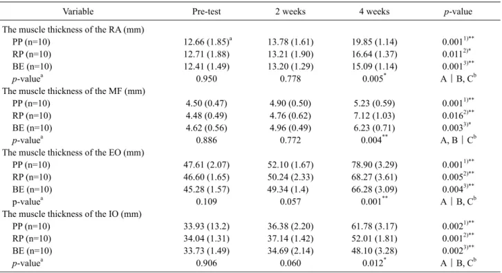
Differences in muscle activity based on exercise groups are listed in (Table 3). Muscle thickness of the RA, MF, EO, and IO was significantly increased in the changes on effect over time points within all groups (p<0.05). Muscle thick- ness of the RA, MF, EO, and IO was significantly different between all groups and interaction between time and groups (p<0.05). We further analyzed the difference between groups using a one-way analysis of variance for each time.
There was no statistically significant difference in the
pre-test and after 2 weeks results, but there was a significant
difference after 4 weeks (p<0.05). The post-hoc analysis
showed that RA, EO, and IO was significantly different in
the prone plank exercise group compared with those in the
Table 3. Changes in muscle thickness according to the exercise methods (N=30)
Variable Pre-test 2 weeks 4 weeks p-value
The muscle thickness of the RA (mm)
PP (n=10) 12.66 (1.85)a 13.78 (1.61) 19.85 (1.14) 0.0011)**
RP (n=10) 12.71 (1.88) 13.21 (1.90) 16.64 (1.37) 0.0112)*
BE (n=10) 12.41 (1.49) 13.20 (1.29) 15.09 (1.14) 0.0013)**
p-valuea 0.950 0.778 0.005* A|B, Cb
The muscle thickness of the MF (mm)
PP (n=10) 4.50 (0.47) 4.90 (0.50) 5.23 (0.59) 0.0011)**
RP (n=10) 4.48 (0.49) 4.76 (0.62) 7.12 (1.03) 0.0162)**
BE (n=10) 4.62 (0.56) 4.96 (0.49) 6.23 (0.71) 0.0033)*
p-valuea 0.886 0.772 0.004** A, B|Cb
The muscle thickness of the EO (mm)
PP (n=10) 47.61 (2.07) 52.10 (1.67) 78.90 (3.29) 0.0011)**
RP (n=10) 46.60 (1.65) 50.24 (2.33) 68.27 (3.61) 0.0052)**
BE (n=10) 45.28 (1.57) 49.34 (1.4) 66.28 (3.09) 0.0043)**
p-valuea 0.109 0.057 0.001** A|B, Cb
The muscle thickness of the IO (mm)
PP (n=10) 33.93 (13.2) 36.38 (2.20) 61.78 (3.17) 0.0021)**
RP (n=10) 34.04 (1.31) 37.14 (1.42) 52.01 (1.81) 0.0012)**
BE (n=10) 33.73 (1.49) 34.69 (2.14) 48.10 (3.28) 0.0023)**
p-valuea 0.906 0.060 0.012* A|B, Cb
Values are expressed as mean (SD).
Repeated measure analysis of variance: 1)within-subjects factors, 2)between-subjects factors, 3)interaction.
RA: rectus abdominis, PP: prone plank exercise group, RP: reverse plank exercise group, BE: bridge exercise group, MF: multifidus, EO: external oblique, IO: internal oblique.
aOne-way analysis of variance, bpost hoc.
*p<0.05, **p<0.01.
other core exercise groups (p<0.05). The MF was sig- nificantly different in the reverse plank and prone plank groups compared with that in the bridge exercise group (p<0.05).
Discussion
We compared the effect of prone plank, reverse plank, and bridge exercises on muscle activity and thickness of the RA, MF, EO, and IO.
The main purpose of performing core exercises is to pro- tect the spine from pain caused by repeated micro-damage, prevent instability and degenerative changes of the spine, as well as stabilize the segments of the spine by stabilizing the trunk [14]. Muscle strengthening and promoting stability through core exercises is used for the treatment and pre- vention of patients with musculoskeletal disorders, and to improve the ability to exercise [6].
The prone plank exercise selectively mobilizes the spinal flexors while at the same time activating the anterior stabil-
izers that are easily affected in the direction of gravity.
Reverse plank and bridge exercises selectively mobilize back muscles that are easily affected by the direction of gravity due to the supine posture and the posterior stabilizers priority activate. Depending on the posture of each exercise, the cross-sectional area and activity of the muscles used against gravity are increased. The RA, EO, and IO were more affected by the exercises than MF, which is an anti- gravity muscle. Previous studies have reported that plank exercises increased the activity of stable core muscles by movement of the limb joints according to the degree of diffi- culty of the motions [15-17]. Barker et al. [18] reported that bridge exercises significantly increased muscle activity of the erector spinae, including the MF. Czaprowski et al. [19]
found that bridge exercises showed lower muscle activity
compared to the prone plank exercise. Lehman et al. [20] re-
ported that bridge exercises showed higher muscle activa-
tion of the MF than the prone plank exercises, while the RA,
EO, and IO had higher muscle activation during the prone
plank exercises than bridge exercises.
The significant difference in core exercises observed be- tween groups during the fourth week was due to active, not passive, movement, and repetitive training is reprogrammed at the muscle. Saal and Saal [21] reported that repeated ex- ercises provide sensory feedback and stimulate the in- tegration of the spine to maintain normal function. In addi- tion, simultaneous contraction in the core muscle is stored in the motor center and occurs automatically in daily activities and habitual postures without conscious control. A mini- mum of 4 weeks is necessary for this effect to appear.
Although there was no significant difference between the bridge and reverse plank exercises, the reverse plank ex- ercises increased muscle activity and thickness after 4 weeks compared to the bridge exercises. It is important that the base is widened or that the center of gravity is located at the base to improve posture stability, but the center of gravity is also very important. This is because a lower center of gravity increases stability, while a higher center of gravity reduces it. Reverse plank exercises, which support the arm, increase instability by providing a higher center of gravity than bridge exercises. Therefore, core muscles are used more to maintain body stability.
We acknowledge that our study population is a limitation of the study. We selected males and females in their early twenties, therefore, it is difficult to generalize and apply our results to patients with low back pain and elderly individuals. Follow-up studies should include patients and subjects of various ages. Long-time and ongoing studies are warranted to evaluate core exercises and carryover effects.
Altogether, our results suggest that prone plank exercises are effective in strengthening the RA, EO and IO muscles, while reverse plank and bridge exercises are effective in strengthening the erector spinae.
Conflict of Interest
The authors declared no potential conflicts of interest with respect to the authorship and/or publication of this article.
References
1. Bendix T, Bendix AF, Busch E, Jordan A. Functional restoration in chronic low back pain. Scand J Med Sci Sports 1996;6:88-97.
2. Hodges PW. Core stability exercise in chronic low back pain.
Orthop Clin North Am 2003;34:245-54.
3. Brill PW. The core program: fifteen minutes a day that can change your life. New York, NY: Bantam Books; 2001.
4. Hibbs AE, Thompson KG, French D, Wrigley A, Spears I.
Optimizing performance by improving core stability and core strength. Sports Med 2008;38:995-1008.
5. Akuthota V, Nadler SF. Core strengthening. Arch Phys Med Rehabil 2004;85(3 Suppl 1):S86-92.
6. O’Sullivan PB, Phyty GD, Twomey LT, Allison GT. Evaluation of specific stabilizing exercise in the treatment of chronic low back pain with radiologic diagnosis of spondylolysis or spondylolisthesis. Spine (Phila Pa 1976) 1997;22:2959-67.
7. Imai A, Kaneoka K, Okubo Y, Shiina I, Tatsumura M, Izumi S, et al. Trunk muscle activity during lumbar stabilization exercises on both a stable and unstable surface. J Orthop Sports Phys Ther 2010;40:369-75.
8. Panjabi MM. Clinical spinal instability and low back pain. J Electromyogr Kinesiol 2003;13:371-9.
9. Nadler SF, Malanga GA, Bartoli LA, Feinberg JH, Prybicien M, Deprince M. Hip muscle imbalance and low back pain in ath- letes: influence of core strengthening. Med Sci Sports Exerc 2002;34:9-16.
10. Saghaei M. Random allocation software for parallel group randomized trials. BMC Med Res Methodol 2004;4:26.
11. Cram JR, Kasman GS, Holtz J. Introduction to surface electromyography. New York, NY: Aspen Publishers; 1998. p.
360-71.
12. Hodges PW. The role of the motor system in spinal pain: im- plications for rehabilitation of the athlete following lower back pain. J Sci Med Sport 2000;3:243-53.
13. Stokes M, Rankin G, Newham DJ. Ultrasound imaging of lum- bar multifidus muscle: normal reference ranges for measure- ments and practical guidance on the technique. Man Ther 2005;10:116-26.
14. Stevens VK, Bouche KG, Mahieu NN, Coorevits PL, Vanderstraeten GG, Danneels LA. Trunk muscle activity in healthy subjects during bridging stabilization exercises. BMC Musculoskelet Disord 2006;7:75.
15. O’Sullivan PB, Twomey L, Allison GT. Dynamic stabilization of the lumbar spine. Boca Raton, FL: CRC Press; 1997. p. 315-30.
16. Marshall PW, Murphy BA. Core stability exercises on and off a Swiss ball. Arch Phys Med Rehabil 2005;86:242-9.
17. Stevens VK, Coorevits PL, Bouche KG, Mahieu NN, Vanderstraeten GG, Danneels LA. The influence of specific training on trunk muscle recruitment patterns in healthy subjects during stabilization exercises. Man Ther 2007;12:271-9.
18. Barker KL, Shamley DR, Jackson D. Changes in the cross-sec- tional area of multifidus and psoas in patients with unilateral back pain: the relationship to pain and disability. Spine (Phila Pa 1976) 2004;29:E515-9.
19. Czaprowski D, Afeltowicz A, Gębicka A, Pawłowska P, Kędra A, Barrios C, et al. Abdominal muscle EMG-activity during bridge exercises on stable and unstable surfaces. Phys Ther Sport 2014;15:162-8.
20. Lehman GJ, Hoda W, Oliver S. Trunk muscle activity during bridging exercises on and off a Swiss ball. Chiropr Osteopat 2005;13:14.
21. Saal JA, Saal JS. Nonoperative treatment of herniated lumbar in- tervertebral disc with radiculopathy: an outcome study. Spine 1989;14:431-7.