Comparison of Supraspinatus Muscle Architecture During Three Different Shoulder Strengthening Exercises Using Ultrasonography
Il-young Moon1,2, BHSc, PT, One-bin Lim3, PhD, PT, Heon-seock Cynn4,5, PhD, PT, Chung-hwi Yi4,5, PhD, PT
1Dept. of Rehabilitation Medicine, Wonju Severance Christian Hospital
2Dept. of Physical Therapy, The Graduate School, Yonsei University
3Dept. of Physical & Occupational Therapy, Rehabilitation Hospital, National Rehabilitation Center
4Dept. of Physical Therapy, College of Health Science, Yonsei University
5Dept. of Ergonomic Therapy, The Graduate School of Health Science, Yonsei University
Abstract
1)Background: Strengthening the supraspinatus is an important aspect of a rehabilitation program for subacromial impingement and tendinopathy. Many authors recommended empty-can (EC), full-can (FC), and prone full-can (PFC) exercises to strengthen the supraspinatus. However, no ultrasonography study has yet investigated supraspinatus muscle architecture (muscle thickness; MT, pennation angle; PA, fiber bundle length; FBL) in relation to supraspinatus strengthening exercises.
Objects: The purpose of this study was to compare the architecture (MT, PA, and FBL) of the supraspinatus muscle during three different types of exercises (EC, FC, and PFC) using diagnostic ultrasound.
Methods: Participants performed three different exercises: (A) EC; the arm was maintained at 60°
abduction with full internal rotation in the sitting position, (B) FC; the arm was maintained at 60°
abduction with full external rotation in the sitting position, and (C) PFC; the arm was maintained at 60°
abduction with full external rotation in the prone position. Ultrasonography was used to measure the MT, PA and FBL of the supraspinatus. One-way repeated analysis of variance with Bonferroni’s post-hoc test was used to compare between the three exercises and the initial position of each exercise.
Results: Compared with each initial position, the FC exercise showed the greatest mean difference in muscle architecture properties and the PFC exercise showed the least mean difference.
Conclusion: The findings suggest that the FC exercise position may have an advantage in increasing the amount of contractile tissue or producing muscle power and the PFC exercise position may be useful in a rehabilitation program because it offers the advantage of maintaining the muscle architecture properties.
Key Words: Rehabilitation; Rotator cuff; Shoulder; Sonography.
Introduction
Rotator cuff muscles play an important role in the stabilization of the glenohumeral joint and the trans- lation of the humeral head during shoulder strength- ening exercises (Escamilla et al, 2009). Glenohumeral joint instability and rotator cuff tears lead to sub-
acromial impingement syndrome contributing to the most common cause of shoulder pain (Burke et al, 2002). Several authors have found that weakness and imbalance in the rotator cuff muscles resulted in me- chanical compression of the subacromial structures between the coracoacromial arch and the greater tu- berosity of the humeral head (Burke et al, 2002;
Corresponding author: Chung-hwi Yi [email protected]
This research was supported by Basic Science Research Program through the National Research Foundation of Korea (NRF) funded by the Ministry of Education (2015R1D1A1A01057620).
Takeda et al, 2002). Among the rotator cuff muscles, the supraspinatus is an effective shoulder abductor muscle during arm elevation, and it is involved in subacromial impingement (Burke et al, 2002; Gates et al, 2010). Strengthening the supraspinatus is an important aspect of a rehabilitation program for subacromial impingement and tendinopathy (Malanga et al, 1996).
Many authors recommended empty-can (EC), full-can (FC), and prone full-can (PFC) exercises to strength- en the supraspinatus (Malanga et al, 1996; Takeda et al, 2002). The supraspinatus showed a similar amount of activity during these three types of shoulder strengthening exercises, as measured by electromyography (EMG) (Reinold et al, 2007). While all three exercises produced similar amounts of su- praspinatus activity, Reinold et al (2007) reported that the FC exercise produced significantly less ac- tivity of the deltoid muscles and may be the optimal position to recruit the supraspinatus muscle for re- habilitation and testing. However, several studies have found inconsistent EMG activity results among EC, FC, and PFC exercises. Townsend et al (1991) reported that the EC exercise shows high EMG ac- tivity in comparison to the FC and PFC exercises, but Blackburn et al (1990) found that the PFC ex- ercise showed high EMG activity and was beneficial for strengthening the supraspinatus. In addition, Kelly et al (1996) reported similar EMG activity be- tween the EC and FC exercises, but not the PFC exercise. These inconclusive EMG activity results can be attributed to several factors, such as the subjects’
characteristics, the testing procedure used, and the EMG analysis. Therefore, other contributing factors should be considered to determine which of these three types of exercises are most appropriate for su- praspinatus rehabilitation.
Muscle architecture properties, such as muscle thickness (MT), fiber bundle length (FBL), and pen- nation angle (PA), can be used to determinate muscle function and force production, which form the basis for physiological movement (Fukutani and Kurihara, 2015; Lieber and Friden, 2001). Morse et al (2008)
suggested that physiological muscle architecture, es- pecially PA, is a better index for determining muscle force than anatomical muscle architecture. Thus, in the fields of physical therapy, structural analysis of the physiological muscle architecture is an important fac- tor for enhancing human performance. The use of ultrasonography to measure changes in the architec- tural parameters of human skeletal muscles is well established (de Boer et al, 2008; Ikai and Fukunaga, 1968). However, no ultrasonography study has yet in- vestigated supraspinatus muscle architecture (MT, PA, and FBL) in relation to supraspinatus strengthen- ing exercises.
This present study aimed to compare the archi- tecture (MT, PA, and FBL) of the supraspinatus mus- cle during three different types of exercises (EC, FC, and PFC) using diagnostic ultrasound. We hypothe- sized that EC, FC, and PFC would influence the mean difference of MT, PA, and FBL.
Methods
Subjects
Based on pilot data, a priori power analysis with G-Power software ver. 3.1.5 (Franz Faul, University of Kiel, Kiel, Germany) was performed to calculate the sample size. A pilot study with five subjects was used to achieve a effect size of 1.72, an alpha level of 5%, and a statistical power of 80%. In the present study, the estimated sample size was 10. Sixteen healthy male subjects participated in the study.
Inclusion criteria for this study was active, right-hand- ed participants so as to optimize the data processing time (by asking participants their preferred hand for writing) (de Castro et al, 2014). Exclusion criteria were the presence of any pain, any previous or cur- rent rotator cuff pathology (test for subacromial im- pingement), glenohumeral joint instability (tests for anterior and posterior apprehension to determine in- stability) (Kim et al, 2015), and engaging in regular physical activity more than three times a week (to
Characteristics Mean±SDa
Age (year) 24.5±1.7
Height (㎝) 175.3±3.7
Weight (㎏) 74.4±11.4
BMIb (㎏/㎡) 24.1±3.3
astandard deviation, bbody mass index.
Table 1. General characteristics of the study subjects (N=16) avoid a possible influence of physical training on the participants’ performance) (de Castro et al, 2014). The study protocol and informed consent document were approved by the Yonsei University Wonju Institutional Review Board (Approval number:
#1041849-201502-BM-014-02). The characteristics of the subjects are shown in Table 1.
Ultrasonography
An ultrasound scanner with 7.5 ㎒ linear trans- ducer (SonoAce x8, Medison Co, Ltd, Seoul, Korea) was used to measure the MT, PA, and FBL of the supraspinatus muscle. Using the protocols recom- mended by Kim et al (2010), supraspinatus archi- tecture images were taken. Kim et al, (2010) reported a strong correlation for the intra- and inter-rater re- liability of FBL measurements (above .85, respectively) and PA measurements (above .80, respectively), with no significant difference between the two (p<.001).
As the present study used a different type of ultra- sound machine than was used by Kim et al (2010), intra-reliability was calculated to compare this study’s findings with the results from the previous study.
We used the intra-class correlation coefficient [ICC (3,1)] to calculate the intra-rater reliability of MT, PA, and FBL in three different contracted positions (EC, FC, and PFC exercises), and we found excellent intra-rater reliability (ICC=.98, .99, .85, respectively).
To ensure the consistency of the measurements, one physical therapist (O.B.L.), who was trained to ad- minister the supraspinatus ultrasound imaging proto- col, took all of the images. The mid-point muscle belly and the anterior part of the supraspinatus were im- aged in two different positions: relaxed 0° abduction
(the arm resting at the side and the palm facing in- ward or outward) and isometric contracted 60° ab- duction (Kim et al, 2015). An inclinometer was used to ensure an accurate 60° abduction position. The mid-point of the muscle belly (sagittal scans) was ob- served to capture MT (Katayose and Magee, 2001).
The anterior part of the supraspinatus (longitudinal panoramic scan) was used to capture PA and FBL (Kim et al, 2015). All of the images were saved for the analyses.
Procedures
All of the volunteer subjects were randomly as- signed an order for the three different exercises (EC, FC, and PFC) (Forbush et al, 2013). The exercise order was generated using Randomization.com (accessed at http://www.randomization.com on April 4, 2015).
One researcher (I.Y.M.), who was trained in the pro- tocols for all three types of exercises, instructed all of the subjects in how to perform the exercises. The subjects were asked to do each of the three exercise positions in the order randomized for their session.
Each position was resisted using a dumbbell that weighed approximately 5% of the each individual’s body mass index (BMI) (ranging from 1 ㎏ to 1.5
㎏) (de Castro et al, 2014). When applying resistance using a dumbbell, a proper hold time (5 sec) was used in order to capture three ultrasound images.
Each subject was securely strapped to a chair and/or a table to prevent any compensatory movement. To maximize the data processing time, only the right arm was tested (de Castro et al, 2014). One minute of rest was provided between the exercise sets to prevent the carry-over effect. To investigate the mean difference of muscle architecture (MT, PA, and FBL), the average of three images in resting and iso- metric contracted positions was measured. Resting average is the average of three images shot in the sitting or prone positions before applying the three different exercises (EC, FC, and PFC). The isometric contracted average is the average of three images shot during the three different exercises (EC, FC,
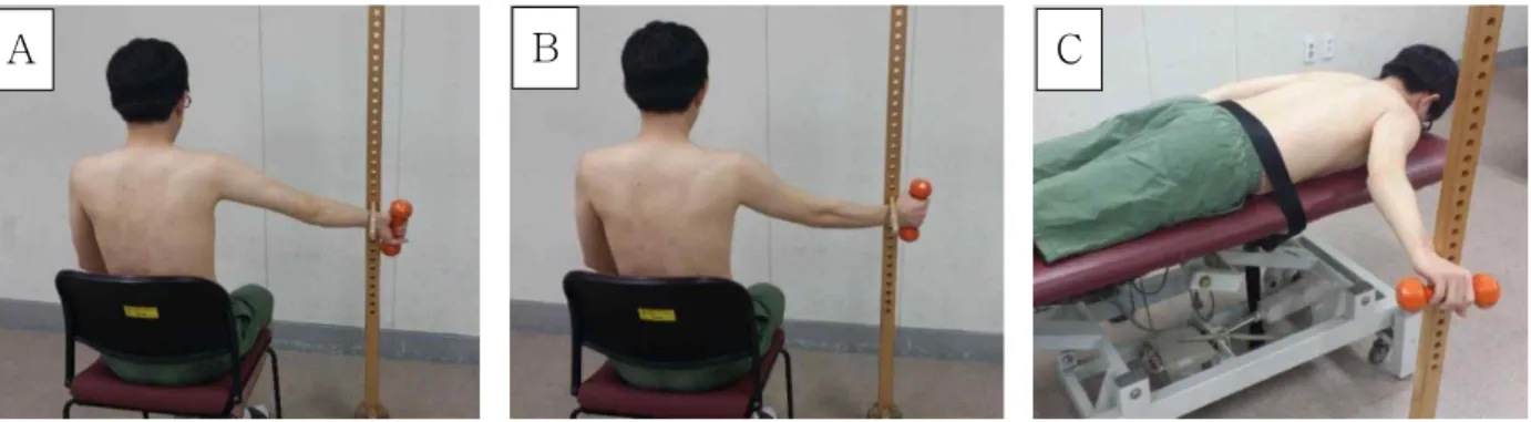
A B C
Figure 1. Three exercise positions (A: empty-can, B: full-can, C: prone full-can).
and PFC). The mean difference of muscle archi- tecture (MT, PA, and FBL) indicates the amount of mean change between the isometric contraction and resting positions, respectively. The three exercise po- sitions are described below.
Empty-can exercise position
The EC exercise position was performed with the subjects sitting in a chair. Each subject’s trunk was securely strapped to the chair to prevent compensa- tory movement. A target bar was located at the lev- el of the 60° abduction position, which was de- termined to be an accurate angle during the con- tracted position. Before elevating their arm, the sub- jects were instructed to rest their arm at their side and position their thumb inward to capture the rest- ing position images. They were then instructed to maintain their arm at 60° abduction in the scapular plane (30° anterior to the frontal plane), with full in- ternal rotation. The thumb was pointing toward the floor (Figure 1A). The subjects maintained this posi- tion as much as possible to capture the images against the downward resistance applied by the dumbbell (5% of BMI) (Jobe and Moynes, 1982).
Full-can exercise position
The FC exercise position was performed with the subjects sitting in a chair. Each subject’s trunk was securely strapped to the chair to prevent compensa- tory movement. A target bar was located at the lev- el of the 60° abduction position, which was de- termined to be an accurate angle during the con-
tracted position. Before elevating their arm, the sub- jects were instructed to rest their arm at their side and position their thumb outward to capture the resting position images. They were then asked to maintain their arm at 60° abduction in the scapular plane (30° anterior to the frontal plane), with full ex- ternal rotation. The thumb was pointing toward the ceiling (Figure 1B). The subjects maintained this po- sition as much as possible to capture the images against the downward resistance applied by the dumbbell (5% of BMI) (Kelly et al, 1996).
Prone full-can exercise position
The PFC exercise position was performed with the subjects lying in a prone position on a bed. Each subject’s trunk was securely strapped to the bed to prevent compensatory movement. A target bar was located at the level of the 60° abduction position, which was determined to be an accurate angle during the contracted position. Before elevating their arm, the subjects were instructed to rest their arm at their side and position their thumb outward to capture the resting position images. They were then asked to maintain their arm at 60° abduction with full external rotation in the horizontal plane. The thumb was pointing toward up (Figure 1C). The subjects main- tained this position as much as possible to capture the images against the downward resistance applied by the dumbbell (5% of BMI) (Blackburn et al, 1990).
Data management
We palpated the supraspinatus muscle examining
Figure 2. Ultrasound probe positioning (right shoulder in the resting position, Acr: acromion; A: sagittal ultrasound probe positioning for the MT images; B: longitudinal panoramic ultrasound probe positioning for the PA and FBL images).
the acromion and the spine of the scapula. These landmarks helped determine the proper probe posi- tioning (Figure 2). MT was measured as the distance between the superficial surface of the muscle and the supraspinatus fossa at the mid-point of the muscle belly (sagittal scan) (Figure 3A). FBL was measured as the linear distance between the medial and the lateral attachment sites, and PA was meas- ured as the angle between the fiber bundle and its at- tachment to the intramuscular tendon (longitudinal panoramic scan) (Kim et al, 2015) (Figure 3B).
Statistical analysis
The one-sample Kolmogorov-Smirnov test was performed to determine if the continuous data ap- proximated a normal distribution; all of the variables were confirmed as normally distributed. A repeated measure one-way analysis of variance was per- formed for all the measured mean variables (MT, PA, and FBL) to compare the variables among the three different shoulder exercises (EC, FC, and PFC). The statistical significance level was set at α
=.05. The variables that showed significant differ- ences were followed up with pairwise comparisons.
A post-hoc analysis was performed using Bonferroni correction to compare the significant difference (.05/3=.017). Statistical analysis was performed using SPSS ver. 21.0 (SPSS Inc., Chicago, IL, USA).
Results
Muscle thickness
Significant difference were found in the MT among the three different shoulder exercises (Wilks’
Lambda=.363, F2,14=12.295, p=.001) (Table 2). The FC exercise showed significantly greater MT in compar- ison to the EC and PFC exercises (p=.002, p=.001), and the EC exercise showed significantly greater MT in comparison to the PFC exercise (p=.013).
Fiber bundle length
Significant differences in FBL were observed among the three different shoulder exercises (Wilks’
Lambda=.247, F2,14=21.381, p<.001) (Table 2). FBL was significantly shorter for the EC and PFC exercises in comparison to the FC exercise (p<.001, p<.001). However, no significant difference in the FBL was found be- tween the EC and PFC exercises (p=.087).
Pennation angle
Statistically significant differences were found for PA (Wilks’ Lambda=.191, F2,14=29.657, p<.001) (Table 2). The PA was significantly greater for the FC ex- ercise in comparison to the EC and PFC exercises (p<.001, p<.001). Whereas, there were no significant difference in the PA between the EC and PFC ex- ercises (p=.196).
Discussion
This is the first study to quantify MT, PA, and FBL following three different isometric exercises (EC, FC, and PFC) that are used for supraspinatus rehabilitation. Significant differences among MT, PA, and FBL were observed in the FC exercise position;
the PFC exercise position showed the least difference among the three muscle architecture properties. These findings support our hypotheses that these three types of exercises can have an effect on the mean difference of the supraspinatus muscle.
A B
Figure 3. Ultrasound scans of the right supraspinatus in the resting position. (A) sagittal scan, (B) longitudinal scan (MT: muscle thickness, SF: supraspinous fossa, TP: trapezius, AM: middle region of anterior part, PA: pennation angle, FBL: fiber bundle length).
Variables Exercises
Empty-can Full-can Prone full-can
MT (㎝) 3.48±1.48a,* 4.26±1.37 1.99±1.06*,†
FBL (㎝) 2.27±1.11* 3.43±1.47 1.66±1.02*
PA (°) 4.65±2.80* 7.70±3.50 3.70±2.20*
amean±standard deviation, *p<.017, significant difference from the full-can exercise, †p<.017, significant difference from the empty-can exercise.
Table 2. Mean difference between isometric contraction and resting positions among the three different exercises Among the three exercise positions, the FC ex-
ercise was found to have the greatest increase in MT, while the least increase in MT was observed in the PFC exercise, and a statistically significant dif- ference was found for each of the other exercise positions. MT is commonly used to predict muscle hypertrophy, which is defined by muscle activity (Farthing and Chilibeck, 2003). Changes in MT can be used to indicate changes in the electrical activity of a muscle. McMeeken et al (2004) showed a positive linear relationship between MT and electrical activity in the transversus abdominis muscle. O’Hagan et al (1995) reported that the type of isometric resistance training increases MT and muscle strength. Thus, EC, FC, and PFC exercises allowed for the con- traction of the supraspinatus muscle, and MT in- creased in comparison to the initial (resting) position.
In the EC exercise position, the scapula gradually tilted anteriorly (sagittal plane) and internally rotated (transverse plane), which contributes to scapular
protraction and reduces the subacromial space width (Thigpen et al, 2006). Smith et al (2006) reported that this scapular kinematic difference reduced the activity and strength of the supraspinatus muscle. In addition, the internal rotation of the humeral head causes greater tuberosity under acromion impacts, thereby decreasing supraspinatus activity (Escamilla et al, 2009). In our data, the prone position showed the least amount of supraspinatus contraction. The anterior part of the supraspinatus was found to have a weak rotator force at 60° during glenohumeral ele- vation (Otis et al, 1994). This change may be ideal for PFC as a low-impact exercise. In contrast, Worrell et al (1992) found the greatest EMG activity in the PFC exercise position, and Reinold et al (2007) showed a similar amount of muscle activity among the three types of exercises. To address the discrep- ancies of supraspinatus EMG activity, this study chose to examine the glenohumeral elevation angle and the anterior and posterior portions of the
supraspinatus. Many EMG studies have viewed the supraspinatus (anterior and posterior) as a whole, and the images in those studies were taken in the 100° horizontal abduction position.
The FBL showed a significant decrease in the FC exercise position in comparison to the EC and PFC exercise positions; no significant difference in the FBL was observed between the EC and PFC positions. The mean FBL decreased during isometric training with all of the exercise positions. Previous studies demonstrated that the FBL of the vastus lat- eralis decreased in different knee positions after ac- tive isometric contractions (Fukunaga et al, 1997).
The FBL is the most important architectural param- eter; it is affected by a decrease in the muscle con- traction velocity or a decrease in the range of the muscle fiber excursion (Lieber, 1993). A more marked shortening of the FBL indicates an increase in the amount of contractile tissue. A large amount of con- tractile tissue and increased muscle contraction ve- locity were observed in the FC exercise position.
These muscle architecture properties correspond with our previous data (MT), and they are of clinical in- terest to authors supporting the idea that the FC ex- ercise is the best method for strengthening the su- praspinatus muscle (Kelly et al, 1996). Khan et al (1999) suggested that the least or maintenance mus- cle architecture has the benefit of healing mi- cro-trauma. A shortened FBL was found to be more difficult to repair, and it often results in ineffective outcomes for repairing rotator cuff tears (Meyer et al, 2012). Thus, the length of the FBL is an im- portant predictor for surgical outcomes for mi- cro-trauma muscle tears. In our data, the PFC ex- ercise position showed the least amount of contractile tissue. Therefore, for patients that need a low load- ing healing advantage, the PFC exercise positions can be recommended for rehabilitation.
The FC exercise showed a significant increase in the PA in comparison to the EC and PFC exercises;
moreover, no significant difference in PA was ob- served between the EC and PFC exercise positions.
A larger PA provides evidence for effective supra- spinatus muscle activation (Aagaard et al, 2001). The anterior part of the supraspinatus is a circumpennate muscle (Kim et al, 2015), which depends on the fiber length axis (force development) (Lieber, 1993). An increase in the MT and the cross-sectional area cor- relates with a greater FBL and a larger PA. A larg- er PA indicates muscle strengthening, which is re- flected in a greater amount of force development (contractile tissue) (Aagaard et al, 2001). In our PA data, the FC exercise position was found to have a greater amount of force development, which is con- sistent with our previous data (MT and FBL). Thus, the FC exercise position may have an advantage over the EC and PFC exercises in terms of increases in the amount of contractile tissue (force development), thereby producing more muscle power.
This present study has several limitations. First, the current experiment is a cross-sectional design, which makes it difficult to observe the long-term ef- fect of exercise. For example, Seynnes et al (2007) reported that muscle size increase might have oc- curred within three weeks of training. Second, the general characteristics of the subjects were limited to healthy males in their twenties. In future studies, a variety of subjects, such as people with shoulder pain, and subjects using their non-dominant arm, are needed to evaluate differences in muscle architecture properties. Finally, for within-subject designs there is a possibility of muscle fatigue during repetitive movements. Thus, a longer resting time between ex- ercise sessions is needed to prevent a possible car- ry-over effect.
Conclusion
The present study investigated muscle architecture properties (MT, PA, and FBL) following three differ- ent isometric exercises (EC, FC, and PFC). During these three different isometric exercises, the mean difference of MT and PA showed a significant in-
crease, and FBL showed a significant decrease.
Furthermore, the FC exercise showed the greatest in- crease in MT and PA, and the PFC exercise showed the least increase in MT and PA. The greatest dif- ference in the FBL data was observed for the FC exercise position, which is consistent with our pre- vious data (MT and FBL), and FBL was found to decrease the least in the PFC exercise. From this finding, the FC exercise position may have an ad- vantage in increasing the amount of contractile tissue or producing muscle power, and the PFC exercise position may be useful in a rehabilitation program because it offers the advantage of maintaining the muscle architecture properties.
References
Aagaard P, Andersen JL, Dyhre-Poulsen P, et al. A mechanism for increased contractile strength of hu- man pennate muscle in response to strength train- ing: Changes in muscle architecture. J Physiol.
2001;534(Pt. 2):613-623.
Blackburn T, McLeod WD, White B, et al. EMG analysis of posterior rotator cuff exercises. Athl Train. 1990;25(1):41-45.
Burke WS, Vangsness CT, Powers CM. Strengthening the supraspinatus: A clinical and biomechanical review. Clin Orthop Relat Res. 2002;402:292-298.
de Castro MP, Ribeiro DC, Forte Fde C, et al.
Shoulder kinematics is not influenced by external load during elevation in the scapular plane. J Appl Biomech. 2014;30(1):66-74. http://dx.doi.org/
10.1123/jab.2012-0083
de Boer MD, Seynnes OR, di Prampero PE, et al.
Effect of 5 weeks horizontal bed rest on human muscle thickness and architecture of weight bear- ing and non-weight bearing muscles. Eur J Appl Physiol. 2008;104(2):401-407. http://dx.doi.org/10.1007/
s00421-008-0703-0
Escamilla RF, Yamashiro K, Paulos L, et al. Shoulder muscle activity and function in common shoulder
rehabilitation exercises. Sports Med. 2009;39(8):663-685.
http://dx.doi.org/10.2165/00007256-200939080-00004 Farthing JP, Chilibeck PD. The effects of eccentric
and concentric training at different velocities on muscle hypertrophy. Eur J Appl Physiol. 2003;89(6):
578-586.
Forbush SW, White DM, Smith W. The comparison of the empty can and full can techniques and a new diagonal horizontal adduction test for su- praspinatus muscle testing using cross-sectional analysis through ultrasonography. Int J Sports Phys Ther. 2013;8(3):237-247.
Fukunaga T, Ichinose Y, Ito M, et al. Determination of fascicle length and pennation in a contracting human muscle in vivo. J Appl Physiol (1985).
1997;82(1):354-358.
Fukutani A, Kurihara T. Comparison of the muscle fascicle length between resistance-trained and untrained individuals: Cross-sectional observation.
Springerplus. 2015;4:341. http://dx.doi.org/10.1186/
s40064-015-1133-1
Gates JJ, Gilliland J, McGarry MH, et al. Influence of distinct anatomic subregions of the supra- spinatus on humeral rotation. J Orthop Res. 2010;
28(1):12-17. http://dx.doi.org/10.1002/jor.20947 Ikai M, Fukunaga T. Calculation of muscle strength
per unit cross-sectional area of human muscle by means of ultrasonic measurement. Int Z Angew Physiol. 1968;26(1):26-32.
Jobe FW, Moynes DR. Delineation of diagnostic cri- teria and a rehabilitation program for rotator cuff injuries. Am J Sports Med. 1982;10(6):336-339.
Katayose M, Magee DJ. The cross-sectional area of supraspinatus as measured by diagnostic ultrasound.
J Bone Joint Surg Br. 2001;83(4):565-568.
Kelly BT, Kadrmas WR, Speer KP. The manual muscle examination for rotator cuff strength. An electromyographic investigation. Am J Sports Med.
1996;24(5):581-588.
Khan KM, Cook JL, Bonar F, et al. Histopathology of common tendinopathies. Update and implications for clinical management. Sports Med. 1999;27(6):
This article was received April 1, 2016, was re- viewed April 1, 2016, and was accepted May 3, 2016.
393-408.
Kim S, Bleakney R, Boynton E, et al. Investigation of the static and dynamic musculotendinous ar- chitecture of supraspinatus. Clin Anat. 2010;23(1):
48-55. http://dx.doi.org/10.1002/ca.20896
Kim SY, Ko JB, Farthing JP, et al. Investigation of supraspinatus muscle architecture following con- centric and eccentric training. J Sci Med Sport.
2015;18(4):378-382. http://dx.doi.org/10.1016/j.jsams.
2014.05.007
Lieber RL. Skeletal muscle architecture: Implications for muscle function and surgical tendon transfer.
J Hand Ther. 1993;6(2):105-113.
Lieber RL, Fridén J. Clinical significance of skeletal muscle architecture. Clin Orthop Relat Res. 2001;
(383):140-151.
Malanga GA, Jenp YN, Growney ES, et al. EMG analysis of shoulder positioning in testing and strengthening the supraspinatus. Med Sci Sports Exerc. 1996;28(6):661-664.
McMeeken JM, Beith ID, Newham DJ, et al. The re- lationship between EMG and change in thick- ness of transversus abdominis. Clin Biomech (Bristol, Avon). 2004;19(4):337-342.
Meyer DC, Wieser K, Farshad M, et al. Retraction of supraspinatus muscle and tendon as predictors of success of rotator cuff repair. Am J Sports Med.
2012;40(10):2242-2247.
Morse CI, Tolfrey K, Thom JM, et al. Gastrocnemius muscle specific force in boys and men. J Appl Physiol (1985). 2008;104(2):469-474.
O’Hagan FT, Sale DG, MacDougall JD, et al.
Comparative effectiveness of accommodating and weight resistance training modes. Med Sci Sports Exerc. 1995;27(8):1210-1219.
Otis JC, Jiang CC, Wickiewicz TL, et al. Changes in the moment arms of the rotator cuff and deltoid
muscles with abduction and rotation. J Bone Joint Surg Am. 1994;76(5):667-676.
Reinold MM, Macrina LC, Wilk KE, et al.
Electromyographic analysis of the supraspinatus and deltoid muscles during 3 common re- habilitation exercises. J Athl Train. 2007;42(4):
464-469.
Seynnes OR, de Boer M, Narici MV. Early skeletal muscle hypertrophy and architectural changes in response to high-intensity resistance training. J Appl Physiol (1985). 2007;102(1):368-373.
Smith J, Dietrich CT, Kotajarvi BR, et al. The effect of scapular protraction on isometric shoulder ro- tation strength in normal subjects. J Shoulder Elbow Surg. 2006;15(3):339-343.
Takeda Y, Kashiwaguchi S, Endo K, et al. The most effective exercise for strengthening the supra- spinatus muscle evaluation by magnetic resonance imaging. Am J Sports Med. 2002;30(3):374-381.
Thigpen CA, Padua DA, Morgan N, et al. Scapular kinematics during supraspinatus rehabilitation exercise: A comparison of full-can versus emp- ty-can techniques. Am J Sports Med. 2006;34(4):
644-652.
Townsend H, Jobe FW, Pink M, et al. Electromyographic analysis of the glenohumeral muscles during a baseball rehabilitation program. Am J Sports Med. 1991;19(3):264-272.
Worrell TW, Corey BJ, York SL, et al. An analysis of supraspinatus EMG activity and shoulder iso- metric force development. Med Sci Sports Exerc.
1992;24(7):744-748.