Tuberc Respir Dis 2009;67:303-310
CopyrightⒸ2009. The Korean Academy of Tuberculosis and Respiratory Diseases. All rights reserved.
비소세포 폐암에서 Cyclooxygenase-2와 Polo-like Kinase-1의 상관관계
서울대학교 의과대학 호흡기내과학교실 폐연구소
이규화, 양석철Relation between Cyclooxygenase-2 and Polo-like Kinase-1 in Non-Small Cell Lung Cancer
Kyu-Hwa Lee, M.S., Seok-Chul Yang, M.D.
Division of Pulmonary and Critical Care Medicine, Department of Internal Medicine and Lung Institute, Seoul National University College of Medicine, Seoul, Korea
Background: Elevated expression of cyclooxygenase-2 (COX-2) and Polo-like kinase-1 (PLK-1) is observed in a wide variety of cancers. Augmented expression of COX-2 and enhanced production of prostaglandin E2 (PGE2) are associated with increased tumor cell survival and malignancy; COX-2 has been implicated in the control of human non-small cell lung carcinoma (NSCLC) cell growth. PLK-1 siRNA induced the cell death of lung cancer cells and the systemic administration of PLK-1 siRNA/atelocollagen complex inhibited the growth of lung cancer in a liver metastatic murine model. COX-2 and PLK-1 are involved in proliferation and in cell cycle regulation, and there is a significant correlation between their interaction in prostate carcinoma.
Methods: In this study, we investigated the pattern of COX-2 and PLK-1 expression in NSCLC, after treatment with IL-1β, COX-2 inhibitor and PLK-1 siRNA.
Results: Expression of PLK-1 was decreased in A549 COX-2 sense cells, and was increased in A549 COX-2 anti-sense cells. Knock out of PLK-1 expression by PLK-1 siRNA augmented COX-2 expression in A549 and NCl-H157 cells.
When A549 and NCI-H157 cells were treated with COX-2 inhibitor on a dose-dependent basis, PLK-1 and COX-2 were reduced. However, when the expression of COX-2 was induced by IL-1β, the production of PLK-1 decreased.
Conclusion: These results demonstrate that COX-2 and PLK-1 are regulated and inhibited by each other in NSCLC, and suggest that these proteins have a reverse relationship in NSCLC.
Key Words: Cyclooxygenase 2, Polo-like kinase 1, Lung neoplasms
본 연구는 2008년 결핵 및 호흡기학회 학술 연구비로 진행됨.
Address for correspondence: Seok-Chul Yang, M.D.
Division of Pulmonary and Critical Care Medine, Depart- ment of Internal Medicine and Lung Institute, Seoul National University College of Medicine, 28, Yongon-dong, Chong- no-gu, Seoul 110-744, Korea
Phone: 82-2-2072-0354, Fax: 82-2-762-9662 E-mail: [email protected]
Received: Sep. 9, 2009 Accepted: Oct. 7, 2009
서 론
염증반응에 관여하는 prostaglandin의 생합성 과정은 여러 가지 사이토카인이나 염증성 매개물질이 관여하는 복잡한 과정으로, 이 중 Cyclooxygenase (COX)가 중요한
역할을 담당한다1. COX는 두 가지 isoform으로 존재하며, COX-1은 constitutive form으로 대부분의 정상 조직에서 발현되어 세포나 조직의 항상성을 유지하는 기능을 한다2. 반면, COX-2는 특정세포에서 염증성 전구사이토카인3이 나 lipopolysaccharide2,4, 성장인자 등의 자극에 의해 유도 되는 inducible form으로, 염증이나 종양이 발생된 조직에 서 발현이 증가하며, 비소세포 폐암 조직의 약 30%, 폐선 암의 40%에서 COX-2가 발현됨이 보고되었다5. COX-2는 metalloproteinase의 합성, 미세혈관 형성 등에 관여하여 암세포의 침윤 및 성장에 영향을 미치고, PGE2를 생성하 여 면역세포인 림프구와 대식구에서 IL-10의 합성을 자극 함으로써 종양세포에 대한 인체의 면역반응을 억제한다6. Arachidonic acid에서 COX에 의해 생성된 prosta-
glandin은 그 자체가 직접적으로 세포분열, 혈관 신생 자 극, 침습 및 전이를 유도하며, 세포 자기 사멸사를 억제시 키는 역할을 할 수 있다7,8. 따라서 COX 억제제는 COX arachidonic acid 대사산물들에 의한 종양성을 직접적으 로 억제하는 기능을 가진다. 그러나 최근 일련의 연구들 에 의하면 COX-2 억제제의 항종양효과는 이러한 COX-2 의존적 경로 이외에도 다양한 COX-2 비의존적 경로를 통 해 종양을 억제시킨다고 보고되었다7.
Polo-like kinases (PLK)는 진핵세포에서 serine/threo- nine 잔기를 인산화시키는 단백질로9, PLK family 중에서 PLK-1이 가장 잘 알려져 있다. PLK-1은 정상세포와 악성 종양세포에서 유사분열의 조절에 관여하여10,11 G2/M tra- sition에 중요한 역할을 한다12. PLK-1의 발현에 관한 연구 는 폐암13, 식도암과 위암14, 피부암15, 유방암16, 뇌종양17, 자궁암과 난소암18,19에서 수행되었다. 정상조직과 비교했 을 때 각각의 암 조직에서 PLK-1의 발현이 증가하며, PLK-1 siRNA의 처리가 암의 전이를 예방할 수 있다는 결 과가 보고되었다20.
여러 종류의 암에서 COX-2와 PLK-1의 과발현은 좋지 않은 예후와 직접적인 연관성을 가진다. 최근에는 전립선 암에서 COX-2와 PLK-1이 함께 과발현되어 세포의 증식과 세포주기의 조절에 관여하며, 이 두 단백질이 전립선 암의 예방과 치료에 있어서 중요한 역할을 할 것이라고 보고된 바 있다21.
COX-2는 폐암의 발병과 예후에 깊이 관여하며, PLK-1 또한 폐암의 전이에 관여한다는 연구가 최근 발표되었다.
이에 저자들은 COX-2와 PLK-1이 폐암에서도 서로 상관관 계를 가질 것으로 예상하였고, 여러 종류의 암에서 과 발 현되는 이 두 단백질의 이러한 상관관계는 어떤 알지 못하 는 경로나 인자에 의하여 조절될 것으로 생각하며 실험을 수행하였다.
본 연구에서는 비소세포 폐암세포주인 A549와 NCI- H157 세포주에 Interleukin (IL)-1β 혹은 COX-2 inhibitor celecoxib를 처리하였을 때 COX-2와 PLK-1의 발현양 변 화를 관찰하였으며, A549 COX-2 sense 세포주와 A549 COX-2 anti-sense 세포주에서의 PLK-1의 변화와, PLK-1 siRNA를 처리하였을 때 COX-2의 변화를 관찰하여 두 단 백질의 연관 관계를 조사하였다.
대상 및 방법
1. 세포 배양 및 처리
비소세포 폐암세포주인 A549 세포(human lung ad- enocarcinoma, American Type Culture Collection, Rock- ville, MD, USA), NCI-H157세포(squamous cell carcino- ma, American Type Culture Collection)를 37oC, 5% CO2
조건하에 10% fetal bovine serum (FBS) (WelGENE, Daegu, Korea), penicillin (60 μg/mL) - streptomycin (100 μg/mL) (Gibco-BRL, Grand Island, NY, USA)이 첨 가된 RPMI 1640 (WelGENE) 배지에 배양하였다. COX-2 억제제인 celecoxib 처리를 위하여 A549, NCI-H157를 1×10⁶개/well가 되도록 6-well에 접종하여 24시간 동안 RPMI 1640 배양액에서 배양하였다. Phosphate buffered saline (PBS) (WelGENE)로 세척한 후 celecoxib를 0, 10, 20, 30, 40, 50 μg/mL의 농도로 투여하고 37oC에서 24시 간 동안 배양하였다.
IL-1β (R&D Systems, Minneapolis, MN, USA)를 처리 하기 위하여 A549, NCI-H157를 1×10⁶개/well가 되도록 6-well에 접종하여 24시간 동안 RPMI 1640 배양액에서 배양하였다. PBS로 세척한 후 IL-1β를 0, 0.015, 0.05, 0.1, 1, 1.5 ng/mL의 농도로 투여하고 37oC에서 24시간 동안 배양하였다. 시간에 따른 IL-1β 처리를 위하여 동일 한 방법으로 6-well에 접종하고 A549, NCI-H157 세포에 각각 0.1 ng/mL, 1 ng/mL의 농도로 투여하여 37oC에서 시간의 변화에 따른 COX-2와 PLK-1 단백의 발현 양상을 분석하였다.
2. A549 COX-2 sense 세포주 및 A549 COX-2 anti- sense 세포주 배양
A549에 COX-2를 retroviral transfection하여 COX-2가 항상 발현되도록 제조한 A549 sense (A549 COX-2 S) 세 포주와, COX-2의 발현이 항상 억제되도록 제조된 A549 anti-sense (A59 COX-2 AS) 세포주를 배양하여 사용하였 다. 이들 세포주들의 COX-2 발현 양상에 따른 PLK-1 발현 정도 변화는 western blot으로 분석하였다.
3. PLK-1 small interference RNA transfection
A549, NCI-H157 세포주를 6-well에 접종하여 70%가 찰 때까지 키우고, serum이 없는 RPMI 1640 배지에 50 nmol과 100 nmol PLK-1 siRNA (Santa Cruz, CA, USA), Control siRNA-A (Santa Cruz, CA, USA)를 Lipofectamin
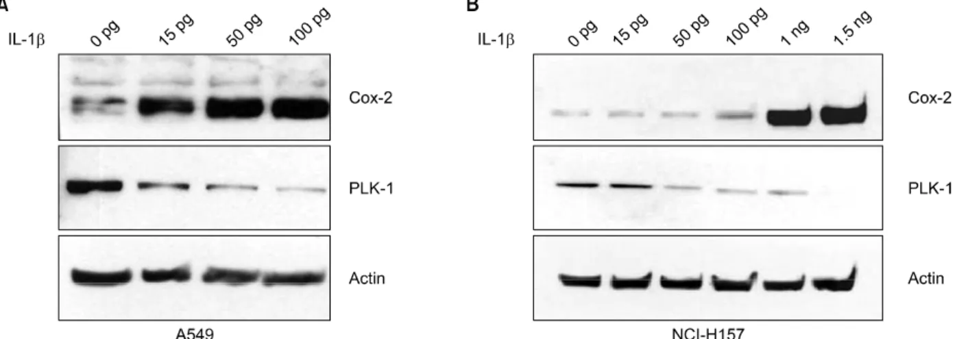
Figure 1. Expression of COX-2 and PLK-1 according to concentration of Interleukin (IL)-1β treatment. A549 (A) and NCI-H157 (B) cells were treated with 0, 0.015, 0.05, 0.1, 1, 1.5 ng/mL for 24 hours. Analysis of COX-2 and PLK-1 expression was done by Western blot. COX-2 protein expression was increased in dose dependent manner, but the expression of PLK-1 was decreased.
2000 (Invitrogen, Carlsbad, CA, USA)과 섞으면서 부드럽 게 세포에 주입하였다. 24시간 후에 serum과 항생제가 포 함된 배지로 교환해주고 48시간 배양한 후에 PLK-1과 COX-2의 발현 양상을 western blot을 통해서 분석하였다.
4. Western blot 분석
COX-2와 PLK-1 단백질의 측정을 위하여 western blot 방법을 사용하였다. 각 세포를 lysis buffer (0.1% NP-40, 5 mM EDTA, 50 mM Tris [pH7.5∼8.0], 250 mM NaCl, 50 mM NaF)로 단백질을 추출·정량하여, 30 μg의 세포 단백질을 10% SDS-polyacrylamide gel에 20 mA에서 2시 간 동안 전기영동하였다. Gel상의 단백질을 nitrocel- lulose membrane으로 이동시키고, 이 membrane을 bloc- king solution (5% skim milk in 1×PBS/Tween 20) (BD Biosciences, Sparks, MD, USA)으로 1시간 동안 block시 킨 후 COX-2 (sc-1745, Santa Cruz, CA, USA)와 PLK-1 (sc-55504, Santa Cruz, CA, USA), Actin (sc-1616, Santa Cruz, CA, USA)에 대한 1차 항체를 4oC에서 16시간 동안 반응시켰다. 세척 후에 2차 항체를 반응시킨 뒤, ECL Western blotting detection system (GenDEPOT, Hous- ton, TX, USA)을 이용하여 면역신호를 검출하였다.
결 과
1. IL-1β에 유도된 COX-2 발현 양상에 따른 PLK-1 단 백질 발현양의 변화
COX-2는 평상시에는 발현되지 않다가 여러 세포 자극 인자에 의해 빠르게 유도되어 발현되므로, IL-1β에 의하 여 COX-2의 발현을 유도하여 PLK-1의 발현양이 변화하는 지 관찰하였다. 먼저 A549 세포주에 IL-1β를 0∼100 pg 의 농도로 24시간 동안 투여하였을 때, IL-1β의 농도가 증 가할수록 COX-2의 발현 양이 증가함에 따라 PLK-1 발현 은 감소하였고(Figure 1A), NCI-H157 세포주에는 IL-1β 를 0∼1.5 ng의 농도로 24시간 동안 투여한 결과 IL-1β 의 농도가 1 ng 이상이 되자 COX-2의 발현 양이 크게 증 가함에 따라 PLK-1 발현은 크게 감소하였다(Figure 1B).
또한 IL-1β 처리 시간에 따른 COX-2와 PLK-1 발현 정 도의 변화를 관찰하였다. A549 세포주에 100 pg의 IL-1β 를 0, 1, 2, 4, 6, 8, 16, 20, 24시간 동안 처리하여 각 처리 시간에 따른 COX-2와 PLK-1의 발현 정도를 관찰하였다.
COX-2는 처리시간이 길어질수록 그 발현양이 점차 증가 하다가 4시간 동안 처리했을 때 최대가 되었으며 이후 조 금씩 감소하였다. 또한 PLK-1은 IL-1β처리 시간이 길어짐 에 따라 발현 양이 점차 감소하다가 4시간에서 8시간 사 이에 최소가 되었으며 16시간이 지남에 따라 점차 증가하 였다(Figure 2A). NCI-H157 세포주에서는 1 ng의 IL-1β 를 0, 1, 2, 4, 6, 8, 16, 20, 24시간 동안 처리하여 각 처리
Figure 2. Expression of COX-2 and PLK-1 according to concentration of Interleukin (IL)-1β treatment. A549 and NCI-H157 cells were treated with 0.1, 1 ng/mL respectively for 0, 1, 2, 4, 8, 16, 20, 24 hours. Analysis of COX-2 and PLK-1 expression was done by Western blot. (A) In A549 cells, the maximum level of COX-2 expression was detected at 4 hour of exposure to IL-1β, and the minimum level of PLK-1 expression was detected from 4 hour to 8 hour of exposure to IL-1β. (B) In NCI-H157 cells, the maximum level of COX-2 expression was detected at 24 hour of exposure to IL-1β, and the minimum level of PLK-1 expression was detected at 16 hour of exposure to IL-1β.
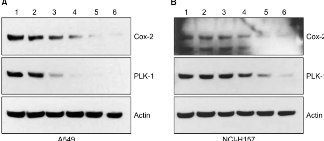
Figure 3. Expression of COX-2 and PLK-1 according to concentration of celecoxib treatment. A549 (A) and NCI-H157 (B) cells were treated with 0, 10, 20, 30, 40, 50 μM (Land 1∼6) for 24 hours. Analysis of COX-2 and PLK-1 expression was done by western blot. Expression of PLK-1, as well as COX-2, was decreased in dose dependent manner.
시간에 따른 COX-2와 PLK-1의 발현 정도를 분석하였다.
COX-2는 IL-1β 처리시간이 길어질수록 그 발현양이 점차 증가하였고, PLK-1은 점차 감소하여 처리시간 16시간에 최소량이 되었다가 점차 증가하였다(Figure 2B). 두 세포 주에서 IL-1β를 24시간 처리했을 때 COX-2의 발현양은 처리하지 않았을 때보다 증가하였고, PLK-1은 감소하였 다. 따라서 자극에 따른 COX-2와 PLK-1의 발현양 변화는 역 상관관계를 가진다는 것을 확인할 수 있었다.
2. Celecoxib 처리에 따른 PLK-1 단백질의 변화
Celecoxib는 항 염증효과를 가지는 대표적인 COX-2 억 제제로, celecoxib를 처리하여 COX-2의 발현양이 감소하
였을 때 PLK-1의 변화를 관찰하고자 하였다. A549 세포주 (Figure 3A), NCI-H157 세포주(Figure 3B)에 celecoxib를 0∼50 μM이 되도록 각각 투여하고, 24시간 동안 배양하 여 COX-2와 PLK-1의 발현 정도 변화를 western blot으로 분석하였다. 그 결과 celecoxib 처리 농도가 높아짐에 따 라, COX-2뿐만 아니라 PLK-1의 발현양도 감소하였다. 이 러한 결과로부터 COX-2와 PLK-1의 상관관계는 celecoxib 에 의하여 COX-2가 억제되는 기전과는 독립적이며, PLK- 1은 celecoxib에 의한 경로와는 다른 경로를 통하여 억제 될 가능성을 예측하였다.
Figure 5. Expression of COX-2 after PLK-1 inhibi- tion in non-small cell lung cancer. Transfection with PLK-1 siRNA and control siRNA (+: 50 nM; ++:
100 nM), to A549 cells (A) and NCI-H157 cells (B) ac- tivated COX-2 expression.
si-cont: control siRNA; si- PLK-1: PLK-1 siRNA.
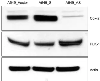
Figure 4. Expression of COX-2 and PLK-1 in COX-2 gene modified human adenocarcinoma cell lines. According to the expression of COX-2 is increase, PLK-1 expression is decreased. A549-vector: A549 COX-2 vector only cell line; A549-S: A549 COX-2 sense cell line; A549-AS:
A549 COX-2 anti-sense cell line.
3. A549 COX-2 sense 세포주와 A549 COX-2 anti- sense 세포주에서 PLK-1의 발현
COX-2가 과발현되도록 제조한 A549 COX-2 S 세포주 와, COX-2의 억제되도록 제조한 A549 COX-2 AS 세포주 에서 COX-2의 발현 정도 변화에 따른 PLK-1의 발현 양상 을 western blot을 통하여 분석하였다. 그 결과 COX-2의 발현이 증가되어 있는 A549 COX-2 S 세포주에서는 PLK-1 의 발현양이 감소하였으며, COX-2의 발현이 억제되어있 는 A549 COX-2 AS 세포주에서는 PLK-1의 발현양이 증가 하였다(Figure 4).
4. PLK-1 발현 감소에 따른 COX-2 발현의 변화
PLK-1의 발현 정도 변화에 의해서도 COX-2의 발현이 달라지는지 확인하기 위하여 siRNA를 이용하여 PLK-1을 억제하여 실험을 진행하였다. A549 세포주(Figure 5A)와 NCI-H157 세포주(Figure 5B)에 각각 PLK-1 siRNA 50 nM, 100 nM을 각각 transfection하여 PLK-1의 발현이 억제되 었을 때 COX-2 발현의 변화를 관찰하였다. 그 결과, PLK- 1의 발현양이 줄어들면 COX-2의 발현이 증가하는 것을 확인할 수 있었다.
고 찰
종양은 복잡한 유전자의 변화에 의해 발생되며, 암을 발생시키는 종양발생유전자와 기능이 없어지면 암을 발 생시키는 종양억제유전자, 세포고사에 관여하여 세포의 수명을 조절하는 유전자들의 변화가 종양 발생에 중요한 기전으로 인식되고 있다.
Arachidonic acid 대사에서 cyclooxygenase 경로의 최 종산물로 생성되는 prostaglandin, 특히 prostaglandin E 계열의 경우 과발현 시 여러 인간 종양에서 세포증식과 신생혈관생성을 촉진시키고, 악성세포에 대한 면역반응 과 세포고사를 억제하며, 종양의 침윤과 전이에 관여한다 고 알려져 있다22,23. 따라서 prostaglandin 생성에 관여하 는 cyclooxygenase (COX)에 대해 많은 연구가 보고되고 있다. 종양에서 COX-2의 발현에 대한 연구는 여러 종류의 암, 특히 대장암에서 많이 이루어졌고, 피부암, 간암, 유방 암 그리고 방광암 등에서도 연구되고 있다. 비소세포 폐 암에서 COX-2의 과발현은 세포사멸에 대한 내성 증가, 혈 관신생의 증가, 암 조직의 침범과 전이의 증가, 그리고 숙
주 면역 기능의 저하를 초래한다고 밝혀졌다. 또한 비소 세포 폐암에서 COX-2에 의하여 생성되는 PGE2는 면역세 포인 림프구와 대식구에서 잘 알려진 면역억제물질 IL-10 의 합성을 자극한다고 밝혀졌다24.
PLK-1의 발현에 관한 연구는 폐암을 포함한 다양한 종 양에서 수행되어왔다. 여러 암 조직에서 PLK-1의 발현이 증가되어 있으며, PLK-1 siRNA의 처리가 암의 전이를 예 방할 수 있다는 결과가 보고되었다20. 최근에는 전립선암 에서 COX-2와 PLK-1의 상호작용이 알려졌으며, 이 두 단 백질은 세포의 증식과 세포주기의 조절에 함께 관여한다 고 보고되었다21. 또한 PLK-1이 폐암의 전이에 관여한다 는 보고가 있었으며, 이에 저자들은 COX-2와 PLK-1이 전 립선암뿐만 아니라 폐암에서도 상관관계가 있을 것이라 고 예상하였다.
COX-2는 평상시에는 발현되지 않다가 여러 세포 자극 인자에 의해 빠르게 유도되어 발현되기 때문에, IL-1β를 처리하여 COX-2의 발현을 유도하였다. IL-1β는 급, 만성 상기도 감염에서 중요한 역할을 하는 염증성 전구 사이토 카인 중 하나이며 천식이나 기관지염, 기관지 확장증과 부비동염 등과 같은 염증성 질환의 병인에 직·간접적으 로 관여한다25,26. A549 세포주에서 PGE2의 생성은 IL-1β 투여 1시간째부터 일어나며 6시간째에 최고치에 이른다 는 이전의 보고가 있었으며27, 사람의 치주조직28과 기도
평활근28-30, 원숭이의 기질내 섬유아세포31 등에서 이루어
진 IL-1β와 COX-2의 관계에 관한 연구결과에서 IL-1β에 의해 COX-2의 발현이 증가된다는 것이 확인되었다. 또한 IL-1β의 용량과 시간에 비례한 COX-2의 발현의 증가는 전사단계에서 조절된다고 보고되었다32. 비소세포 폐암 세포주에서 IL-1β에 의해 유도된 COX-2와 PLK-1의 발현 양상 변화를 분석한 결과, 이 두 단백질이 역상관관계를 가진다는 것을 확인할 수 있었다.
이러한 결과를 재확인하기 위하여, COX-2의 발현이 억 제되었을 때 PLK-1의 발현이 어떻게 달라지는가 확인하기 위하여, 임상적으로 사용되는 선택적 COX-2 억제제인 celecoxib를 투여하고 PLK-1의 발현양 변화를 관찰한 결 과 COX-2와 함께 PLK-1의 발현이 감소되는 것을 확인할 수 있었다. COX 억제제는 COX arachidonic acid 대사산 물들에 의한 종양성을 직접적으로 억제하는 기능을 가지 지만, 최근 일련의 연구들에 의하면 celecoxib가 COX-2 의존적 경로 이외에도 다양한 COX-2 비의존적 경로를 통 해 종양을 억제시킬 수 있음이 보고되었다. 이것은 사용 된 선택적 COX-2 억제제의 항종양효과가 COX-2 활성을
억제시킬 수 있는 농도 이상의 고용량에서 관찰된다는 점, 이러한 효과가 prostaglandin의 투여로 역전되지 않는 점, celecoxib보다 더 강력한 COX-2 억제제인 rofecoxib에서 는 이러한 효과가 나타나지 않는다는 점 그리고 COX-2 억제효과가 없는 celecoxib 유사체인 2,5-dimethyl-cele- coxib (DMC)가 종양억제효과를 가지고 있는 점 등으로 설명된다8,33.
비소세포 폐암 세포주 A549로부터 제조된 A549 COX-2 S 세포주 및 COX-2 AS 세포주에서 PLK-1의 발현을 분석 한 결과, 이 두 단백질의 발현은 역으로 관계되는 것을 알게 되었다. 따라서 COX-2의 발현이 달라짐에 따라 PLK-1의 양이 변할 뿐만 아니라 PLK-1의 발현 양 변화에 따라 COX-2의 생성이 달라지는지 확인하기 위하여, 비소 세포 폐암세포주인 A549와 NCI-H157 세포주에 PLK-1 siRNA를 transfection하여 두 단백질의 발현양상을 비교하 였다. 그 결과 PLK-1이 억제되었을 때 COX-2의 발현이 증가하였는데, 이러한 결과는 PLK-1과 COX-2는 어느 한 쪽에 의해 일방적으로 조절되는 것이 아니라, 서로를 억제 하면서 조절하고 있음을 시사한다.
사실상 COX-2와 PLK-1은 암세포의 생장과 전이를 활 성화시키며, 전립선 암에서 COX-2와 PLK-1이 함께 과 발 현되어 세포의 증식과 세포주기의 조절에 관여한다고 알 려졌다. 이 두 단백질은 비슷한 기능을 하는 것으로 여겨 지며 COX-2와 PLK-1의 역상관 관계에 관한 보고는 아직 없으나, 실제로 이 두 단백질은 서로 억제하며 조절하는 것으로 보인다. 따라서 COX-2와 PLK-1이 어떠한 경로를 통하여 서로의 발현을 조절하는지, 그리고 이 과정에 관계 된 신호전달체계와 세포의 증식과 분화에 미치는 영향에 관한 연구가 이어져야 할 것이다.
참 고 문 헌
1. Chen CC, Sun YT, Chen JJ, Chiu KT. TNF-alpha-in- duced cyclooxygenase-2 expression in human lung epi- thelial cells: involvement of the phospholipase C-gam- ma 2, protein kinase C-alpha, tyrosine kinase, NF-kap- pa B-inducing kinase, and I-kappa B kinase 1/2 path- way. J Immunol 2000;165:2719-28.
2. Mitchell JA, Akarasereenont P, Thiemermann C, Flower RJ, Vane JR. Selectivity of nonsteroidal antiinflam- matory drugs as inhibitors of constitutive and inducible cyclooxygenase. Proc Natl Acad Sci U S A 1993;90:
11693-7.
3. Maier JA, Hla T, Maciag T. Cyclooxygenase is an imme-
diate-early gene induced by interleukin-1 in human en- dothelial cells. J Biol Chem 1990;265:10805-8.
4. Lee SH, Soyoola E, Chanmugam P, Hart S, Sun W, Zhong H, et al. Selective expression of mitogen-in- ducible cyclooxygenase in macrophages stimulated with lipopolysaccharide. J Biol Chem 1992;267:25934-8.
5. Hasturk S, Kemp B, Kalapurakal SK, Kurie JM, Hong WK, Lee JS. Expression of cyclooxygenase-1 and cyclo- oxygenase-2 in bronchial epithelium and nonsmall cell lung carcinoma. Cancer 2002;94:1023-31.
6. Yoon JM, Lim JJ, Yoo CG, Lee CT, Han SK, Shim YS, et al. The role of uteroglobin in the immunomodul- ation of nonsmall cell lung cancer cells. Tuberc Respir Dis 2004;57:336-44.
7. Grosch S, Maier TJ, Schiffmann S, Geisslinger G. Cyclo- oxygenase-2 (COX-2)-independent anticarcinogenic ef- fects of selective COX-2 inhibitors. J Natl Cancer Inst 2006;98:736-47.
8. Cervello M, Montalto G. Cyclooxygenases in hepatocel- lular carcinoma. World J Gastroenterol 2006;12:5113- 21.
9. Clay FJ, McEwen SJ, Bertoncello I, Wilks AF, Dunn AR.
Identification and cloning of a protein kinase-encoding mouse gene, Plk, related to the polo gene of Dros- ophila. Proc Natl Acad Sci U S A 1993;90:4882-6.
10. Sunkel CE, Glover DM. polo, a mitotic mutant of Drosophila displaying abnormal spindle poles. J Cell Sci 1988;89:25-38.
11. Nigg EA. Polo-like kinases: positive regulators of cell division from start to finish. Curr Opin Cell Biol 1998;
10:776-83.
12. Donaldson MM, Tavares AA, Hagan IM, Nigg EA, Glover DM. The mitotic roles of Polo-like kinase. J Cell Sci 2001;114:2357-8.
13. Wolf G, Elez R, Doermer A, Holtrich U, Ackermann H, Stutte HJ, et al. Prognostic significance of polo-like kin- ase (PLK) expression in non-small cell lung cancer.
Oncogene 1997;14:543-9.
14. Tokumitsu Y, Mori M, Tanaka S, Akazawa K, Nakano S, Niho Y. Prognostic significance of polo-like kinase expression in esophageal carcinoma. Int J Oncol 1999;
15:687-92.
15. Kneisel L, Strebhardt K, Bernd A, Wolter M, Binder A, Kaufmann R. Expression of polo-like kinase (PLK1) in thin melanomas: a novel marker of metastatic disease.
J Cutan Pathol 2002;29:354-8.
16. Wolf G, Hildenbrand R, Schwar C, Grobholz R, Kaufm- ann M, Stutte HJ, et al. Polo-like kinase: a novel marker of proliferation: correlation with estrogen-receptor ex-
pression in human breast cancer. Pathol Res Pract 2000;196:753-9.
17. Dietzmann K, Kirches E, von B, Jachau K, Mawrin C.
Increased human polo-like kinase-1 expression in gliomas. J Neurooncol 2001;53:1-11.
18. Takai N, Miyazaki T, Fujisawa K, Nasu K, Hamanaka R, Miyakawa I. Expression of polo-like kinase in ovar- ian cancer is associated with histological grade and clinical stage. Cancer Lett 2001;164:41-9.
19. Takai N, Miyazaki T, Fujisawa K, Nasu K, Hamanaka R, Miyakawa I. Polo-like kinase (PLK) expression in en- dometrial carcinoma. Cancer Lett 2001;169:41-9.
20. Kawata E, Ashihara E, Kimura S, Takenaka K, Sato K, Tanaka R, et al. Administration of PLK-1 small interfer- ing RNA with atelocollagen prevents the growth of liver metastases of lung cancer. Mol Cancer Ther 2008;7:
2904-12.
21. Denkert C, Thoma A, Niesporek S, Weichert W, Koch I, Noske A, et al. Overexpression of cyclooxygenase-2 in human prostate carcinoma and prostatic intra- epithelial neoplasia-association with increased ex- pression of Polo-like kinase-1. Prostate 2007;67:361-9.
22. Fosslien E. Molecular pathology of cyclooxygenase-2 in neoplasia. Ann Clin Lab Sci 2000;30:3-21.
23. Jang JW. Anti-tumor mechanisms and regulation of sur- vivin by selective cyclooxygenase-2 inhibitor. Korean J Hepatol 2008;14:305-8.
24. Peri A, Cordella-Miele E, Miele L, Mukherjee AB.
Tissue-specific expression of the gene coding for hu- man Clara cell 10-kD protein, a phospholipase A2-inhi- bitory protein. J Clin Invest 1993;92:2099-109.
25. Yoon JH, Kim KS, Kim HU, Linton JA, Lee JG. Effects of TNF-alpha and IL-1 beta on mucin, lysozyme, IL-6 and IL-8 in passage-2 normal human nasal epithelial cells. Acta Otolaryngol 1999;119:905-10.
26. Shelhamer JH, Levine SJ, Wu T, Jacoby DB, Kaliner MA, Rennard SI. NIH conference. Airway inflammation.
Ann Intern Med 1995;123:288-304.
27. Lin CH, Sheu SY, Lee HM, Ho YS, Lee WS, Ko WC, et al. Involvement of protein kinase C-gamma in IL- 1beta-induced cyclooxygenase-2 expression in human pulmonary epithelial cells. Mol Pharmacol 2000;57:
36-43.
28. Morton RS, Dongari-Bagtzoglou AI. Cyclooxygenase-2 is upregulated in inflamed gingival tissues. J Periodon- tol 2001;72:461-9.
29. Laporte JD, Moore PE, Panettieri RA, Moeller W, Heyder J, Shore SA. Prostanoids mediate IL-1beta-in- duced beta-adrenergic hyporesponsiveness in human
airway smooth muscle cells. Am J Physiol 1998;275:
L491-501.
30. Pang L, Knox AJ. Effect of interleukin-1 beta, tumour necrosis factor-alpha and interferon-gamma on the in- duction of cyclo-oxygenase-2 in cultured human airway smooth muscle cells. Br J Pharmacol 1997;121:579-87.
31. Strakova Z, Srisuparp S, Fazleabas AT. Interleukin- 1beta induces the expression of insulin-like growth fac- tor binding protein-1 during decidualization in the primate. Endocrinology 2000;141:4664-70.
32. Kim YD, Song SY, Kwon EJ, Baek SH, Cho GS, Kim HS, et al. IL-1beta mediated COX-2 expression in hu- man airway epithelial cells. Korean J Otolaryngol - Head Neck Surg 2002;45:132-36.
33. Pyrko P, Soriano N, Kardosh A, Liu YT, Uddin J, Petasis NA, et al. Downregulation of survivin expression and concomitant induction of apoptosis by celecoxib and its non-cyclooxygenase-2-inhibitory analog, dimethyl- celecoxib (DMC), in tumor cells in vitro and in vivo.
Mol Cancer 2006;5:19.