원광대학교 의과대학 내과학교실
정종훈, 김학렬, 김은정, 황기은, 김소영, 박정현, 김휘정, 양세훈, 정은택
The Role of Heme Oxygenase-1 in Lung Cancer Cells
Jong-Hoon Jung, M.D., Hak-Ryul Kim, M.D., Eun-Jung Kim, B.S., Ki-Eun Hwang, M.D., So-Young Kim, M.D., Jung-Hyun Park, M.D., Hwi-Jung Kim, M.D., Sei-Hoon Yang, M.D. and Eun-Taik Jeong, M.D.
Department of Internal Medicine, College of Medicine Wonkwang University, Iksan,, Korea
Background : Heme oxygenase-1 (HO-1) is an inducible enzyme that catalyzes the oxidative degradation of heme to form biliverdin, carbon monoxide (CO), and free iron. The current evidence has indicated a critical role of HO-1 in cytoprotection and also in other, more diverse biological functions. It is known that the high expression of HO-1 occurs in various tumors, and that HO-1 has an important role in rapid tumor growth because of its antioxidative and antiapoptotic effects. Therefore, the role of HO-1 was analyzed in human lung cancer cell lines, and especially in the A549 cell line.
Material and Methods : Human lung cancer cell lines, i.e., A549, NCI-H23, NCI-H157 and NCI-H460, were used for this study. The expression of HO-1 in the untreated state was defined by Western blotting. ZnPP, which is the specific HO inhibitor we used, and the viability of cells were tested for by conducting MTT assaysy. The HO enzymatic activity, as determined via the bilirubin level, was also indirectly measured. Moreover, the generation of intracellular hydrogen peroxide (H2O2) was monitored fluorimetrically with using a scopoletin-horse radish peroxidase (HRP) assay and 2’,7’-dichlorofluorescein diacetate (DCFH-DA). We have also transfected small HO-1 interfering RNA (siRNA) into A549 cells, and the apoptotic effects were evaluated by flow cytometric analysis and Western blotting.
Results : The A549 cells had a greater expression of HO-1 than the other cell lines, whereas ZnPP significantly decreased the viability of the A549 cells more than the viability of the other lung cancer cells in a dose-dependant fashion. Consistent with the viability, the HO enzymatic activity also was decreased. Moreover, intracellular H2O2 generation via ZnPP was induced in a dose-dependent manner. Apoptotic events were, then induced in the HO-1 siRNA transfected A549 cells.
Conclusion : HO-1 provides new important insights into the possible molecular mechanism of the antitumor therapy in lung cancer. (Tuberc Respir Dis 2006; 60: 304-313)
Key words : Heme oxygenase-1, Lung cancer
‡ 본 연구는 2004년도 원광대학교 교내 연구비 지원에 의해 이루어짐
Address for correspondence : Hak-Ryul Kim, M.D., Department of Internal Medicine, Wonkwang University Hospital, 344-2 Shinyong-dong, Iksan, Jeonbuk, 570-711, Korea.
Phone : 063-850-1328 Fax : 063-855-2025 E-mail : [email protected]
Received : Jan. 2. 2006 Accepted : Feb. 17. 2006
서 론
폐암은 전세계적으로 증가 추세에 있으며, 국내에 서도 이미 암 사망률 제 1위를 차지하고 있다1. 폐암 을 정복하기 위한 여러 노력에도 불구하고 전체적으 로 5년 생존율이 14%에 불과할 정도로 폐암의 예후
는 매우 불량하여 기존의 수술, 방사선 요법, 항암화 학요법과 이들을 근간으로 한 병용요법으로는 치료 의 한계를 보여주고 있어2 새로운 제재들을 이용한 다양한 치료방법이 요구되고 있다. 최근에는 종양학 의 분자생물학 분야의 발달로 인해 규명된 새로운 세 포표적을 목표로 하여 암의 전이, 신생혈관형성 및 신 호전달과정에 적용하여 항암효과를 나타내는 치료방 법이 각광받고 있으며 단독치료뿐만 아니라 기존의 치료방법과의 병합요법 등이 시도되고 있다3,4.
Heme oxygenase (HO)는 heme의 분해 대사과정 에 관여하는 속도 조절효소로 heme을 분해하여 bili- verdin, free iron, 및 일산화탄소 등을 생성시킨다5. Biliverdin은 즉각적으로 환원효소에 의해 빌리루빈 이 되는데 강력한 항산화작용으로 허혈성 심장질환
과 산화적 손상 모델에서 세포보호작용을 한다고 밝 혀져 있다6. HO는 HO-1, HO-2, HO-3라는 3개의 이 형체를 가지고 있고5,7, 이들은 서로 다른 유전자에서 발현되고, 이들의 발현은 세포의 형태, 조직의 분포와 그 조절에 의해 각각 다르게 일어난다.
HO-1은 HSP계의 한 종류로 32kDa의 분자량을 갖 는 유도성 이형체로서 생리적 상황에서 모든 조직에 서 발현되나 특히 간이나 비장에서 높게 발현된다.
HO-1의 발현은 저산소상태8, UV 방사선9, 카드뮴, 납, 수은과 같은 중금속이나10,11, 활성산소종12,13 등의 다양한 스트레스성 자극에 반응하여 생체방어 기능 을 갖는 것으로 알려져 있는데 세포성장이나 세포사 특히 세포고사를 조절하는 것으로 보고되고 있다14-17. 현재 신장암, 전립선암, 간암, 육종 등의 고형암에서 발현됨이 알려져 있고, 실제 HO-1 억제제를 투여했 을 때 암성장이 억제됨이 보고되었다9,18-20.
이 연구의 목적은 폐암세포주들에서 HO-1의 발현 유무와 그 역할을 규명하고 나아가 HO-1 억제제의 치료제로서의 가능성을 알아보는데 있다.
재료 및 방법 1. 재 료
A549(선암), NCI-H23(선암), NCI-H157(편평상피 암), NCI-H460(대세포암)은 사람의 폐암조직에서 얻 은 세포주로서 한국 세포주 은행(Korean cell line bank, 서울대학교)으로부터 분양 받아 계대배양하면 서 실험을 실시하였다. 실험에 필요한 RPMI 1640, 항 생제, trypsin 및 우태아 혈청(fetal bovine serum:
FBS)은 GIBCO BRL사(Grand Island, NY, USA)에 서, ZnPP는 Porphyrin Products사(Logan, UT, USA) 제품을 구입하였다. Methylthiazol-2-yl-2,5-diphen- yl, tetrazolium bromide(MTT)는 Sigma사(St. Lou- is, USA)에서, H2DCF-DA는 Molecular Probes사 (Leiden, Netherlands) 제품을 사용하였다. 또한 ca- spase-3 protease의 기질인 Ac-DEVD-7-amino-4- -methylcoumarin(AMC)은 Calbiochem사(SanDieg- o, USA)에서, anti-rabbit IgG conjugated horse-r-
adish peroxidase와 enhanced chemiluminescence k- it(ECL kit)는 Amersham사(Buckinghamshire, UK) 에서 구입하였다.
2. 방 법
1) 세포배양 및 시약처리
세포배양은 37℃, 5% CO2 배양기에서 10% 우태아 혈청이 포함된 RPMI 1640(GIBCO BRL, UK) 배양액 으로 배양하였으며, 24시간 간격으로 배양액을 교체 하여 log phase에 있는 세포에 ZnPP를 각각 농도별 로 처리한 후에 48시간 후에 세포 생존율을 관찰하였다.
2) 세포 생존율 측정
폐암세포주들의 각각의 세포수는 5X10⁴/mL 정도 로서 세포 배양판(24-well plate)에 1mL씩 분주하여 12시간이상 CO2 세포배양기 안에서 안정시킨 후, 실 험에 필요한 시약을 처리한 다음, MTT 용액 (5mg/ml)을 배양액 최종부피의 1/10이 되게 첨가하 였다. 4시간 후 살아있는 세포에 의해 생성된 보라색 formazan의 검출은 MTT가 들어있는 배양액을 모두 버리고 DMSO 500ml를 넣어 충분히 녹인 후 96 well plate에 100ml를 넣고 분광광도계(ELISA reader, Molecular Devices Co., Sunnyvale, USA)를 이용하 여 595nm파장에서 흡광도를 측정하였다.
3) Western blotting
세포를 포집하여 차가운 Hank's balanced salt solution(HBSS, pH 7.4)으로 2회 세척 후, 얻어진 세 포는 파쇄용액(50mM HEPES pH 7.4, 150mM Nacl, 1% deoxy-cholate, 1mM EDTA, 1mM PMSF, 1μg/ml aprotinin)과 4℃에서 30분간 반응시켰다. 세포 파쇄 액은 13,000rpm에서 20분간 원심분리 한 후 상층액을 BCA 용액을 이용하여 단백질을 정량하였다. 동량의 세포 파쇄액(단백질: 200μg)은 2×sample buffer와 혼 합하여 100℃에서 5분간 가열한 후 12.5% SDS-P- AGE를 시행하였다. 전기영동이 끝난 gel의 단백질은 semi-dry 방법으로 실온에서 단위 면적당 0.8mA를 2 시간 동안 부하시켜 nitrocellulose membrane상에 이
동시켰다. Nitrocellulose membrane은 blocking buf- fer(5% skim milk)와 상온에서 1시간 반응하여 비특 이적 항체결합을 예방하였다. HO-1과 caspase-3에
대한 항체는 0.01%(v/v)의 Tween-20이 포함된 3%
skim milk/TBS에 1:1000으로 희석하여 상온에서 3시 간 반응 후 이차항체인 anti-mouse IgG conjugated horse-radish peroxidase와 1시간 반응하였다. Nitro- cellulose membrane은 TBS로 3번 세척한 후 ECL kit를 사용하여 ECL 필름에 노출하였다.
4) HO-1 효소활성측정
시약처리 되어 포집된 세포로부터 얻어진 micro- some들을 NADPH, biliverdine 환원효소의 공급원으 로서의 rat liver cytosol, hemin이 포함된 반응성 혼 합물에 차광상태로 37℃에서 1시간 동안 반응시킨 후 chloroform 1ml를 첨가하였다. 추출된 빌리루빈은 464와 530nm의 흡광도 차이에 의해 계산되었다. H- O-1의 효소활성은 시간당 세포단백질의 mg당 형성 된 빌리루빈의 양으로 나타내었다.
5) 활성산소종(reactive oxygen species, ROS) 생성 측정
세포 내 H2O2의 생성은 두 가지 방법을 이용하였 다. 먼저 horse radish peroxidase(HRP)에 의한 산화 작용 동안에 scopoletin의 형광감소 정도를 측정하였 다. 시약처리 한 세포(5X10⁴ 세포수/mL)의 상층액을 얻어 1U/ml의 HRP을 첨가한 후, 암실에서 scopoletin 5μM을 10분간 반응시킨 후, 형광측정기(Molecular Devices Co, U.S.A.)로 350nm(excitation wavelengt- h)와 460nm(emission wavelength)의 파장에서 측정 하였다. 또한 2’,7’-dichlorofluorescein diacetate(DC- FH-DA, Eastman Kodak, NY, U.S.A)를 이용한 방 법으로, 산화체가 존재하면 DCFH가 강한 형광물질 인 2’,7’-dichlorofluorescein(DCF)로 전환되어 붉은색 을 띠게 된다. 시약처리 된 세포는 phenol red가 없는 RPMI 배양액으로 3회 세척하고 여기에 1 ml의 동일 배양액을 넣고 50μm의 DCF-DA와 30분간 반응시킨 후 PBS로 세척하여 형광현미경(Leica MPS 60, Ger- many)으로 관찰하였다.
6) HO-1 small interfering RNA(siRNA) 주입 HO-1의 억제 효과를 관찰하기 위해 HO-1 mRNA 612-630 염기서열을 표적으로 하는 21-nucleotide d- uplex small interfering RNA(siRNA)를 이용하여 특 이적으로 억제하였다. 이용된 ribonucleotide의 순서 는 5’-rGACUGCGUUCCUGCUCAACdTdT-3’와 5
’-rGUUGAGCAGGAACGCAGUCdTdT-3’(Roche 사, Germany)였다. A549세포를 세포 배양판(6-well plate)에 각각 분주한 후 12시간 동안 배양하여 well 전체바닥의 30-50%의 면적을 차지하도록 하였다. 그 리고 LipofectAMINE 2000(Introgen, Carlsbad, CA) 을 이용하여 제조회사에서 제공하는 방법에 따라서 각 well당 2μg siRNA를 세포에 주입하였다. Silencer Negative Control siRNA(Roche, Germany)는 음성대 조군으로 사용되었고 같은 방법으로 세포에 주입하 였다.
7) 역전사 중합효소 연쇄반응(RT-PCR)을 통한 HO-1의 확인
A549세포에 HO-1 siRNA의 성공적인 주입은 역 전사 중합효소 연쇄반응을 통해 확인하였다. Total RNA는 70℃에서 10분간 변성시킨 후, 4℃에서 급냉 후 사용하였다. 역전사 반응은 total RNA (3-5μg), oligo d(T) (1μg), 2μl dNTP (10mM), MMLV rev- erse transcriptase (200U), DTT (10mM), RNasin (1 μl; Promega, USA)을 20 μl 완충용액(50mM Tr- is-Cl pH 8.3, 75mM KCl, 3mM MgCl2)에 용해하여 42℃에서 60분간 반응하였다. 2μl 역전사 반응액, 2μl dNTP (2.5mM), 3μl primer (5 μM) 및 Taq DNA polymerase (0.6 U; TAKARA)가 함유된 30μl 반응 액 (20mM Tris-Cl pH 8.0, 100mM KCl, 0.1 mM EDTA, 1mM DTT)을 94℃에서 5분 전변성(preden- aturation) 시킨 후, 변성(denaturation, 94℃, 45초), 결합(annealing, 58℃, 45초), 연장(elongation, 74℃, 60초) 반응을 33회 반복 수행하였다. PCR 반응 후 DNA 존재는 1.5% 아가로스 젤에 전개하여 확인하였 다. 이때 사용한 HO-1 유전자에 대한 sense primer 는 5’-ACA TCT ATG TGG CCC TGG AG-3’와 antisense primer는 5’-GTT GAG CAG GAA CGC
* p < 0.05
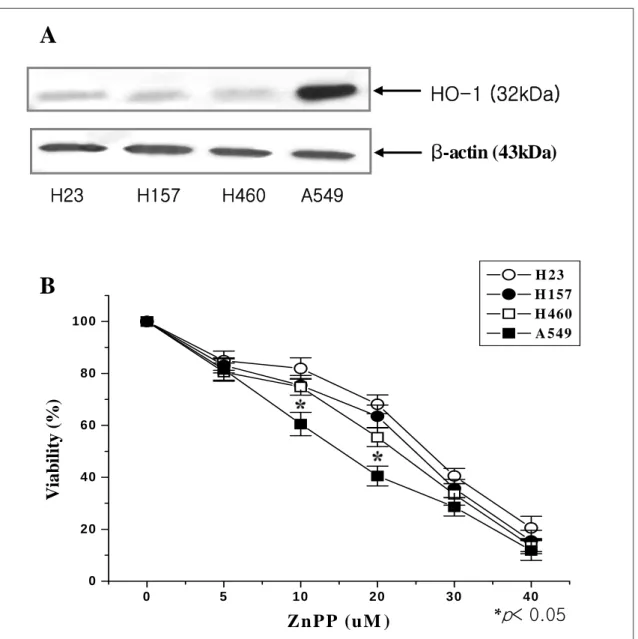
Figure 1. (A) Under endogenous state, HO-1 was expressed highly in A549 cells than other lung cancer cells. Cell lysates of endogenous state were subjected to 12.5% SDS-PAGE to measure the expression of HO-1 protein. (B) ZnPP significantly decreased the viability of A549 cells than other lung cancer cells in a dose-dependent fashion. Cells were treated with different concentrations of ZnPP for 48 hrs, and viability was determined by MTT assay. The data represent the mean±S.D. of three independent experiments. *, p<0.05 compared to control.
0 5 10 20 30 40
0 20 40 60 80 100
V iability (% )
Zn P P (u M )
H 23 H 157 H 460 A 549 AGT CT-3’을 사용하였고, PCR 반응여부를 확인하
기 위한 내부조건으로 이용한 GAPDH에 대한 sense primer는 5’-CAT GGA GAA GGC TGG GGC TC- -3’와 antisense primer는 5’-CAC TGA CAC GTT GGC AGT GC-3’ 염기서열 oligonucleotide을 합성하 여 사용하였다.
8) 유식세포 분석
세포고사의 현상의 확인을 위해 propidium iodide (PI)로 DNA를 염색한 후 형광의 세기를 측정하였다.
A549 세포에 HO-1 siRNA 리포솜을 이용하여 A549 세포에 운반시킨 후 24시간 후에 포집하여 PBS로 두 번 세척하였다. 세척된 세포에 PBS 300μl을 넣고 세
B
HO-1 (32kDa) β-actin (43kDa)
A
H23 H157 H460 A549
ZnPP(M)
Figure 2. ZnPP significantly decreased the enzymatic activity of HO-1 in A549 cells. Cells were treated wit h different concentrations of ZnPP for 48 hrs. Microsom- al extracts were prepared and assayed for HO activity by measurement of bilirubin generation as described in Materials and Methods. HO activity was expressed as nmol·mg protein-1·h-1. The data represent the mean-
±S.D. of three independent experiments.
0 10 20 30 40 50 60
0.0 0.5 1.0 1.5 2.0 2.5 3.0 3.5
HO activity (nmol bilirubin/mg/h)
포의 DNA는 PI solution(0.1% Triton X-100, 20μ g/ml PI, 200μg/ml Rnase) 600μl을 넣어 20분간 반응 시킨 후 FACS Vantage flow cytometer를 이용하여 sub-G0/G1으로 나타나는 세포고사를 분석하였으며, 그 정보의 분석은 Cell Quest softwave(Becton Dick- inson)을 이용하였다.
9) 결과 산출 방법
표시된 결과는 3번 이상의 독립적인 실험결과이며 이들의 평균(mean)과 표준편차(standard deviation, S.D.)를 산출하여 표시하였다. 실험결과의 통계처리 는 Student's t-test에 준하여 처리하였다.
결 과
1. 비처리 상태에서 폐암세포주의 HO-1 발현
비처리 상태에서 각각의 폐암세포주의 HO-1 발현
을 Western blot을 통해 확인한 결과 다른 세포주에 서 미미하게 발현된 32kDa의 HO-1이 A549 세포주 에서 발현이 증가되었다 (Fig. 1A).
2. ZnPP 처리 후 폐암세포주의 생존율 변화
이러한 결과에 근거하여 A549, NCI-H23, NCI-H- 157, NCI-H460 폐암세포주에서 HO-1 활성억제제인 ZnPP의 농도별 변화에 의한 생존율의 변화를 알아보 기 위해 48시간 후에 생존율을 MTT 방법으로 측정 하였다. 생존율의 결과는 10μM일 때 NCI-H23, NCI-H157, NCI-H460 세포주에서 각각 81.9%, 75.
4%, 74.8%인데 비해 A549 세포주에서 60.5%였고, 20 μM일 때 각각 68.1%, 63.5%, 55.4%인데 비해 40.5%
로서 ZnPP에 대하여 다른 폐암세포주에 비해 의의 있게 감소됨을 확인하였다 (Fig. 1B).
3. ZnPP 처리 후 A549 세포에서 HO-1 효소활성의 변화
ZnPP처리 후 HO-1의 효소활성 변화를 시간당 세 포단백질의 mg당 형성된 빌리루빈의 양으로 측정하 였다. 대조군의 빌리루빈 양인 3.10 nmol bilirubin/- mg/h을 100%로 하였을 때, ZnPP 10M 71.7%, 20M 59.4%, 30M 50.0%, 40M 34.8%로서 농도 의존적으로 효소활성의 감소가 확인되었다 (Fig. 2).
4. ZnPP 처리 후 A549 세포에서 활성산소종의 생성
ZnPP 처리시의 의미 있는 생존율의 감소가 활성산 소종의 생성과 관련이 있는지 알아보기 위해 H2O2를 측정하였다. 먼저 HRP에 의한 산화작용 동안에 scopoletin의 형광감소 정도를 측정한 결과 대조군에 비해 ZnPP 10μM 1.3배, 20μM 1.4배, 30μM 1.65배, 40 μM 2.2배로 농도 의존적으로 의의 있게 증가된 H2O2
의 생성을 보였다 (Fig. 3A). 또한 DCF-DA와 반응 시켰을 때 세포 내 H2O2에 의해 탈아세틸화되어 형광 을 나타내는 물질로 전환되어 붉은 색을 띠게 되는데 역시 농도 의존적으로 붉은색의 음영이 증가됨을 확
0 10 20 30 40 50
100 150 200 250
ROS production (% of control)
ZnPP (μM)
A
B
*p< 0.05
ZnPP 30uM ZnPP 40uM
ZnPP 20uM ZnPP 10uM
Cont Cont
Figure 3. ZnPP induced the generation of intracellular H2O2 in A549 cells. (A) Cells were treated with dif- ferent concentrations of ZnPP for 48 hrs. H2O2 gen- eration was determined by monitoring the decrease in fluorescence of scopoletin (5 μM) during its oxidation catalyzed by HRP (1 U/ml). Fluorescence value of sco- poletin is representative counts and relative fluoresc- ence values are mean±S.D. of three independent expe- riments. *, p<0.05 compared to control. (B) Cells were treated with different concentrations of ZnPP for 48 hrs, and were harvested, washed with media, and then stained with oxidant-sensitive fluorescence dye DCF- DA (50 μM) for 30 mins in the dark. After washing unbound dye out, the generation of H2O2 were visu- alized under fluorescent microscope.
cont nonsense HO-1 siRNA
0 10 20 30 40
Apoptosis (% of control)
A
B
C
control nonsense HO - 1 siRNA
HO - 1
GAPDH
*P<0.05
Procaspase- 3(35 kDa)
β - a c t i n (43 kDa) nonsense HO - 1 siRNA
control
Figure 4. A549 cells were transfected by HO-1 siRNA induced apoptosis. (A) Cells were transfected with siR- NA for HO-1 mRNA. The nonsense HO-1 siRNA was used as a negative control. RT-PCR for the HO-1 mRNA shows expression of the cells sense or nonsense siRNA transfection. Total RNA of transfected cells was iso- lated and HO-1 gene was amplified by RT-PCR detailed in the materials and methods. (B) HO-1 siRNA incr- eased sub-G0/G1 fraction in A549 cells. Cellular DNA were stained with PI staining solution and analyzed by flow cytometry . (C) HO-1 siRNA induced the activation of caspase-3. Cell lysate was subjected to 12.5%
SDS-PAGE to measure the expression of pro-casp- ase-3.
인하였다 (Fig. 3B).
5. A549 세포의 HO-1 siRNA 주입에 의한 세포고 사 유도
HO-1 활성억제제인 ZnPP의 효과를 HO-1 siRNA 를 이용하여 HO-1 mRNA를 억제시킴으로서 유전자 수준에서의 효과를 측정하였다. HO-1 siRNA를 리포 솜을 이용하여 A549 세포주에 운반시킨 후 24시간 후에 성공적인 주입여부는 역전사 중합효소 연쇄반 응을 통해 관찰하였다. 비처리군과 음성대조군에서 보이던 HO-1의 발현이 HO-1 siRNA 주입에 의해 HO-1의 knock-down의 발생을 확인하였다 (Fig.
4A). 이때 유식세포분석을 이용하여 세포주기를 분석 한 결과 세포고사가 일어난 세포들의 낮은 분자량을 가진 DNA 단편들이 세포막의 변화로 이동하여 G1
주기 전에 나타나는 sub-G0/G1 DNA 분획은 비처리 A549 세포군은 4.7%였고 nonsense siRNA 군은 5.8%인데 반해 HO-1 siRNA 군에서는 22.4%로 현저 하게 증가함을 확인하였다 (Fig. 4B). 또한 세포고사 와 관련된 단백질의 발현을 알아보기 위해 caspase-3 protease의 활성을 Western blot을 통해 확인하였다.
35kDa의 procaspase-3가 대조군으로 치료하지 않은 A549 세포군과 nonsense siRNA 군에 비해 HO-1 siRNA군에서 절단되어 감소됨을 관찰하였다 (Fig. 4C).
고 찰
유도성 효소인 HO-1은 산화 스트레스를 증가시키 는 조건인 lipopolysaccharides, 저산소증, 과산소증, 산화질소, heat shock, UV 방사선 등에 의해 발현되 어 산화 스트레스, 염증반응, 세포고사, 이식거부에 대한 보호기능을 하는 것으로 알려져 있다21-23. 호흡 기 영역에서 과산소증에 의한 손상에 대해 폐상피세 포와 섬유아세포에서 HO-1의 과발현이 보고되었고24,25, HO-1의 유도가 다발성 장기부전에 대한 보호작용을 한다고 하였다26. 또한 HO-1을 knockout시켰을 때 산 화 스트레스에 더 민감한 것으로 보고되었다27.
최근의 보고들은 HO-1이 세포성장과 세포의 다양
한 형태의 증식을 자극하는 것으로 알려지고 있다.
HO-1의 발현이 표피의 각질세포의 성장을 증가시켰 고28, HO-1 유전자를 각막 내피세포에 주입하였을 때 모세양 관상 구조물들이 형성되어 혈관생성의 역할 가능성을 제시하였다29. 또한 HO-1은 가장 빠르게 증 식하는 세포인 선암, 간암, 육종, 흑색종, 그리고 상피 암세포 등의 다양한 암 세포들에서 광범위하게 발현
된다9,18,30-32. Fang 등은 HO-1 활성억제제인 ZnPP를
대장암과 간암에 주입하였을 때 종양의 성장을 의미 있게 억제하여 HO-1이 세포성장 특히 암세포 증식 에 중요한 역할이 있음을 제시하였다9,33,34. 이러한 효 과는 산화스트레스에 의한 세포고사에 대하여 HO-1 의 보호효과 때문인 것으로 알려져 있는데35, 분자생 물학적인 기전으로 세포 내 전산화제의 양을 감소시 키고 빌리루빈의 양을 감소시키며 일산화탄소의 생 성증가가 알려져 있다. Heme은 다양한 활성산소종의 생성을 유도하고, 증가된 세포 내 산화제는 다양한 종 류의 세포에서 세포고사를 초래하는데 36,37, HO-1은 이러한 heme을 분해시켜 ferritin의 합성을 증가시킴 으로써 전산화제의 양을 감소시킨다38. 또한 heme의 대사과정에서 HO과 biliverdin reductase에 의해 생 성된 빌리루빈은 강력한 활성산소종의 제거기능을 가져서 세포고사에 대한 보호효과를 보인다.
본 연구에서 비처리 상태에서 A549 세포주는 다른 폐암세포주에 비해 HO-1의 발현이 증가되어 있음을 Western blot을 통해 확인하였다. 또한 HO-1 활성억 제제인 ZnPP 처리 후 A549 세포주에서 의의 있는 생 존율의 감소를 보여 HO-1이 세포독성에 대한 일부 보호작용이 있을 것으로 추정하였다. 한편 ZnPP 처 리에 의한 이러한 효과는 HO-1 활성의 감소를 통해 세포 내 활성산소종의 생성증가와 빌리루빈의 생성 감소를 통한 기전에 의한 것임을 확인하였다. 본 연구 에서 A549세포에서 HO-1 활성억제제인 ZnPP의 효 과를 HO-1 siRNA를 이용하여 HO-1 mRNA를 kno- ck-down 시켜 유전자 수준에서의 효과를 측정하였 는데 HO-1 siRNA가 처리된 세포에서 sub-G0/G1
DNA 분획이 현저하게 증가함을 확인하였고, Weste- rn blot 방법을 통해 caspase-3 protease의 활성을 증 가시켜 세포고사가 유도됨을 확인하였다.
HO-1의 높은 발현이 다양한 암종에서 알려져 있 고, 항산화와 항세포고사효과 때문에 빠른 암종의 성 장에 중요한 역할이 확인되고 있어 이것을 표적으로 하는 ZnPP 등의 활성억제제나 siRNA를 통한 kno- ck-down방법이 새로운 항암치료로의 이용가능성을 보여주고 있다. 이러한 기전은 HO-1의 억제를 통해 세포 내 활성산소양 증가와 빌리루빈의 생성감소가 세포고사를 유도할 것으로 보인다. 따라서 폐암의 치 료에 있어서 항암화학요법과 이들을 근간으로 한 병 용요법으로는 치료의 한계를 보여주고 있는 상황에 서 이러한 접근은 새로운 표적치료로서 가능성을 보 여줄 것으로 기대된다.
요 약
연구배경 :
Heme oxygenase-1 (HO-1)은 heme의 분해 대사 과정에 관여하는 유도성 효소로 heme을 분해하여 biliverdin, free iron, 및 일산화탄소 등을 생성시킨다.
HO-1의 발현은 다양한 스트레스성 자극에 반응하여 생체방어 기능을 갖는 것으로 알려져 있는데 세포성 장이나 세포사 특히 세포고사를 조절하는 것으로 보 고되고 있다. 현재 신장암, 전립선암, 간암, 육종 등의 고형암에서 발현됨이 알려져 있고, 실제 HO-1 억제 제를 투여했을 때 암성장이 억제됨이 보고되었다. 저 자들은 폐암세포주들에서 HO-1의 발현유무와 그 역 할을 규명하고 나아가 HO-1 억제제의 치료제로서의 가능성을 알아보고자 하였다.
방 법 :
비소세포폐암세포주인 A549, H23, NCI-H157, N- CI-H460을 이용하였다. 세포독성은 MTT 방법으로 구하였고, HO-1의 발현은 Western blotting으로 확 인하였다. HO의 효소활성은 시간당 세포단백질의 mg당 형성된 빌리루빈의 양을 이용하여 측정하였다.
또한 H2O2의 생성은 horse radish peroxidase(HRP) 와 형광물질인 2’,7’-dichlorofluorescein(DCF)를 이용 한 두 가지 방법을 이용하였다. A549세포에 HO-1 small interfering RNA(siRNA)을 주입하여 유식세포 분석과 caspase-3에 대한 Western blotting을 통하여
세포고사유무를 확인하였다.
결 과 :
비처리 상태에서 다른 세포주에 비해 A549세포의 HO-1 발현이 증가되었으며 HO-1 활성억제제인 ZnPP를 처리하였을 때 생존율의 의미 있는 감소를 보였다. 이러한 소견과 일치하여 ZnPP는 용량의존적 으로 HO 의 효소활성 감소와 세포 내 H2O2 생성의 증가를 초래하였다. 또한 HO-1 siRNA로 주입된 A- 549세포는 세포고사를 유도하였다.
결 론 :
HO-1은 폐암의 치료에 있어서 새로운 분자생물학 적 기전의 가능성을 제시하여 HO-1에 대한 표적치 료의 가능성을 보여줄 것으로 기대된다.
참 고 문 헌
1. Bae JM, Won YJ, Jung KW, Suh KA, Yun YH, Shin MH, et al. SurvivalKorean cancer patients diagnosed in 1995. Cancer Res Treat 2002;34:319-25.
2. Schiller JH, Harrington D, Belani CP, Langer C, San- dler A, Krook J, et al. Comparison of four chemother- apy regimens for advanced non-small-cell lung cancer.
N Engl J Med 2002;346:92-8.
3. Giaccone G, Herbst RS, Manegold C, Scagliotti G, Resell R, Miller V, et al. Gefitinib in combination with gemcitabine and cisplatin in advanced non-sm all-cell lung cancer: a phase III trial-INTACT 1. J Clin Oncol 2004;22:777-84.
4. Sandler AB, Johnson DH, Herbst RS. Anti-vascular endothelial growth factor monoclonals in non-small cell lung cancer. Clin Cancer Res 2004;10:4258S-62S.
5. Tenhunen R, Marver HS, Schmid R. The enzymatic catabolism of hemoglobin: stimulation of microsomal heme oxygenase by hemin. J Lab Clin Med 1970;75:
410-21.
6. Baranano DE, Rao M, Ferris CD, Snyder SH.
Biliverdin reductase: a major physiologic cytoprote- ctant. Proc Natl Acad Sci U S A 2002;99:16093-8.
7. McCoubrey WK Jr, Huang TJ, Maines MD. Isolation and characterization of a cDNA from the rat brain that encodes hemoprotein heme oxygenase-3. Eur J Biochem 1997;247:725-32.
8. Motterlini R, Foresti R, Bassi R, Calabrese V, Clark JE, Green CJ. Endothelial heme oxygenase-1 induct- ion by hypoxia: modulation by inducible nitric-oxide
synthase and S-nitrosothiols. J Biol Chem 2000;275:1- 3613-20.
9. Doi K, Akaike T, Fujii S, Tanaka S, Ikebe N, Beppu T, et al. Induction of haem oxygenase-1 nitric oxide and ischaemia in experimental solid tumours and im- plications for tumour growth. Br J Cancer 1999;8- 0:1945-54.
10. Elbirt KK, Whitmarsh AJ, Davis RJ, Bonkovsky HL.
Mechanism of sodium arsenite-mediated induction of heme oxygenase-1 in hepatoma cells: role of mito- gen-activated protein kinases. J Biol Chem 1998;
273:8922-31.
11. Eyssen-Hernandez R, Ladoux A, Frelin C. Differential regulation of cardiac heme oxygenase-1 and vascular endothelial growth factor mRNA expressions by hem- in, heavy metals, heat shock and anoxia. FEBS Lett 1996;382:229-33.
12. Keyse SM, Tyrrell RM. Heme oxygenase is the major 32-kDa stress protein induced in human skin fibro- blasts by UVA radiation, hydrogen peroxide, and sod- ium arsenite. Proc Natl Acad Sci U S A 1989;86:99-103.
13. Lautier D, Luscher P, Tyrrell RM. Endogenous glutathione levels modulate both constitutive and UV- A radiation/hydrogen peroxide inducible expression of the human heme oxygenase gene. Carcinogenesis 19- 92;13:227-32.
14. Wagner M, Cadetg P, Ruf R, Mazzucchelli L, Ferrari P, Redaelli CA. Heme oxygenase-1 attenuates isch- emia/ reperfusion-induced apoptosis and improves su- rvival in rat renal allografts. Kidney Int 2003;63:15- 64-73.
15. Choi BM, Pae HO, Chung HT. Nitric oxide priming protects nitric oxide-mediated apoptosis via heme oxygenase-1 induction. Free Radic Biol Med 2003;3- 4:1136-45.
16. Amon M, Menger MD,Vollmar B. Heme oxygenase and nitric oxide synthase mediate cooling-associated protection against TNF-alpha-induced microcirculato- ry dysfunction and apoptotic cell death. FASEB J 2003;17:175-85.
17. Ke B, Shen XD, Zhai Y, Gao F, Busuttil RW, Volk HD, et al. Heme oxygenase 1 mediates the immu- nomodulatory and antiapoptotic effects of interleukin 13 gene therapy in vivo and in vitro. Hum Gene Ther 2002;13:1845-57.
18. Goodman AI, Choudhury M, da Silva JL, Schwa- rtzman ML, Abraham NG. Overexpression of the heme oxygenase gene in renal cell carcinoma. Proc Soc Exp Biol Med 1997;214:54-61.
19. Maines MD, Abrahamsson PA. Expression of heme oxygenase-1 (HSP32) in human prostate: normal, hyperplastic, and tumor tissue distribution. Urology
1996;47:727-33.
20. Sahoo SK, Sawa T, Fang J, Tanaka S, Miyamoto Y, Akaike T, et al. Pegylated zinc protoporphyrin: a water-soluble heme oxygenase inhibitor with tum- or-targeting capacity. Bioconjug Chem 2002;13:1031-8.
21. Pae HO, Oh GS, Choi BM, Chae SC, Kim YM, Chung KR, et al. Carbon monoxide produced by heme oxyg- enase-1 suppresses T cell proliferation via inhibition of IL-2 production. J Immunol 2004;172:4744-51.
22. Otterbein LE, Bach FH, Alam J, Soares M, Tao Lu H,Wysk M, et al. Carbon monoxide has anti-infl- ammatory effects involving the mitogen-activated pro- tein kinase pathway. Nat Med 2000;6:422-8.
23. Berberat PO, Katori M, Kaczmarek E, Anselmo D, Lassman C, Ke B, et al. Heavy chain ferritin acts as an antiapoptotic gene that protects livers from isc- hemia reperfusion injury. FASEB J 2003;17:1724-6.
24. Lee PJ, Alam J, Wiegand GW, Choi AM. Overex- pression of heme oxygenase-1 in human pulmonary epithelial cells results in cell growth arrest and incr- eased resistance to hyperoxia. Proc Natl Acad Sci U S A 1996;93:10393-8.
25. Dennery PA, Sridhar KJ, Lee CS, Wong HE, Shokoohi V, Rodgers PA, et al. Heme oxygenase-m- ediated resistance to oxygen toxicity in hamster fibro- blasts. J Biol Chem 1997;272:14937-42.
26. Otterbein L, Chin BY, Otterbein SL, Lowe VC, Fessler HE, Choi AM. Mechanism of hemoglobin-ind- uced protection against endotoxemia in rats: a ferriti- n-independent pathway. Am J Physiol 1997;272:L268-75.
27. Yachie A, Niida Y, Wada T, Igarashi N, Kaneda H, Toma T, et al. Oxidative stress causes enhanced endothelial cell injury in human heme oxygenase-1 deficiency. J Clin Invest 1999;103:129-35.
28. Clark JE, Green CJ, Motterlini R. Involvement of the heme oxygenase-carbon monoxide pathway in kerat- inocyte proliferation. Biochem Biophys Res Commun 1997;241:215–20.
29. Deramaudt BM, Braunstein S, Remy P, Abraham NG. Gene transfer of human heme oxygenase into coronary endothelial cells potentially promotes angio- genesis. J Cell Biochem 1998;68:121-7.
30. Tsuji MH, Yanagawa T, Iwasa S, Tabuchi K, Onizawa K, Bannai S, et al. Heme oxygenase-1 expression in oral squamous cell carcinoma as invo- lved in lymph node metastasis. Cancer Lett 1999;1- 38:53-9.
31. Deininger MH, Meyermann R, Trautmann K, Duffner F, Grote EH, Wickboldt J, et al. Heme oxygenase (HO)-1 expressing macrophages/microglial cells accu- mulate during oligodendroglioma progression. Brain Res 2000;882:1-8.
32. Torisu-Itakura H, Furue M, Kuwano M, Ono M.
Co-expression of thymidine phosphorylase and heme oxygenase-1 in macrophages in human malignant ver- tical growth melanomas. Jpn J Cancer Res 2000;9- 1:906-10.
33. Fang J, Sawa T, Akaike T, Akuta T, Sahoo SK, Khaled G, et al. In vivo antitumor activity of pegyl- ated zinc protoporphyrin: targeted inhibition of heme oxygenase in solid tumor. Cancer Res 2003;63:3567-74.
34. Fang J, Akaike T, Maeda H. Antiapoptotic role of heme oxygnease (HO) and the potential of HO as a target in anticancer treatment. Apoptosis 2004;9:27-35.
35. Tanaka S, Akaike T, Fang J, Beppu T, Ogawa M, Tamura F, et al. Antiapoptotic effect of haem oxyg-
enase-1 induced by nitric oxide in experimental solid tumor. Br J Cancer 2003;88:902-9.
36. Dumont A, Hehner SP, Hofmann TG, Ueffing M, DrogeW, Schmitz ML. Hydrogen peroxide-induced apoptosis is CD95-independent, requires the release of mitochondria-derived reactive oxygen species and the activation of NF-kappaB. Oncogene 1999;18:747-57.
37. Slater AF, Stefan C, Nobel I, van den Dobbelsteen DJ, Orrenius S. Signalling mechanisms and oxidative stress in apoptosis. Toxicol Lett 1995;82/83:149-53.
38. Vile GF, Tyrrell RM. Oxidative stress resulting from ultraviolet A irradiation of human skin fibroblasts leads to a heme oxygenase-dependent increase in ferritin. J Biol Chem 1993;268:14678-81.