Copyright © 2017. Anatomy & Cell Biology
non-small cell lung cancer (NSCLC). The molecular patho- genesis of SCLC and NSCLC is different. SCLC accounts for 15% of lung cancer cases and it is a highly malignant and clini cally aggressive tumor. On the other hand, 85% of lung cancers are diagnosed as NSCLC and has three major sub- types: adenocarcinoma, squamous cell carcinoma, and large cell carcinoma [3, 5-7].
A lot of agents have been used for clinical treatment of lung cancer [7]. One of the targets is the cyclooxygenase (COX) which is evaluated in the treatment of lung cancer. COX is essential enzyme for the prostaglandins (PGs) synthesis.
PGs are involved in various physiological and pathogenetic processes such as inflammatory reaction, gastro-intestinal cytoprotection, ulceration, hemostasis, and thrombosis [8-14].
Introduction
Lung cancer is a common cancer and life-threatening dis- ease in Korea. The mortality of lung cancer is highest than colon, breast, or prostate cancers [1-4]. Lung cancer is classi- fied as two types which are small cell lung cancer (SCLC) and
Corresponding author:
Yeong Seok Kim
Department of Anatomy and Research Center for Tumor Immunology, Inje University College of Medicine, 75 Bokji-ro, Busanjin-gu, Busan 47392, Korea
Tel: +82-51-890-6634, Fax: +82-51-896-6634, E-mail: newsoft@inje.ac.kr
*These two authors contributed equally to this work.
Celecoxib induces cell death on non-small cell lung cancer cells through endoplasmic reticulum stress
Bomi Kim
1,*, Jayoung Kim
2,*, Yeong Seok Kim
21Department of Pathology, Inje University Haeundae Paik Hospital, Busan, 2Department of Anatomy and Research Center for Tumor Immunology, Inje University College of Medicine, Busan, Korea
Abstract: Cyclooxygenase-2 (COX-2) is an enzyme induced by various proinflammatory and mitogenic stimuli. Celecoxib is a selective inhibitor of COX-2 that have been shown to affect cell growth and apoptosis. Lung cancer cells expressing COX-2 is able to be a target of celecoxib, this study focuses on investigating that celecoxib induces apoptosis via endoplasmic reticulum (ER) stress on lung cancer cells. We investigated whether celecoxib induced apoptosis on non-small cell lung cancer cell line, A549 and H460. The 50 μM of celecoxib increased apoptotic cells and 100 μM of celecoxib significantly induced apoptosis. To check involvement of caspase cascade, pretreatment of z-VAD-fmk blocked celecoxib-induced apoptosis. However, caspase-3, -8, and -9 were not activated, but cleavage of non-classical caspase-4 was detected using western blot. As checking ER stress associated molecules, celecoxib did not increase expressions of growth arrest and DNA damage inducible protein 34, activating transcription factor 4, and spliced X-box binding protiens-1, but increase of both glucose-regulated protein 78 (GRP78) and C/
EBP homologous transcription factor were detected. Salubrinal, inhibitor of eIF2 and siRNA for IRE1 did not alter celecoxib- induced apoptosis. Instead, celecoxib-induced apoptosis might be deeply associated with ER stress depending on GRP78 because siRNA for GRP78 enhanced apoptosis. Taken together, celecoxib triggered ER stress on lung cancer cells and celecoxib- induced apoptosis might be involved in both non-classical caspase-4 and GRP78.
Key words: Celecoxib, Lung neoplasms, ER stress, Apoptosis
Received November 27, 2017; Revised November 30; Accepted December 1, 2017
COX enzyme has two isoforms, COX-1 and COX-2. While COX-1 is consistutively expressed in most cells and tissues, COX-2 is expressed in central nervous system, kidney, and seminal vesicles. However, it can be induced by various proin- flammatory and mitogenic stimuli [8-10].
To regulate function of COX, non-steroidal anti-inflamma- tory drugs (NSAIDs) have been used traditionally for several decades. NSAIDs are agents for treatment of pain, inflam- mation, and fever [12, 15]. NSAIDs has been used to man- age pain and suppress inflammation in arthritis, but these days they are also used as the agents for cancer therapeutics.
NSAIDs have contributed to anti-proliferation and pro-apop- tosis on a various cancer cells, such as colorectal cancer, breast cancer, or gastric cancer [8, 11-13, 16-18]. NSAIDs block a production of PGs by the inhibition of COX-1 and COX-2 in normal and inflammatory tissues. There are side effects such as gastrointestinal tract bleeding and kidney failure when NSAIDs are treated for a long time [12, 15, 18, 19]. These side effects are known by inhibition of COX-1 [12, 19]. On the other hand, COX-2 inhibitors are widely used because they have little side effect. There are several selective COX-2 in- hibitors, for example rofecoxib, etoricoxib, and celecoxib. The celecoxib makes little effects for COX-1, and inhibits tumor initiation and tumor cell growth. In past studies, celecoxib was reported which decreases risk of colon, breast, esophagus, and gastric cancer, it is also effective for chemotherapy and radiotherapy of cancer patients [16]. Celecoxib induces cell cycle arrest, inhibits tumor growth, suppresses tumor angio- genesis and induces apoptosis in several types of cancers [14, 16, 18]. Moreover, celecoxib induces apoptotic cell death in tumor cells and endothelial cells [11, 16, 19]. Therefore, in- vestigation of mechanism on celecoxib-induced apoptosis is important to understand anti-cancer activity.
Recently, endoplasmic reticulum (ER) stress has been re- ported as a major mechanism that celecoxib can induce cell death in cancer [20-24]. Unfolded protein response (UPR) is a pro-survival mechanism and remains homeostasis of cells in ER stress state. However, if this adaptation is not proven to be sufficient, the apoptosis starts [20, 21, 25, 26].
Several solid tumors undergo ER stress and are going to fall a fate, such as adaptation or apoptosis. Three types of ER transmembrane proteins are important: protein-kinase and site-specific endoribonuclease inositol-requiring enzyme 1 (IRE1), protein-kinase R-like ER kinase (PERK)/pancreatic eIF2 kinase and activating transcription factor 6 (ATF6) [20, 21, 26]. C/EBP homologous transcription factor (CHOP) is a
major transcription factor of pro-apoptosis and regulates ER stress-induced apoptosis. CHOP induces cell death through controlling the expression of other genes [20, 21]. ER stress is induced by NSAIDs treatment which causes ER membrane protein PERK to phosphorylate eIF2α. CHOP is up-regulated after the phospho-eIF2α protein activates ATF4. NSAIDs also induces apoptosis through another complicated mechanism as well as ER stress [19].
Taken together, celecoxib affects obviously cell death of lung cancer but its mechanism is unclear. In this study, the involvement of ER stress in cell death is investigated when ce- lecoxib is treated on lung cancer cells.
Materials and Methods
Cell cultureHuman lung carcinoma cell line A549 and H460 (ATCC, Manassas, VA, USA) were maintained in RPMI1640 medium (Hyclone, Logan, UT, USA) supplemented with 10% fetal bovine serum (Hyclone) and 100 U/ml penicillin, and 100 μg/ml streptomycin (Hyclone) at 37°C in humiditied 5% CO2
atmosphere.
Analysis of cytotoxicity
Cell viability was examined through the WST-1 assay (Ta- kara, Shiga, Japan). Cells were seeded in 96-well plates and incubated for 24 hours. Celecoxib (TRC, Toronto, Ontario, Canada) of various concentrations was treated on cells for overnight. WST-1 was added each well and the plates were incubated for 1 hour and the absorbance was measured using microplate reader (Thermo Pierce, Waltham, MA, USA) at 450 nm wavelength.
Annexin V assay
Cells were collected by centrifugation and washed with phosphate buffered saline (PBS). Fluorescein isothiocyanate (FITC)‒labeled annexin V (BD Bioscience, San Jose, CA, USA) was added in dark after cell pellet was resuspended with annexin V binding buffer. Cells were detected by flow cytometry (Miltenyi Biotech, Bergisch Gladhach, Germany) after incubation for 15 minutes. To assess involvement of cas- pase cascade, Z-VAD-fmk (Merck, Darmstadt, Germany) was treated for 1 hour before celecoxib treatment.
RNA interference
A549 cells were transfected with glucose-regulated protein
78 (GRP78), IRE1, or negative control siRNA (GenePharma, Shanghai, China) according to manufacturer’s instruction.
A549 cells were seeded in 6-well plates with growth medium without antibiotics and allowed to reach 50% confluence for transfection. Lipofectamine 2000 (Invitrogen, Carlsbad, CA, USA) and siRNA were respectively diluted in medium with- out serum and incubated for 5 minutes at room temperature.
Two solutions were mixed and incubated for 20 minutes at room temperature. The mixture was treated on cells for 16 hours.
Reverse transcription polymerase chain reaction Total RNA was isolated using TRIzol (Invitrogen) accord- ing to the manufacturer’s protocol. Briefly, harvested cells were mixed TRIzol and added chloroform. The mixture was centrifugated at 13,000 rpm for 15 minutes at 4°C. Upper solution of the separated two layer was transferred new tube and combined with isopropanol. After incubation for 10 minutes, combined was centrifugated for 15 minutes at 4°C.
Supernatant was discarded and washed with 75% ethanol in diethylpyrocarbonate (DEPC) treated water. RNA containing pellet was resuspended with DEPC treated water. RT-premix (Bioneer, Daejeon, Korea) was used for cDNA synthesis from total RNA. cDNA was amplified using primers and Tenuto PCR premix (Enzynomics, Daejeon, Korea) in PCR Thermal Cycler (Takara). PCR products were analyzed by gel electro- phoresis in TAE buffer through agarose gel.
Western blot analysis
Cells were lysed in lysis buffer (Invitrogen) supplemented
with protease inhibitor cocktail (Sigma, St. Louis, MO, USA) after the cells were harvested. Samples were incubated on ice for 10 minutes and centrifugated for 10 minutes at 13,000 rpm. Proteins were determined by the bicinchoninic acid assay (Thermo Pierce) and were separated in 10% sodium dodecyl sulfate gel overlaid with a 4% polyacrylamide staking gel. Proteins were transferred to a nitrocellulose membrane after separation. The membrane was blocked with 5% skim milk for 1 hour at room temperature and washed with Tris- buffered saline containing 0.1% Tween 20 (TBST, pH 7.4) for 10 minutes each time for three times. The membrane was incubated with primary antibodies of caspase 3 (Cell Signal- ing, Beverly, MA, USA), caspase-4 (Abcam, Cambridge, MA, USA), caspase 8 (Cell Signaling), caspase 9 (Cell Signaling), GRP78 (Santa Cruz Biotechnology, Santa Cruz, CA, USA), CHOP (Cell Signaling), and β-actin (Santa Cruz Biotechnol- ogy) in 0.1% TBST at 4°C overnight. The membrane was incubated with secondary antibody (anti-goat IgG for GRP78;
anti-rabbit IgG for caspase 3 and caspase 9; anti-mouse IgG for caspase 8, CHOP and β-actin at 1:5,000 dilutions) for 1 hour at room temperature after the membrane was washed with 0.1% TBST three times for 10 minutes. Following incu- bation, membrane was washed with 0.1% TBST three times.
The band signals were detected by chemiluminescence (Fuji Photo Film Co., Tokyo, Japan).
Results
Effect of celecoxib on cell death of lung cancer cells Cytotoxicity was examined when celecoxib was treated
150 H460
100
50
0
Viability(%)
0 25 50 100 200
A A549
Celecoxib ( M)
150
100
50
0
Viability(%)
0 25 50 100 200
B
Celecoxib ( M)
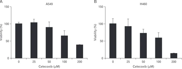
Fig. 1. Viability of celecoxib-treated A549 (A) and H460 (B). A549 and H460 were treated with celecoxib (0, 25, 50, 100, and 200 μM) for 16 hours. Cell viability was measured by WST-1 assay as described in materials and methods.
on lung cancer cells. A549, H460 lung cancer cell lines were incubated with various concentration of celecoxib (0, 25, 50, 100, and 200 μM). Cell viability was observed by WST-1 assay after overnight treatment of celecoxib.
Approximately, the half maximal inhibitory concentration
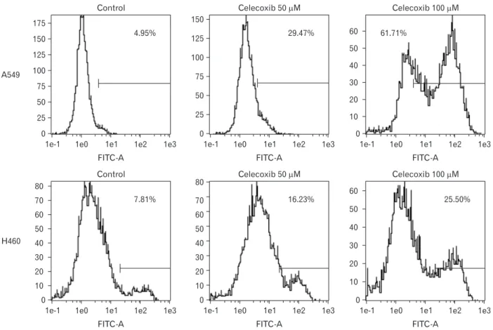
(IC50) of celecoxib was between 50 and 100 μM (Fig. 1). To determine whether celecoxib induces apoptosis, celecoxib (50 and 100 μM) was treated on A549 and H460 cells for over- night and then analyzed by flow cytometry after staining with FITC-labeled annexin V. Apoptosis was induced on A549 and
A549
H460
60 50 40 30 20 10 0
1e-1 1e0 1e1 1e2 1e3 FITC-A
61.71%
Celecoxib 100 M
60 50 40 30 20 10 0
1e-1 1e0 1e1 1e2 1e3 FITC-A
25.50%
Celecoxib 100 M Control
150 125 100 75 50 25 0
1e-1 1e0 1e1 1e2 1e3 175
FITC-A 4.95%
Control
0
1e-1 1e0 1e1 1e2 1e3 80
70 60 50 40 30 20 10
FITC-A 7.81%
Celecoxib 50 M 150
125 100 75 50 25 0
1e-1 1e0 1e1 1e2 1e3 FITC-A
29.47%
Celecoxib 50 M
0
1e-1 1e0 1e1 1e2 1e3 FITC-A
16.23%
80 70 60 50 40 30 20 10
Fig. 2. Flow cytometry analysis for celecoxib-induced apoptosis on A549 and H460. A549 and H460 were treated with 50 and 100 μM of celecoxib for 16 hours. Then, cells were harvested and analyzed using flow cytometry after staining with fluorescein isothiocyanate (FITC)‒labeled annexin V. Indicated percentile numbers mean cell proportion in gated region. A representative example of five independent experiments is shown.
Control 125
100 75 50 25 0
1e-1 1e0 1e1 1e2 1e3 FITC-A
4.06%
Z-VAD-fmk /celecoxib 100 M 80
70 60 50 40 30 20 10 0
1e-1 1e0 1e1 1e2 1e3 FITC-A
34.93%
Celecoxib 100 M 50
40 30 20 10 0
1e-1 1e0 1e1 1e2 1e3 FITC-A
46.15%
Z-VAD-fmk 125
100 75 50 25 0
1e-1 1e0 1e1 1e2 1e3 FITC-A
4.09%
Fig. 3. Effect of z-VAD-fmk on celecoxib-induced apoptosis. Z-VAD-fmk (Merck, Darmstadt, Germany) was treated on A549 for 1 hour before celecoxib treatment. A549 was washed with phosphate buffered saline, and then treated with 100 μM of celecoxib for 16 hours. Cells were harvested and analyzed using flow cytometry after staining with fluorescein isothiocyanate (FITC)‒labeled annexin V. Indicated percentile numbers mean cell proportion in gated region. A representative example of three independent experiments is shown.
H460 cells which were treated 100 μM of celecoxib. The rate of apoptosis by celecoxib was 61.7% on A549 and 25.5% on H460 respectively when 100 μM of celecoxib was treated (Fig. 2).
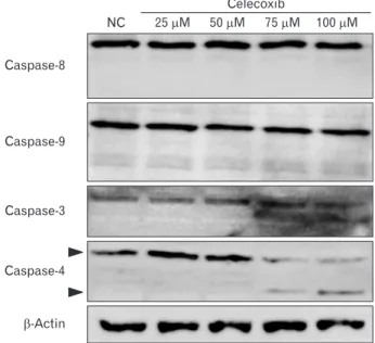
Involvement of caspases in celecoxib-induced apoptosis Celecoxib-induced apoptosis on lung cancer cells (Fig.
2). To investigate that celecoxib induces apoptosis through caspases, celecoxib was treated on A549 cells. It was observed whether pan-caspase inhibitor, z-VAD-fmk reduced celecox- ib-induced apoptosis. The z-VAD-fmk was treated for 1 hour before treatment of celecoxib. Celecoxib-induced apoptosis was decreased at pretreatment with z-VAD-fmk than only treatment with celecoxib (Fig. 3). Then, caspases were de- tected using western blot after celecoxib treatment on A549.
Caspase-3, -8, and -9 did not be activated, but caspase-4 was cleaved with treatment of 75 and 100 μM celecoxib (Fig. 4).
Involvement of ER stress in celecoxib-induced apoptosis To examine whether celecoxib-induced apoptosis is in- volved in ER stress, markers of ER stress were investigated.
These markers include GRP78/immunoglobulin heavy chain binding protein (BiP), CHOP/growth arrest and DNA dam- age inducible protein (GADD) 153, ATF4, GADD34, and spliced X-box binding protiens-1 (sXbp-1). According to the results of reverse transcription polymerase chain reaction, expression of GADD34 and CHOP was increased but GRP78, sXbp-1, and ATF4 was not in A549 cells. GRP78, sXBP-1, GADD34, and CHOP was increased in H460 but ATF4 was not increased (Fig. 5A). Also, GRP78 and CHOP was in-
creased in protein level in A549 (Fig. 5B).
UPR induces transcription of folding chaperones. How- ever, ER stress signaling pathways trigger cell apoptosis when UPR is continued. These signaling pathway-related molecules are IRE1, PERK, and ATF6. GRP78 regulates pro-survival pathway in UPR and is important to protein folding and as-
Caspase-8
Caspase-9
Caspase-3
Caspase-4
-Actin
NC 25 M 50 M 75 M 100 M Celecoxib
Fig. 4. Western blot for caspase-3, 4, 8, and 9 in celecoxib-treated A549.
A549 was treated with celecoxib (0, 25, 50, 75, and 100 μM) for 16 hours. After washing with phosphate buffered saline, cell lysates were obtained using lysis buffer. Western blot for caspase-3, -4, -8, and -9 were performed using antibodies against each caspase, and visualized as described in Materials and Methods. A representative example of six independent experiments is shown. NC, negative control.
A549 H 604
GRP78
sXBP-1
GADD34
ATF4
CHOP
-Actin
GRP78
CHOP
-Actin Control Celecoxib
A B
Control Celecoxib Control Celecoxib Control Celecoxib
A549 H 604 Fig. 5. Expressions of endoplasmic reti- culum stress markers in A549 and H460 after celecoxib treatment. Celecoxib 75 μM was treated on A549 and H460 cells for 16 hours and RNA and protein were isolated respectively. (A) Glucose- regulated protein 78 (GRP78), spliced X-box binding protiens-1 (sXBP-1), growth arrest and DNA damage in- ducible protein 34 (GADD34), acti va- ting transcription factor 4 (ATF4), and C/EBP homologous transcription fac tor (CHOP) mRNA were measured by reverse transcription polymerase chain reaction. A representative example of three independent experiments is shown.
(B) GRP78 and CHOP pro tein were measured using western blot. A re pre- sentative example of four inde pen dent ex periments is shown.
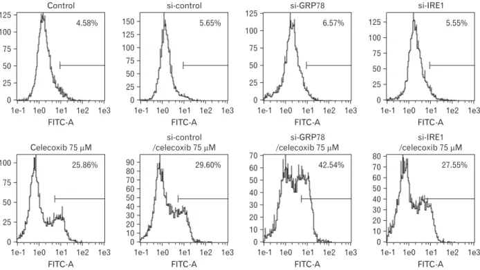
sembly. To detect whether GRP78 is related to celecoxib-in- duced apoptosis, GRP78 was inhibited using by siRNA. Pro- portion of apoptotic cells induced by 75 μM of celecoxib was 25.86% and apoptosis was increased (42.54%) when GRP78 siRNA and celecoxib were combined (Fig. 6). When sXBP-1 as a downstream effector of IRE1α was inhibited by siRNA, the rate of apoptosis was not changed (27.55%).
PERK induces ATF4 and CHOP after eIF2α is phosphory-
lated. To examine whether PERK pathway was played by celecoxib, salubrinal, inhibitor of eIF2, was pre-incubated for 1 hour and Annexin V was detected by flow cytometry after 100 μM of celecoxib treatment. The rate of apoptosis was little changed on celecoxib treatment A549 cells when eIF2 was in- hibited (Fig. 7).
Control 125
100 75 50 25 0
1e-1 1e0 1e1 1e2 1e3 FITC-A
4.58%
si-IRE1
0
1e-1 1e0 1e1 1e2 1e3 FITC-A
5.55%
si-GRP78 125
100 75 50 25 0
1e-1 1e0 1e1 1e2 1e3 FITC-A
6.57%
si-control 150
100 75 50 25 0
1e-1 1e0 1e1 1e2 1e3 FITC-A
5.65%
Celecoxib 75 M 100
75 50 25 0
1e-1 1e0 1e1 1e2 1e3 FITC-A
25.86%
si-IRE1 /celecoxib 75 M 80
70 60 50 40 30 20 10 0
1e-1 1e0 1e1 1e2 1e3 FITC-A
27.55%
si-GRP78 /celecoxib 75 M
50 40 30 20 10 0
1e-1 1e0 1e1 1e2 1e3 FITC-A
42.54%
si-control /celecoxib 75 M 90
80 70 60 50 40 30 20 10 0
1e-1 1e0 1e1 1e2 1e3 FITC-A
29.60%
125
125 100 75 50 25
60 70
Fig. 6. Analysis for celecoxib-induced apoptosis after transfection of siRNA for glucose-regulated protein 78 (GRP78) or IRE1 in A549. Two hundred pmol of siRNA for GRP78 or inositol-requiring enzyme 1 (IRE1) was transfected in A549 cells and stabilized overnight. And then celecoxib 75 μM was treated for 16 hours. Cells were harvested and analyzed using flow cytometry after staining with fluorescein isothiocyanate (FITC)‒labeled annexin V. Indicated percentile numbers mean cell proportion in gated region. A representative example of four independent experiments is shown.
Control 100
75 50 25 0
1e-1 1e0 1e1 1e2 1e3 FITC-A
4.14%
Salubrinal /celecoxib 100 M 60
50 40 30 20 10 0
1e-1 1e0 1e1 1e2 1e3 FITC-A
30.02%
Celecoxib 100 M 50
40 30 20 10 0
1e-1 1e0 1e1 1e2 1e3 FITC-A
35.98%
Salubrinal 125
100 75 50 25 0
1e-1 1e0 1e1 1e2 1e3 FITC-A
2.24%
Fig. 7. Analysis for celecoxib-induced apoptosis after treatment of salubrinal in A549. The 10 μM of salubrinal, eIF2 inhibitor was preincubated with A549 for 1 hour and then then celecoxib 75 μM was treated for 16 hours. Cells were harvested and analyzed using flow cytometry after staining with fluorescein isothiocyanate (FITC)‒labeled annexin V. Indicated percentile numbers mean cell proportion in gated region. A representative example of three independent experiments is shown.
Discussion
NSAIDs are most common drugs that reduce pain and inhibit synthesis of PGs. NSAIDs have been contributed anti- proliferation and pro-apoptosis on a various cancer cells. This study found out how lung cancer cells undergo celecoxib- induced apoptosis. It was confirmed that ER stress is a key role in celecoxib-induced apoptosis in lung cancer cells.
Evidences that NSAIDs induce cell death are reported in many studies. Other studies observe that aspirin and indo- methacin induce apoptosis on gastric cancer [27], aspirin induces apoptosis on ovarian cancer [28], and NS398 induces apoptosis on colon cancer [29]. As shown in Fig. 1, celecoxib among NSAIDs induced cell death on H460 by dose-depen- dent manner. However, when celecoxib was treated on A549 cells, cell viability was slightly increased at low concentration of celecoxib (25 μM). In other study, cell death was observed when 30 μM of celecoxib was treated on A549 cells [30]. Al- though WST-1 assays for celecoxib was retried on A549 cells several time, similar results that viability was increased at low concentration of celecoxib were detected. The low concentra- tion of celecoxib might regulate other signal pathways and then temporarily increase cell viability. To find solution to this problem, other investigations are needed.
This study focused the mechanism of celecoxib-induced apoptosis on lung cancer. Several studies suggest that NSAIDs-induced cell death is related with ER stress in vari- ous cancer cells [22, 26, 31-34]. UPR induces transcription of genes encoding ER resident chaperones. However, if ER stress is continuously persisted, signaling pathways including PERK, IRE1, and ATF6 trigger apoptosis. In this study, two of three pathways involved in ER stress-induced apoptosis were determined. Celecoxib-induced apoptosis might be less related to PERK pathway because salubrinal, eIF2α inhibitor did not block apoptosis (Fig. 7). The rate of apoptosis was also not changed by inhibition of IRE1 on lung cancer cells (Fig.
6). siRNA for CHOP also did not diminish celecoxib-induced apoptosis (data not shown), although CHOP as an important effector of ER stress was increased after celecoxib treatment.
In contrast, inhibition of GRP78 increased apoptosis of lung cancer cells. Continuous ER stress consumes GRP78 and sub- sequently increases GRP78 production; thus, down-regulation of GRP78 by siRNA might aggravate ER stress conditions.
This result suggests that celecoxib-induced apoptosis is deeply associated with ER stress depending on GRP78.
However, other studies suggest that celecoxib-induced
apoptosis is involved in caspase cascade [17, 18, 34]. Liu et al.
[34] investigated that celecoxib treatment appears to activate the caspase cascade in lung cancer cells. They showed that apoptosis with celecoxib was involved in cleavage of upstream and downstream of caspase cascades, caspase-3, -8, -9, and poly(ADP-ribose) polymerase, so celecoxib is regarded to in- duce apoptosis through ER stress and caspases cascade. Actu- ally, we investigated that z-VAD-fmk, pan-caspase inhibitor, reduced celecoxib-induced apoptosis but celecoxib did not activate caspase-3, -8, and -9 on lung cancer cells (Fig. 4). ER stress is also associated with non-classical caspases such as caspase-4 [35]. We found that celecoxib activated caspase-4 on A549 (Fig. 4).
Taken together, celecoxib triggered ER stress on lung can- cer cells and celecoxib-induced apoptosis might be involved in both non-classical caspase-4 and GRP78. These finding could provide an insight of a therapeutic approach using cele- coxib as an adjuvant with anti-cancer drugs.
Acknowledgements
This study was supported by Basic Science Research Pro- gram through NRF funded by the Ministry of Education (NRF-2015R1D1A1A01060152).
References
1. Hirsch FR, Lippman SM. Advances in the biology of lung cancer chemoprevention. J Clin Oncol 2005;23:3186-97.
2. Keith RL. Lung cancer chemoprevention. Proc Am Thorac Soc 2012;9:52-6.
3. Dela Cruz CS, Tanoue LT, Matthay RA. Lung cancer: epidemiol- ogy, etiology, and prevention. Clin Chest Med 2011;32:605-44.
4. Sandler AB, Dubinett SM. COX-2 inhibition and lung cancer.
Semin Oncol 2004;31(2 Suppl 7):45-52.
5. Salgia R, Skarin AT. Molecular abnormalities in lung cancer. J Clin Oncol 1998;16:1207-17.
6. Collins LG, Haines C, Perkel R, Enck RE. Lung cancer: diagnosis and management. Am Fam Physician 2007;75:56-63.
7. Dy GK, Adjei AA. Novel targets for lung cancer therapy: part I. J Clin Oncol 2002;20:2881-94.
8. Gasparini G, Longo R, Sarmiento R, Morabito A. Inhibitors of cyclo-oxygenase 2: a new class of anticancer agents? Lancet On- col 2003;4:605-15.
9. Breyer MD, Harris RC. Cyclooxygenase 2 and the kidney. Curr Opin Nephrol Hypertens 2001;10:89-98.
10. Frungieri MB, Calandra RS, Mayerhofer A, Matzkin ME. Cyclo- oxygenase and prostaglandins in somatic cell populations of the testis. Reproduction 2015;149:R169-80.
11. Vosooghi M, Amini M. The discovery and development of
cyclooxygenase-2 inhibitors as potential anticancer therapies.
Expert Opin Drug Discov 2014;9:255-67.
12. Ghosh N, Chaki R, Mandal V, Mandal SC. COX-2 as a target for cancer chemotherapy. Pharmacol Rep 2010;62:233-44.
13. Rizzo MT. Cyclooxygenase-2 in oncogenesis. Clin Chim Acta 2011;412:671-87.
14. Grösch S, Maier TJ, Schiffmann S, Geisslinger G. Cyclooxygen- ase-2 (COX-2)-independent anticarcinogenic effects of selective COX-2 inhibitors. J Natl Cancer Inst 2006;98:736-47.
15. Kismet K, Akay MT, Abbasoglu O, Ercan A. Celecoxib: a potent cyclooxygenase-2 inhibitor in cancer prevention. Cancer Detect Prev 2004;28:127-42.
16. Jendrossek V. Targeting apoptosis pathways by celecoxib in can- cer. Cancer Lett 2013;332:313-24.
17. Ko SH, Choi GJ, Lee JH, Han YA, Lim SJ, Kim SH. Differential effects of selective cyclooxygenase-2 inhibitors in inhibiting proliferation and induction of apoptosis in oral squamous cell car cinoma. Oncol Rep 2008;19:425-33.
18. Wu T, Leng J, Han C, Demetris AJ. The cyclooxygenase-2 in- hibitor celecoxib blocks phosphorylation of Akt and induces apop tosis in human cholangiocarcinoma cells. Mol Cancer Ther 2004;3:299-307.
19. Liggett JL, Zhang X, Eling TE, Baek SJ. Anti-tumor activity of non-steroidal anti-inflammatory drugs: cyclooxygenase-inde- pendent targets. Cancer Lett 2014;346:217-24.
20. Wang WA, Groenendyk J, Michalak M. Endoplasmic reticulum stress associated responses in cancer. Biochim Biophys Acta 2014;1843:2143-9.
21. Chevet E, Hetz C, Samali A. Endoplasmic reticulum stress-acti- vated cell reprogramming in oncogenesis. Cancer Discov 2015;5:
586-97.
22. Huang KH, Kuo KL, Chen SC, Weng TI, Chuang YT, Tsai YC, Pu YS, Chiang CK, Liu SH. Down-regulation of glucose-regulat- ed protein (GRP) 78 potentiates cytotoxic effect of celecoxib in human urothelial carcinoma cells. PLoS One 2012;7:e33615.
23. Tsutsumi S, Namba T, Tanaka KI, Arai Y, Ishihara T, Aburaya M, Mima S, Hoshino T, Mizushima T. Celecoxib upregulates en- doplasmic reticulum chaperones that inhibit celecoxib-induced apoptosis in human gastric cells. Oncogene 2006;25:1018-29.
24. Sano R, Reed JC. ER stress-induced cell death mechanisms. Bio- chim Biophys Acta 2013;1833:3460-70.
25. McGuckin MA, Eri RD, Das I, Lourie R, Florin TH. ER stress
and the unfolded protein response in intestinal inflammation.
Am J Physiol Gastrointest Liver Physiol 2010;298:G820-32.
26. Tsutsumi S, Gotoh T, Tomisato W, Mima S, Hoshino T, Hwang HJ, Takenaka H, Tsuchiya T, Mori M, Mizushima T. Endoplas- mic reticulum stress response is involved in nonsteroidal anti- inflammatory drug-induced apoptosis. Cell Death Differ 2004;
11:1009-16.
27. Zhou XM, Wong BC, Fan XM, Zhang HB, Lin MC, Kung HF, Fan DM, Lam SK. Non-steroidal anti-inflammatory drugs in- duce apoptosis in gastric cancer cells through up-regulation of bax and bak. Carcinogenesis 2001;22:1393-7.
28. Duncan K, Uwimpuhwe H, Czibere A, Sarkar D, Libermann TA, Fisher PB, Zerbini LF. NSAIDs induce apoptosis in nonprolif- erating ovarian cancer cells and inhibit tumor growth in vivo.
IUBMB Life 2012;64:636-43.
29. Li M, Wu X, Xu XC. Induction of apoptosis in colon cancer cells by cyclooxygenase-2 inhibitor NS398 through a cytochrome c- dependent pathway. Clin Cancer Res 2001;7:1010-6.
30. Ramer R, Walther U, Borchert P, Laufer S, Linnebacher M, Hinz B. Induction but not inhibition of COX-2 confers human lung cancer cell apoptosis by celecoxib. J Lipid Res 2013;54:3116-29.
31. Gentz SH, Bertollo CM, Souza-Fagundes EM, da Silva AM. Im- plication of eIF2alpha kinase GCN2 in induction of apoptosis and endoplasmic reticulum stress-responsive genes by sodium salicylate. J Pharm Pharmacol 2013;65:430-40.
32. Chen ST, Thomas S, Gaffney KJ, Louie SG, Petasis NA, Schön- thal AH. Cytotoxic effects of celecoxib on Raji lymphoma cells correlate with aggravated endoplasmic reticulum stress but not with inhibition of cyclooxygenase-2. Leuk Res 2010;34:250-3.
33. Mandegary A, Torshabi M, Seyedabadi M, Amirheidari B, Sharif E, Ghahremani MH. Indomethacin-enhanced anticancer effect of arsenic trioxide in A549 cell line: involvement of apoptosis and phospho-ERK and p38 MAPK pathways. Biomed Res Int 2013;2013:237543.
34. Liu X, Yue P, Zhou Z, Khuri FR, Sun SY. Death receptor regula- tion and celecoxib-induced apoptosis in human lung cancer cells. J Natl Cancer Inst 2004;96:1769-80.
35. Hitomi J, Katayama T, Eguchi Y, Kudo T, Taniguchi M, Koyama Y, Manabe T, Yamagishi S, Bando Y, Imaizumi K, Tsujimoto Y, Tohyama M. Involvement of caspase-4 in endoplasmic reticulum stress-induced apoptosis and Abeta-induced cell death. J Cell Biol 2004;165:347-56.